Topic 1Crystallization routes during amorphous dissolution1 of 4
A wetted amorphous drug loses its supersaturation either by crystallizing in the solid or by crystallizing from the solution.
Amorphous felodipine at 37 °C, nifedipine and celecoxib crystallized in the wetted solid within minutes and dissolved no higher than the crystal. Amorphous indomethacin at 25 °C stayed amorphous while crystals grew in the liquid and the concentration fell alonzo2010 R2, R1, R11, R19, R7; raina2014b R6; xie2015 R8, R7.
moderateModerate to strong where in situ Raman and polarized light separate the two routes directly; weaker where the route was inferred, as for felodipine at 25 °C and resveratrol at pH 6.8 alonzo2010 K1, X6; raina2014b K1; wegiel2015 K6.
Wetted neat amorphous drugs followed by Raman or polarized light; only indomethacin kept its amorphous solid, while needles grew in the liquid. alonzo2010 R2, R11, R19; raina2014b R6; xie2015 R8
Nifedipine is in both lists: the neat glass crystallized in the solid, its 50:50 dispersions from the solution. alonzo2010 R2, R11, R19; raina2014b R2, R13; xie2015 R8; wegiel2015 R18; elkhabaz2019a R16
For your drug
- Wet the neat amorphous drug with the dissolution medium at 37 °C and follow the solid by Raman, PXRD or polarized light for 15–30 min while the concentration is read in a non-sink test.
- If the solid crystallizes within minutes, the polymer has to be in the dispersion; if the solid stays amorphous while the concentration falls, a solution crystallization inhibitor is the lever.
alonzo2010 P5, P8, C33, C40; raina2014b P4
How it works and key numbers
- Water lowers the Tg of the wetted solid: dry felodipine has a Tg of about 45 °C and just below 35 °C after exposure to 75% RH (cited), so at 37 °C the hydrated solid is above its Tg alonzo2010 C34, M1.
- A solid that "crystallizes more rapidly than it can dissolve" gives the solubility of the crystal, and no supersaturation forms (felodipine at 37 °C) alonzo2010 C8, C33.
- A solid that stays amorphous dissolves past the crystalline solubility, and the crystals then form in the liquid, which limits the concentration (indomethacin at 25 °C) alonzo2010 C31, C40.
- A polymer mixed into the dispersion keeps the hydrated matrix amorphous, so for nifedipine the route "has changed from the matrix to the solution phase" raina2014b C21, C24.
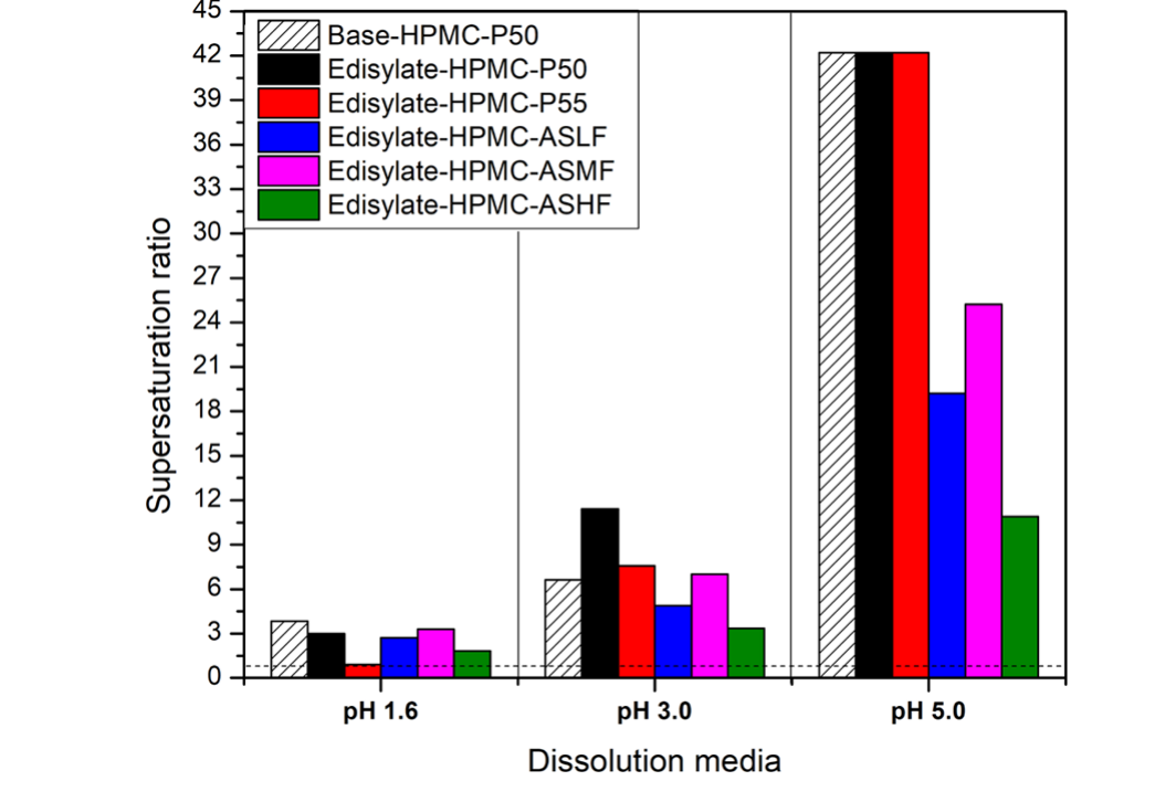
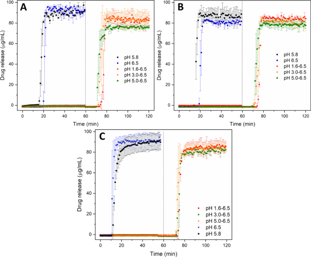
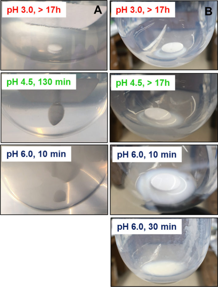
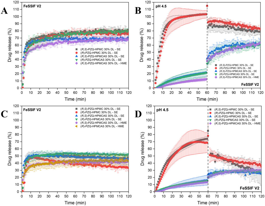
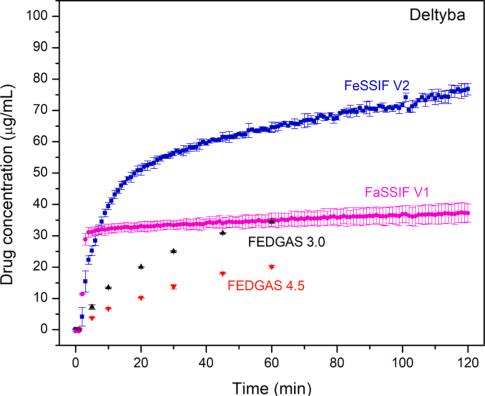
- In gastric fluid, where HPMCAS stays undissolved and the solution is subsaturated, "Solution-phase crystallization cannot occur", so crystals form in the matrix (F24) elkhabaz2019a C34.
Evidence: data, conditions and papers
Finding F1. An amorphous drug can lose its supersaturation during dissolution by crystallizing in the wetted solid or from the solution, and which route acts depends on the drug, the temperature, and which polymer is present and where it sits
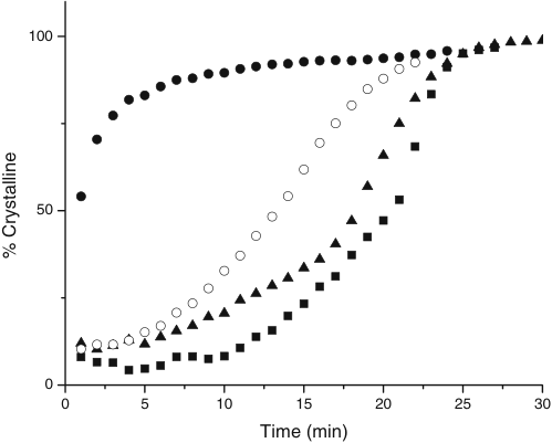
In short. Neat amorphous felodipine slurried in pH 6.8 buffer at 37 °C was ≈54% crystalline at the first Raman reading (1 min) and dissolved no higher than the crystal (≈0.9–1.1 µg/mL); at 25 °C it took about 8 min to reach 50% crystallinity and its solution peaked at ≈2.45 µg/mL alonzo2010 R2, R1. Amorphous indomethacin at 25 °C stayed amorphous for at least 4 h while needles grew in the liquid and the concentration fell from ≈14.9 to ≈2.7 µg/mL alonzo2010 R11, R19, R7. Neat amorphous nifedipine crystallized inside the particles within minutes, with or without polymer in the buffer, whereas in 50:50 dispersions the particles stayed non-birefringent and crystals below 2 µm formed in the solution raina2014b R2, R6, R13. Amorphous celecoxib behaved like nifedipine: it was crystalline in a slurry within an hour, mostly on the particles, and dissolved no higher than the crystal (≈1.70 against ≈1.85 µg/mL at 16 h) xie2015 R4, R8, R7; a 25% resveratrol–HPMCAS dispersion crystallized about equally at pH 6.8 and at pH 1.2, where the polymer does not dissolve, with crystals inside intact particles at pH 1.2 wegiel2015 R17, R18. The authors conclude that the rates of these two transformations relative to dissolution "in large part shape" the concentration–time profile alonzo2010 C32, C47.
Type. observation in 7 drugs (felodipine, indomethacin, nifedipine, celecoxib, resveratrol, posaconazole, delamanid) Strength. to strong for the routes, which in situ Raman and polarized light separate directly for felodipine at 37 °C, indomethacin at 25 °C, neat nifedipine and neat celecoxib, and SHG for posaconazole dispersions in gastric fluid alonzo2010 K1: strong, K5: moderate to strong; raina2014b K1: strong; xie2015 K2: moderate; elkhabaz2019a K2: moderate to strong; weaker where the route was inferred rather than seen (felodipine at 25 °C and with dissolved polymer, the nifedipine dispersions, resveratrol at pH 6.8, where only the similarity to pH 1.2 points to the matrix) alonzo2010 X6, X7; raina2014b K5: moderate, X10; wegiel2015 K6: weak to moderate.
How it was shown
Alonzo and co-workers (2010) followed slurries of melt-quenched felodipine (pH 6.8, 25 and 37 °C) and indomethacin (pH 2) by in situ Raman spectroscopy and polarized light, beside non-sink dissolution with fibre-optic UV alonzo2010 P1, P2, P3, P5. Raina and co-workers (2014) did so for nifedipine glass and 50:50 dispersions, Xie and Taylor (2015) for neat amorphous celecoxib, and Wegiel and co-workers (2015) a 25% resveratrol–HPMCAS dispersion at pH 6.8 and at pH 1.2, where HPMCAS does not dissolve raina2014b P6, P4; xie2015 P3, P7; wegiel2015 P7, A3.
What the data show
Felodipine at 37 °C, nifedipine glass and neat celecoxib crystallized in the wetted solid within minutes, while indomethacin at 25 °C stayed amorphous as needles grew in the liquid alonzo2010 R2, R11, R19; raina2014b R6; xie2015 R8.
| drug and form | conditions | solid | solution | ids |
|---|---|---|---|---|
| felodipine, neat amorphous | pH 6.8, 37 °C | ≈54% crystalline at 1 min | plateau ≈0.9–1.1 µg/mL, like the crystal | alonzo2010 R2, R1 |
| felodipine, neat amorphous | pH 6.8, 25 °C | ≈46% crystalline at 8 min | peak ≈2.45 µg/mL at ≈16 min | alonzo2010 R2, R1 |
| indomethacin, neat amorphous | pH 2, 25 °C | amorphous band kept at 120–240 min; needles in the liquid within 60 min | ≈14.9 → ≈2.7 µg/mL | alonzo2010 R11, R19, R7 |
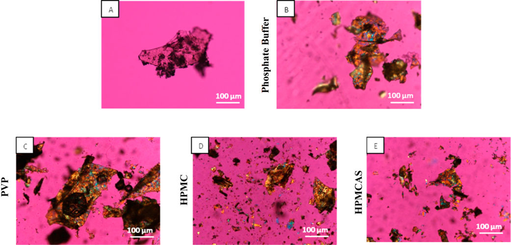
| nifedipine glass; 50:50 PVP dispersion | pH 6.8, 25 °C | glass fraction zero from 6 min; dispersion particles not birefringent | crystals below 2 µm in the solution around the dispersion | raina2014b R6, R13 |
| celecoxib, neat amorphous | pH 6.8, 37 °C | crystallization complete within an hour | ≈1.70 against ≈1.85 µg/mL for the crystal at 16 h | xie2015 R4, R7 |
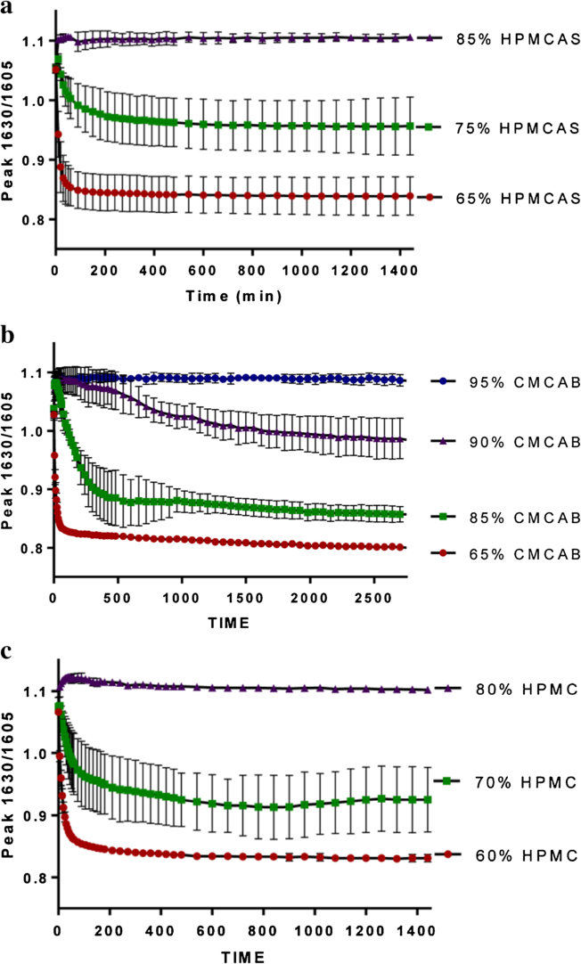
| resveratrol–HPMCAS 25:75 | pH 6.8 and pH 1.2, 37 °C | Raman ratio ≈0.96 and ≈0.93 (crystal 0.76); crystals inside intact particles at pH 1.2 | not measured | wegiel2015 R17, R18, R8 |
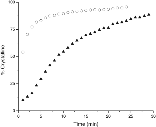
At 37 °C the slurry is above 50% crystalline at the first reading, at 25 °C only after about 8 min alonzo2010 R2.
Why
For felodipine at 37 °C the amorphous solid "crystallizes more rapidly than it can dissolve", which the authors explain by the glass transition: the hydrated solid at 37 °C is above Tg and "presumably" crystallizes faster alonzo2010 C8, C33, C34. For indomethacin at 25 °C crystallization from solution is the limiting factor alonzo2010 C40. In the nifedipine dispersions the polymer keeps the hydrated matrix amorphous, so the route "has changed from the matrix to the solution phase" raina2014b C21, C24.
Limits and transfer
The slurries held about 250–375 mg/mL felodipine against 500 µg/mL in dissolution, so their timings rank systems rather than give the time scale in a dissolution vessel alonzo2010 X5; raina2014b X9. Wet the neat amorphous drug with the medium at 37 °C and follow the solid for 15–30 min beside a non-sink test: a solid that crystallizes within minutes needs the polymer in the dispersion, and an amorphous solid with a falling concentration needs a solution inhibitor alonzo2010 P5, C33, C40.
How it was shown
Alonzo and co-workers (2010) asked which crystallization route limits the concentration reached from an amorphous drug alonzo2010 C5. They melt-quenched felodipine and indomethacin, cryomilled them and kept the 100–300 µm fraction alonzo2010 P1. Felodipine was dissolved at 100, 500 or 1,000 µg solid per mL in 20 mL of 50 mM pH 6.8 phosphate buffer at 37 °C, and at 500 µg/mL at 25 °C, with fibre-optic UV every minute at 500 rpm; indomethacin at 300 µg/mL in 50 mL of 50 mM pH 2 phosphate buffer at 25 and 37 °C alonzo2010 P2, P3. Slurries of about 500–750 mg solid in 2 mL (felodipine) or 5 mL (indomethacin) of the same media were followed by in situ Raman spectroscopy, with a PLS model for felodipine crystallinity and, for indomethacin, the α band at 1,648 cm−1 and an amorphous-only band at 753 cm−1; wetted indomethacin was watched under polarized light alonzo2010 P5, P6, P7, P9. Raina and co-workers (2014) slurried about 500 mg of melt-quenched nifedipine in 2 mL of 50 mM pH 6.8 phosphate buffer, with or without 1 mg/mL PVP, HPMC or HPMCAS dissolved in it, or 1 g of 50:50 dispersions in 4 mL, at 25 and 37 °C; Raman spectra were taken every minute for 15 min and every 5 min up to 3 h and split into glass, γ and α by multivariate curve resolution (MCR), and particles were watched under crossed polarizers raina2014b P6, P7, P10, P4. A dissolution test of the neat glass and of the HPMC dispersion at 5 mg/mL solids and 37 °C gave the solution concentration raina2014b P11. Xie and Taylor (2015) melt-quenched celecoxib, sieved it to 106–250 µm, wetted particles with 3–4 drops of pH 6.8, 100 mM phosphate buffer, with or without 5 µg/mL HPMC or HPMCAS, under polarized light, slurried it in the same buffer at 37 °C with in situ Raman, and dissolved 8.8 mg in 400 mL at 37 °C and 300 rpm with in situ UV for 16 h, in triplicate, against the same mass of crystal xie2015 P2, P7, P3, P5. Wegiel and co-workers (2015) slurried about 300–500 mg of a 25% resveratrol–HPMCAS dispersion in 5 mL of pH 6.8 phosphate buffer, where HPMCAS dissolves, and of pH 1.2 HCl solution, where it does not, at 37 °C, followed the 1630/1605 cm−1 Raman ratio (0.76 for the crystal, about 1.1 for the amorphous dispersions; n = 3) and imaged the particles under polarized light after 24 h wegiel2015 P7, A3, R8, P9.
What the data show
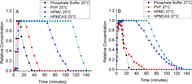
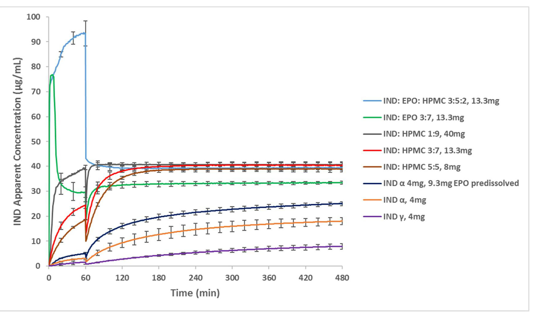
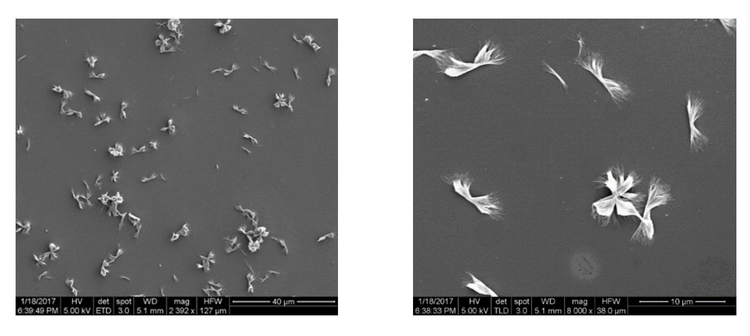
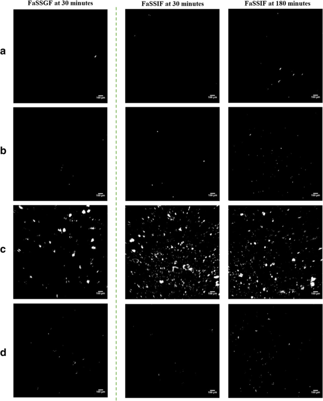
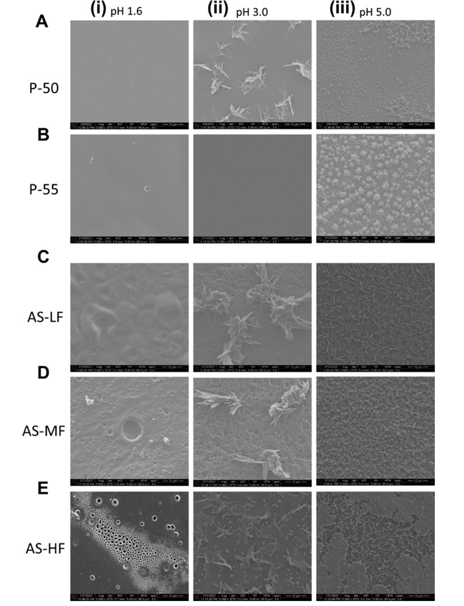
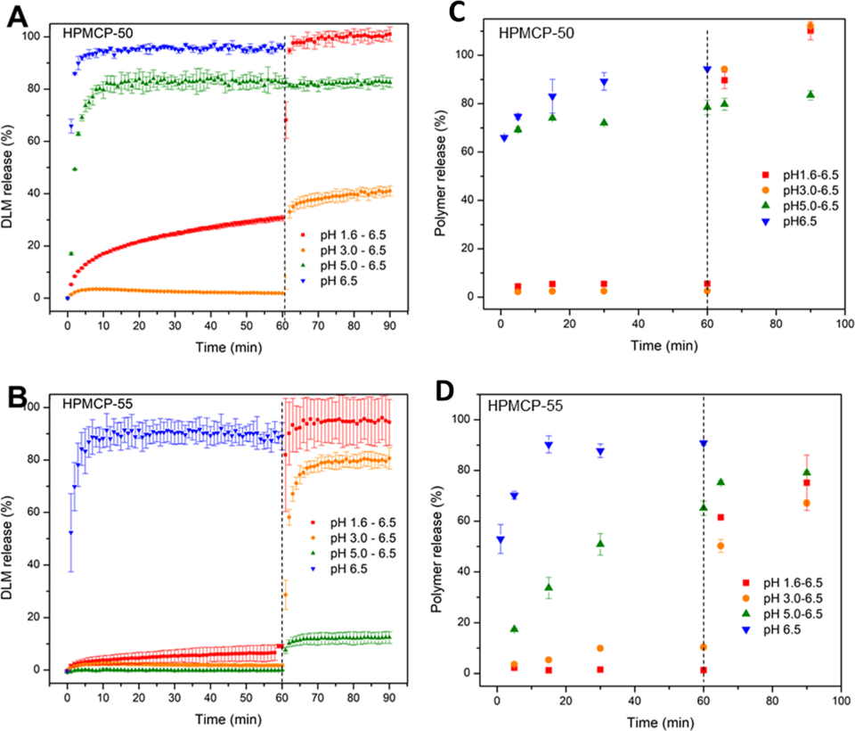
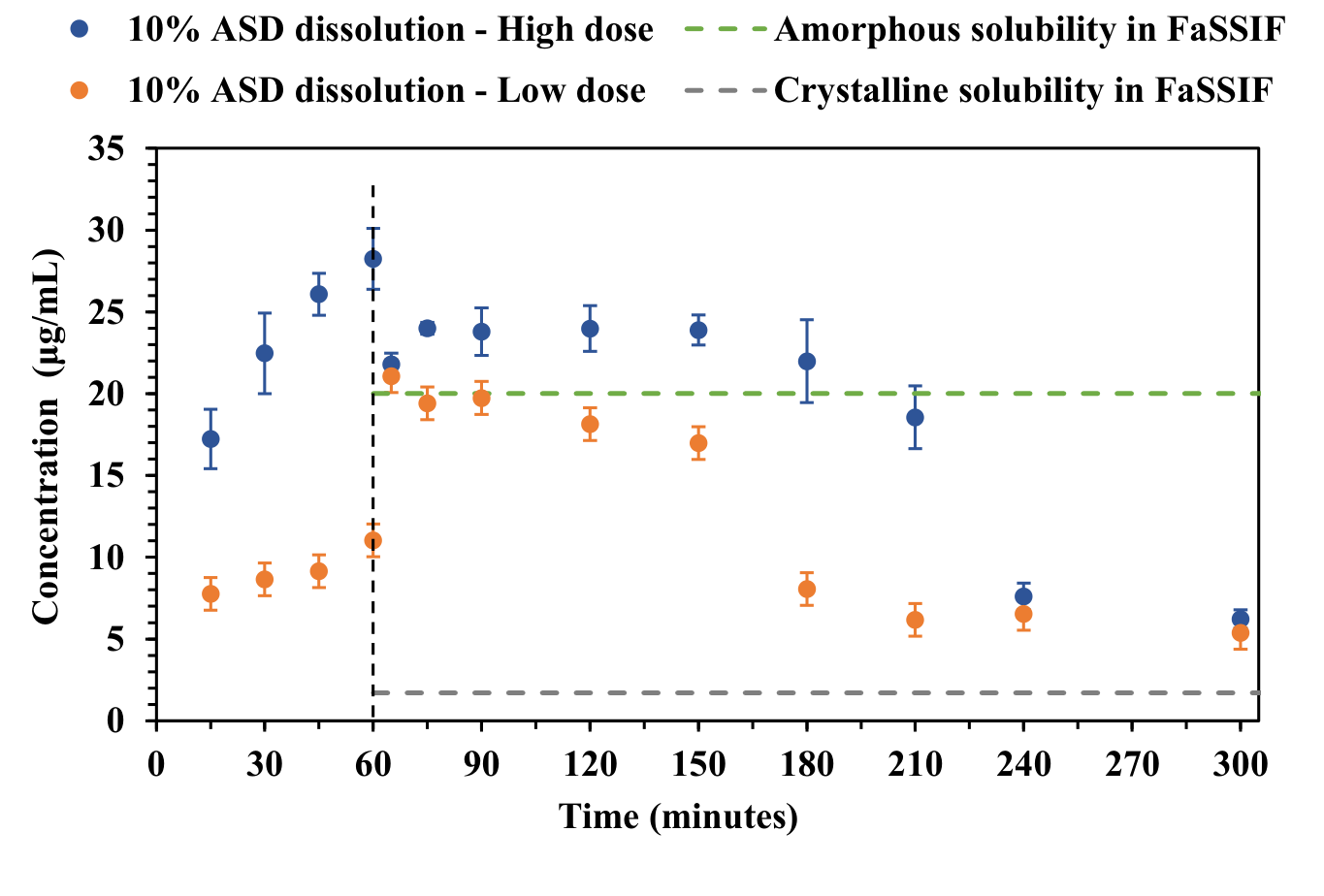
Felodipine at 37 °C. The slurry was ≈54, ≈83 and ≈96% crystalline at 1, 5 and 24 min alonzo2010 R2. 100 µg/mL of amorphous solid followed almost the curve of the same mass of crystal (≈0.85 against ≈1.0 µg/mL near 120 min), and 500 and 1,000 µg/mL dissolved faster but plateaued at only ≈1.1 and ≈0.95–1.0 µg/mL, from ≈30 and ≈10 min alonzo2010 R1. Felodipine at 25 °C. The slurry was ≈10% crystalline at 1 min, ≈46% at 8 min and ≈89% at 29 min, and the solution peaked at ≈2.45 µg/mL at ≈16 min before falling to ≈0.8 µg/mL at 90 min alonzo2010 R2, R1. Indomethacin at 25 °C. Spectra after 120–240 min kept the amorphous band; dendritic needles appeared in the liquid between the particles within 60 min while the particles themselves did not crystallize; the solution peaked at ≈14.9 µg/mL at ≈25 min and was ≈2.7 µg/mL at 130 min alonzo2010 R11, R19, R7. Indomethacin at 37 °C. The slurry was largely amorphous at 20 min, clearly α at 90 min and like pure α at 120 min; the solution peaked at ≈11.2 µg/mL at ≈30 min, near the onset of conversion, and the powder agglomerated into a few large particles alonzo2010 R12, R13, R7, R10. Nifedipine. Particles were covered with birefringent crystals 15 min after wetting at 25 °C, in buffer and with each dissolved polymer; by MCR the glass fraction was ≈0.95, ≈0.61 and ≈0.35 at 3, 4 and 5 min and zero from 6 min; the neat glass gave ≈6 µg/mL in the dissolution test, on the crystalline-solubility line raina2014b R2, R6, R14. In the 50:50 PVP dispersion the particles shrank without turning birefringent and small plate-shaped crystals appeared in the surrounding solution within minutes; the authors state the same for the HPMC and HPMCAS dispersions without images raina2014b R13, X18. The HPMC dispersion reached ≈100 µg/mL at ≈22 min and fell to ≈41 µg/mL by 150 min raina2014b R14. Celecoxib. Wetted particles showed birefringent spots after 2 min and were widely crystalline at 10 min, with needles also in the surrounding liquid; in the slurry crystallization was complete within an hour xie2015 R8, R4. The amorphous solid dissolved faster at first (≈0.49 against ≈0.13 µg/mL at 20 min) but then followed the crystal, ending at ≈1.70 against ≈1.85 µg/mL at 16 h xie2015 R7. Resveratrol. The Raman ratio of the 25% HPMCAS dispersion fell from ≈1.05 to ≈0.96 at pH 6.8 and from ≈1.05–1.08 to ≈0.93 at pH 1.2 over about 1440 min, the curves overlapping within their error bars; at pH 1.2 the particles (≈200–300 µm) stayed intact with birefringent regions inside, at pH 6.8 they dispersed and left small crystalline particles wegiel2015 R17, R18. Posaconazole and delamanid in acid. Posaconazole–HPMCAS dispersions crystallized within 30 min in FaSSGF although the medium stayed below the crystalline solubility, at about 68% of the undissolved drug at 50% drug, and delamanid dispersions crystallized at their surfaces at pH 3.0 and 5.0 while the drug concentration in the acid hardly fell (F24) elkhabaz2019a R16, R20, R1; nguyen2023c R19, R23.
| paper | system | method and conditions | result | ids |
|---|---|---|---|---|
| alonzo2010 | felodipine, neat amorphous, pH 6.8 | Raman slurry and dissolution (100–1,000 µg/mL), 37 °C | ≈54% crystalline at 1 min; plateau ≈0.9–1.1 µg/mL, like the crystal | R2, R1 |
| alonzo2010 | felodipine, 25 °C | same, 500 µg/mL | 50% crystalline after about 8 min; peak ≈2.45 µg/mL at ≈16 min | R2, R1 |
| alonzo2010 | indomethacin, pH 2, 25 °C | Raman, polarized light, dissolution 300 µg/mL | solid amorphous for at least 4 h; needles in the liquid by 60 min; ≈14.9 → ≈2.7 µg/mL | R11, R19, R7 |
| alonzo2010 | indomethacin, 37 °C | same | α by 90 min; peak ≈11.2 µg/mL near the onset of conversion | R12, R13, R7 |
| raina2014b | nifedipine glass, buffer ± 1 mg/mL PVP, HPMC, HPMCAS | polarized light, Raman–MCR, 25 °C | birefringent at 15 min in every medium; glass gone by ≈6 min | R2, R6 |
| raina2014b | nifedipine–PVP 50:50 | polarized light | particles stay dark; crystals below 2 µm form in the solution | R13 |
| raina2014b | neat glass; nifedipine–HPMC 50:50 | dissolution 5 mg/mL, 37 °C | ≈6 µg/mL; peak ≈100 µg/mL, ≈41 µg/mL at 150 min | R14 |
| ilevbare2013c (Crystallization from solution) | amorphous ritonavir, efavirenz, celecoxib | dissolution, pH 6.8, 37 °C | 19.8 of 20.6, 12.3 of 19.8 and 2.5 of 22.6 µg/mL calculated | R2 |
| chen2015a | celecoxib–HPMCAS spray-dried particles, pH 3, Triton X100 | suspension, polarized light, 7 days | birefringence on the particles, no free needles | R20 |
| xie2015 | celecoxib, neat amorphous, pH 6.8 | polarized light; Raman slurry, 37 °C | spots at 2 min, widely crystalline at 10 min; complete within an hour | R8, R4 |
| xie2015 | same; crystal | dissolution 8.8 mg in 400 mL, 37 °C, 16 h | ≈1.70 against ≈1.85 µg/mL | R7 |
| wegiel2015 | resveratrol–HPMCAS 25:75, pH 6.8 and pH 1.2 | Raman ratio, polarized light, 37 °C, 24 h | ≈0.96 and ≈0.93 (crystal 0.76); crystals inside intact particles at pH 1.2 | R17, R18, R8 |
| ilevbare2013c (Crystallization from solution) | amorphous celecoxib; efavirenz | polarized light, pH 6.8 | celecoxib birefringent at ≈1 min; efavirenz shrank without birefringence for 3 h | R4, R5 |
| elkhabaz2019a | posaconazole–HPMCAS 10 / 25 / 50% and Noxafil powder in FaSSGF, solution subsaturated | SHG and TPE-UVF after 30 min, 37 °C | crystals in every system; estimated matrix crystallization ≈0.026 / ≈0.050 / ≈68 / ≈0.047% | R16, R20 |
| nguyen2023c | delamanid salt dispersions, five enteric grades, 1 h at pH 1.6–5.0 | PLM and SEM of films and powders; concentration in acid; PVPVA in the medium | crystals on the dispersion surfaces; concentration held and PVPVA changed nothing | R19, R23, R22 |
Figure
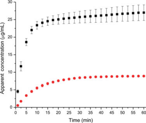
At 37 °C (open circles) the slurry is already above 50% crystalline at the first reading; at 25 °C (triangles) 50% takes about 8 min, the window in which some supersaturation could form alonzo2010 R2.
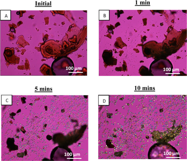
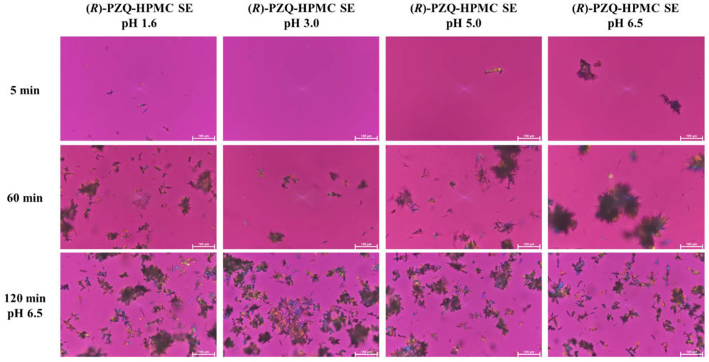
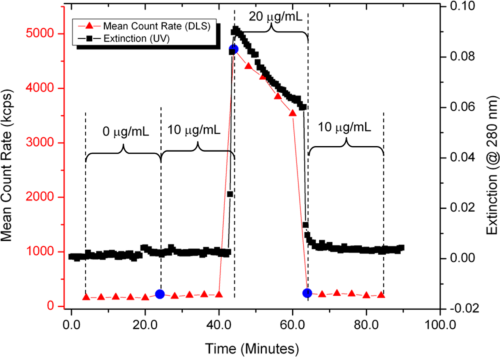
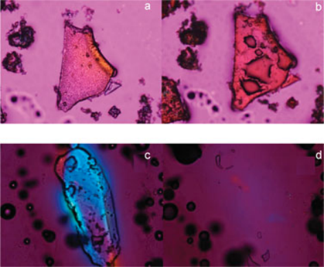
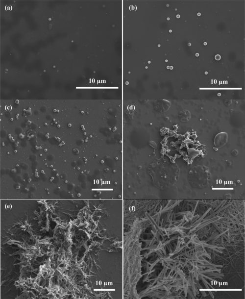
The dispersion particles shrink without birefringence while fine crystals multiply in the liquid around them between 1 and 10 min raina2014b R13.
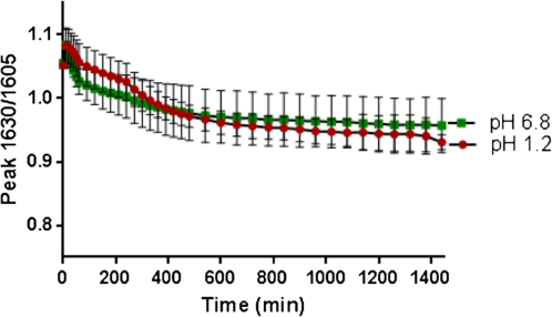
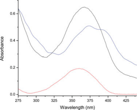
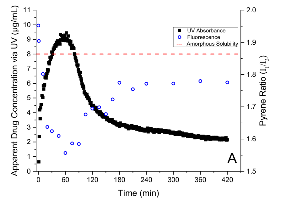
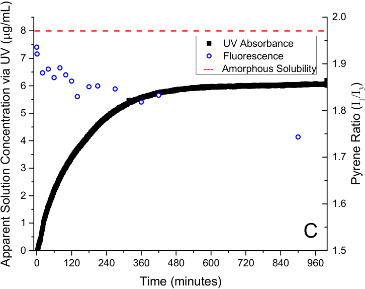
The two curves overlap within their error bars although HPMCAS dissolves only at pH 6.8: the crystals form in or at the matrix whether or not the polymer leaves it wegiel2015 R17, C15.
Why
For felodipine at 37 °C the authors state that the amorphous solid "crystallizes more rapidly than it can dissolve", so that the crystal is what dissolves and no amorphous advantage appears alonzo2010 C8, C33. They explain the difference between 25 and 37 °C by the glass transition: dry felodipine has a Tg of about 45 °C and just below 35 °C after exposure to 75% RH (cited), so the hydrated solid at 37 °C is above Tg and "presumably" crystallizes faster alonzo2010 C34, M1. Indomethacin crystallized much more slowly although its Tg also falls with water and its driving force is larger, which they take as "an indication" that a single parameter such as Tg "will not fully explain" the behaviour alonzo2010 C42. For the falling indomethacin concentration at 25 °C they point to the needles in the liquid: crystallization from solution is the limiting factor there ("These observations indicate") alonzo2010 C31, C40. At 37 °C they combine agglomeration (less surface), conversion of the solid to α and weaker solution inhibition ("Presumably") alonzo2010 C41, C21. Fig. 16 of the paper sets the competition out as a scheme in which dissolution scales with surface area and a concentration difference and nucleation and growth rise with supersaturation alonzo2010 C43, E3, E4, E5. For nifedipine, a relatively fast crystallizer, Raina and co-workers find the matrix route "primarily" for the neat glass and argue that its inherent faster crystallization "appears" to carry over from earlier solid-state work raina2014b C10, C28, R17; in the dispersions the polymer keeps the hydrated matrix amorphous, so the route "has changed from the matrix to the solution phase" ("strongly suggest") raina2014b C21, C24. An alternative reading for felodipine at 25 °C is that the fall after the peak comes partly from loss of the dissolving amorphous phase, since the slurry was ≈70% crystalline at 15 min; the routes were not separated there alonzo2010 X6. For celecoxib, Xie and Taylor conclude that the neat amorphous drug "appears to undergo rapid crystallization", "primarily via the matrix route", which they relate to water lowering the Tg (not measured); the low concentration "indicates either" rapid complete matrix crystallization or rapid growth of seed crystals from the residual amorphous material xie2015 C26, C1, C32, C34. For resveratrol, a rapid crystallizer in the cited classification, Wegiel and co-workers infer from the alike kinetics at pH 6.8 and 1.2 that the route "may also be analogous", the crystals at pH 6.8 "most likely" forming from the matrix rather than from a supersaturated solution wegiel2015 R22, C14, C15; crystallization "appeared to occur in or at the surface of the matrix" wegiel2015 C16. (synthesis: in all five drugs the outcome follows from one comparison, how fast the wetted solid crystallizes against how fast it dissolves; the temperature effect for felodipine and the polymer placement for nifedipine both act through that comparison, and which polymer forms the dispersion decides the solid side of it, see F16.) For posaconazole in gastric fluid the authors argue that "Solution-phase crystallization cannot occur" in a subsaturated medium, so the crystals formed in the matrix elkhabaz2019a C34, and for delamanid the solution route was ruled out by a constant concentration in the acid and an ineffective solution inhibitor nguyen2023c C37, C39.
Conditions and limits
Neat melt-quenched powders and 50:50 dispersions; felodipine and nifedipine in pH 6.8 buffer, indomethacin in pH 2 buffer; 25 and 37 °C; up to 4 h alonzo2010 A1, A2; raina2014b A2, A3. The Raman slurries held about 250–375 mg/mL felodipine against 500 µg/mL in dissolution, so the polymer-to-solid ratio differed about 500–750-fold, and the nifedipine slurries held 125–250 mg/mL drug; slurry timings therefore rank systems rather than give the time scale in a dissolution vessel alonzo2010 X5; raina2014b X9. Replicates are not stated for any slurry run alonzo2010 X8; raina2014b X14. The two drugs of Alonzo were tested in different media and set-ups, so their comparison is qualitative alonzo2010 X9. The celecoxib micrographs do not always show one field, and the amount of neat amorphous celecoxib slurried is not stated xie2015 X13, X11; the resveratrol Raman ratio is uncalibrated, and the pH comparison has no statistical test wegiel2015 X13, C14.
Counterexamples and conflicts
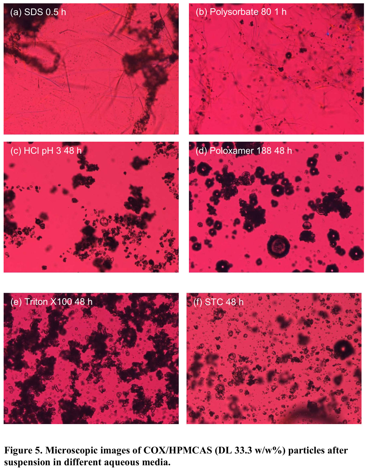
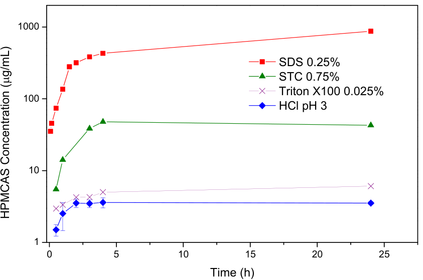
Not every neat amorphous drug loses its advantage on wetting: in the crystallization-from-solution chapter amorphous ritonavir dissolved to 19.8 of a calculated 20.6 µg/mL, while celecoxib reached only 2.5 of 22.6 µg/mL, and amorphous efavirenz dissolved slowly without birefringence for 3 h ilevbare2013c R2, R5, C23, and in the LLPS chapter amorphous films of three weak bases reached 69–110% of their LLPS concentration (Amorphous solubility and LLPS F13). In acidified suspensions of celecoxib–HPMCAS particles, where the polymer barely dissolves, the particles stayed crystal-free for 48 h without surfactant and crystallized on or in the particles only after about 7 days chen2015a R14, R20, C33. No paper in the library contradicts the two-route frame itself.
Transfer check
Wet the neat amorphous drug with the dissolution medium at 37 °C and follow the solid (Raman, PXRD or polarized light) during the first 15–30 min while the concentration is read in a non-sink test alonzo2010 P5, P8; raina2014b P4. Above about 50% crystallinity within minutes, as for felodipine at 37 °C and nifedipine, the neat glass dissolves like the crystal and the polymer has to be in the solid; if the solid stays amorphous while the concentration falls, as for indomethacin at 25 °C, crystallization from solution is the problem and a solution inhibitor is the lever alonzo2010 C33, C40. Test at 37 °C: felodipine supersaturated at 25 °C but not at 37 °C alonzo2010 C6. For a dispersion of an enteric polymer, a slurry at a pH where the polymer stays undissolved (pH 1.2 for HPMCAS) shows matrix crystallization without the solution route wegiel2015 A3, C16.
History
The two ways in which an amorphous form loses its advantage were proposed in cited work alonzo2010 C2. Alonzo and co-workers (2010) were the first in the library to separate them in situ, for felodipine and indomethacin alonzo2010 C47. Raina and co-workers (2014) added nifedipine, the switch from the matrix route of the neat glass to the solution route of the dispersion, and a metastable crystal form on both routes (F3) raina2014b C21, C29. Wegiel and co-workers (2015) added resveratrol and used an undissolving medium to show the matrix route in a dispersion wegiel2015 C16; Xie and Taylor (2015) added celecoxib, whose neat glass crystallized mainly on its particles xie2015 C1. The title was widened from "where the polymer sits" to "which polymer is present and where it sits" when the papers of 2015–2018 showed that the dispersion polymer itself decides whether the wetted matrix crystallizes (F16). Later papers of the library cite Alonzo 2010 for matrix against solution crystallization during dissolution, among them work on celecoxib dispersions, on ezetimibe and posaconazole dispersions and on residual crystallinity (builds_on, as listed in the evidence index of this subcategory). Elkhabaz and co-workers (2019) and Nguyen and co-workers (2023) found the matrix route in gastric media, where an enteric polymer stays undissolved and the solution cannot be the source (F24) elkhabaz2019a C23; nguyen2023c C43.