Topic 1Phase that forms1 of 4
Past a certain concentration, a supersaturated drug solution splits into drug-rich droplets before any crystal appears.
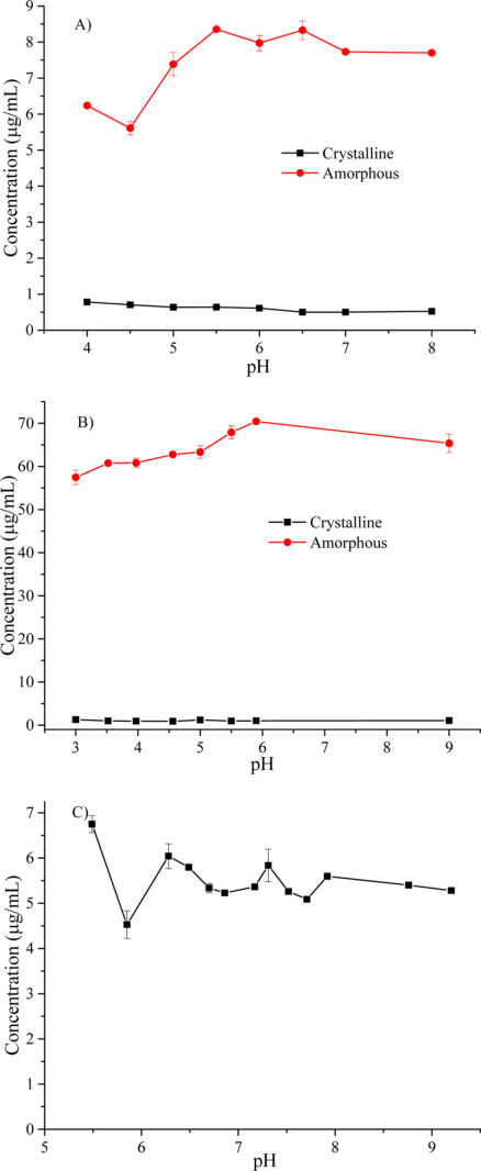
Add a drug from an organic stock solution to water until it is far above its crystal solubility, and at a drug-specific concentration the clear solution turns cloudy. The turbidity is not caused by crystals: the new phase takes up polarity probes, shows no birefringence under polarized light and gave only a broad halo by X-ray diffraction. This was seen for 21 compounds ilevbare2013a C2, R8; almeidaesousa2015 C41; indulkar2015 C6, R19, R20, R21; mosqueragiraldo2015 C21.
strongStrong for the phenomenon, because four kinds of read-out (light scattering, dyes, microscopy or X-ray diffraction, DSC) agree; but the phase itself was isolated and examined for only five drugs ilevbare2013a K1; mosqueragiraldo2015 K1; indulkar2015 K5; almeidaesousa2015 K1, C38.
Onsets measured in different media and at 25 or 37 °C, so the drugs are not strictly comparable; griseofulvin was measured with 100 µg/mL HPMCAS-HF. ilevbare2013a R8; almeidaesousa2015 R4, R5, R6
For your drug
- Raise the concentration slowly in your medium while recording extinction at a wavelength where the drug does not absorb, then repeat with a polarity probe or the drug's own fluorescence.
- Isolate the pellet by ultracentrifugation and check birefringence, XRPD and a DSC Tg. Extinction rise plus probe change plus a halo-only pellet means a drug-rich phase; an extinction rise without a probe change and with birefringent particles means crystallization.
almeidaesousa2015 P1, P2, R4; indulkar2015 P14, P15, P16; ilevbare2013a P7, P9
How it works and key numbers
- The supersaturation is made mainly by a solvent shift, which leaves about 1–2% v/v organic solvent in the medium almeidaesousa2015 X9; mosqueragiraldo2015 X7.
- Ilevbare and Taylor read the turbidity as liquid–liquid phase separation: the solution and the droplets are in equilibrium, so the drug has the same activity in both ilevbare2013a C29, C36.
- A polarity-sensitive dye partitions into the disordered drug-rich phase and changes its emission, whereas crystallization leaves the dye in solution; extinction alone cannot tell the two apart almeidaesousa2015 C12, C16.
- Griseofulvin without polymer is the exception: extinction rose near 150 µg/mL with no probe change and small solid particles, which means crystallization almeidaesousa2015 R4.
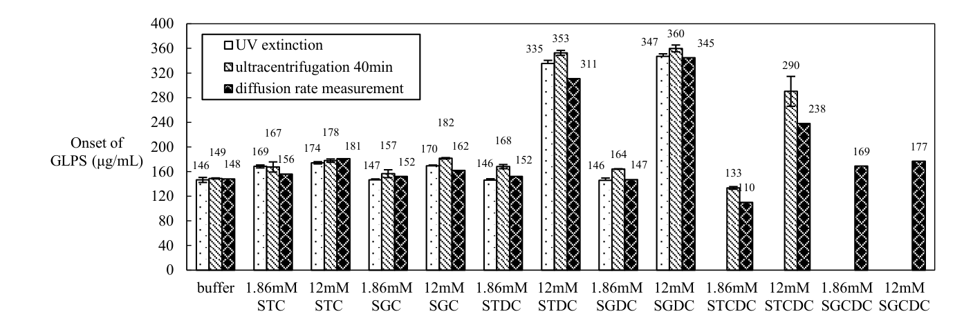
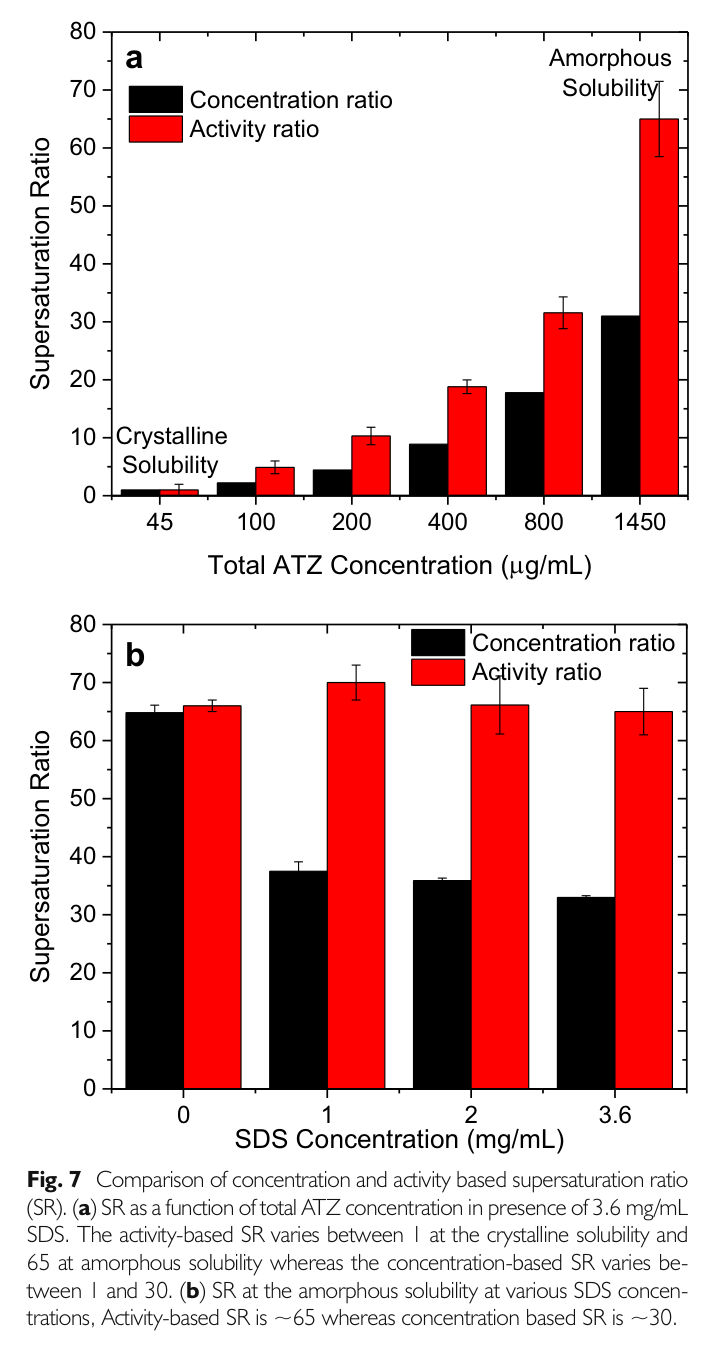
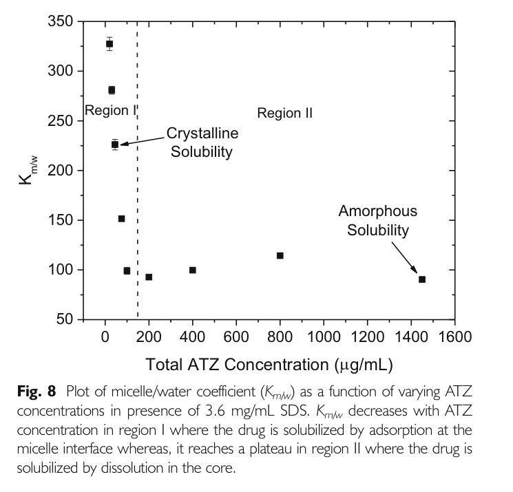
- A caution: in 3.6 mg/mL SDS the 369 nm band appeared from about 200 µg/mL, far below the amorphous solubility of about 1450 µg/mL, so it did not mark a drug-rich phase there indulkar2017a R17, R18, C27.
Evidence: data, conditions and papers
Finding F1. Highly supersaturated solutions of poorly soluble drugs split into a non-crystalline drug-rich phase before crystals appear
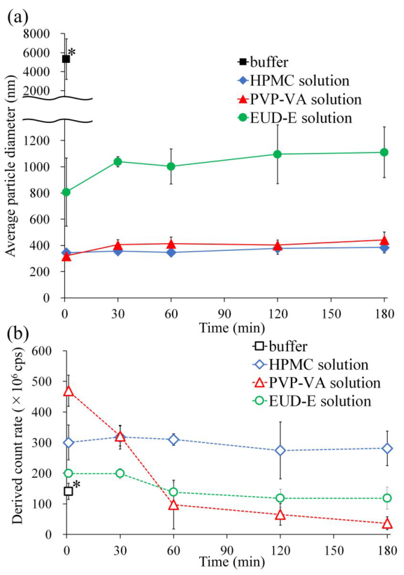
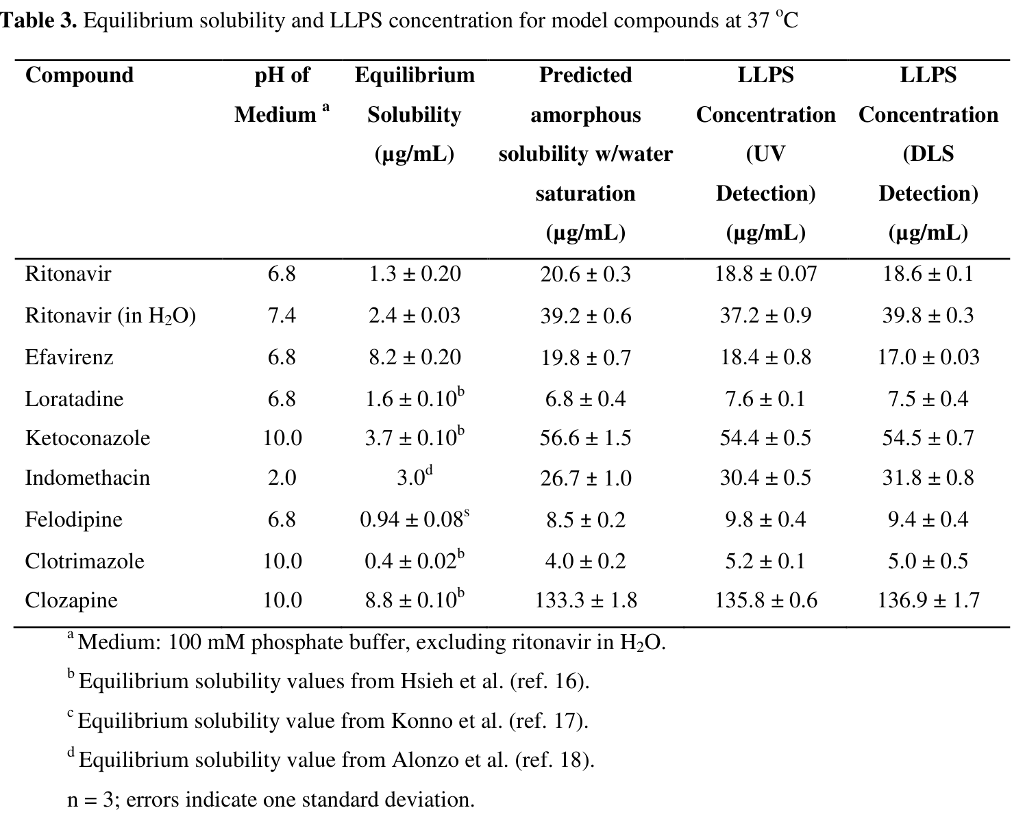
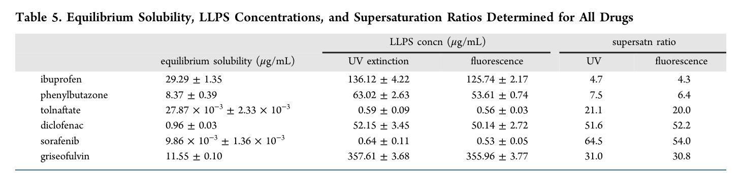
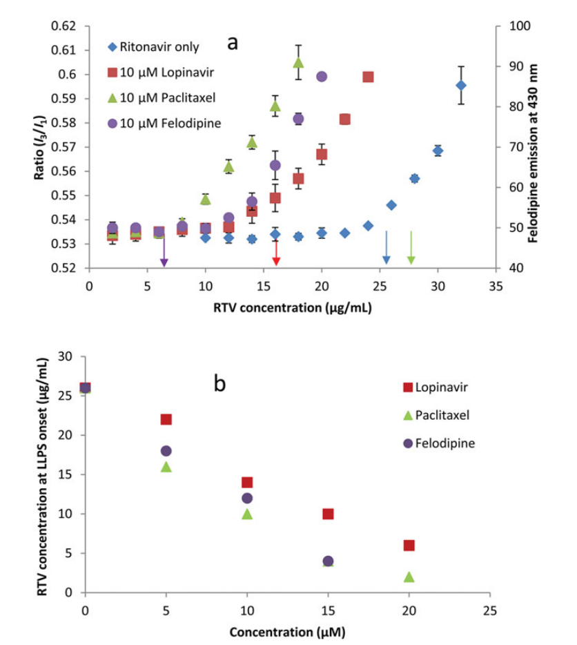
In short. Past a drug-specific concentration the solutions of 21 poorly water-soluble compounds in these twelve papers turned turbid, took up polarity probes and, for the five drugs whose phase was isolated or examined (ritonavir, clotrimazole, atazanavir, nicardipine, telaprevir), gave an amorphous pellet or precipitate, not crystals (synthesis: 8 + 6 + 2 + 1 + 2 + 1 + 1 = 21 distinct compounds, because clotrimazole, ritonavir and felodipine recur and the later atazanavir, telaprevir and ibuprofen studies repeat compounds already counted; the added compound is pazopanib) ilevbare2013a C2, R8; almeidaesousa2015 C41; indulkar2015 C6; mosqueragiraldo2015 C21; trasi2015a C12; almeidaesousa2016 C17. The ritonavir phase held about 95% drug and 4–5% water and grew from about 345 to about 842 nm in 64 min ilevbare2013a R6, R3. Onsets ran from 0.53 µg/mL (sorafenib) to 355.96 µg/mL (griseofulvin, with polymer) almeidaesousa2015 R5, R6. Griseofulvin without polymer was the exception: it crystallized before any probe signal almeidaesousa2015 R4. The 2017–2022 papers add pazopanib, whose HCl salt shifted from pH 1.2 to pH 6.5 gave a milky, bluish solution at once with non-birefringent aggregates and particles of 496 nm (145 nm with HPMC) sugihara2018 R11, R14, R15, and ibuprofen, whose drug-rich droplets measured 3182–5313 nm in buffer and 268–345 nm with HPMC or PVP-VA ueda2020b R5.
Type. observation in 21 compounds Strength. for the phenomenon: four kinds of read-out (light scattering, polarity probes, microscopy or XRPD, DSC) point the same way ilevbare2013a K1: moderate to strong; mosqueragiraldo2015 K1: strong; indulkar2015 K5: moderate to strong; almeidaesousa2015 K1: moderate. The phase itself was isolated and examined for five drugs only, and "disordered" elsewhere rests on probe responses almeidaesousa2015 C38. The pazopanib precipitate was examined by DSC after drying, but the crystalline or amorphous nature of its solids was assigned by appearance under polarized light sugihara2018 R17, X6.
How it was shown
Ilevbare and Taylor (2013) pumped a methanol stock into medium at 37 °C and followed extinction for eight drugs; for ritonavir they added DLS and a pyrene titration and isolated the phase by ultracentrifugation ilevbare2013a P5, P7, P8, P9, P10, P11. Almeida e Sousa and co-workers (2015) added a polarity probe for six fast crystallizers almeidaesousa2015 P1, P2; Indulkar and co-workers (2015) used XRPD, microscopy and DSC for three weak bases indulkar2015 P8, P14, P15, P16.
What the data show
Past a drug-specific concentration the solutions turned turbid, took up the probes and gave non-birefringent droplets; where the phase was isolated it was an amorphous pellet. Onsets ran from 0.53 µg/mL (sorafenib) to 355.96 µg/mL (griseofulvin, with polymer) almeidaesousa2015 R5, R6. Griseofulvin without polymer was the exception: it crystallized before any probe signal almeidaesousa2015 R4.
| system | method | result | ids |
|---|---|---|---|
| ritonavir, water, 37 °C | extinction, DLS, pyrene | non-birefringent droplets; onset 37.2 (UV), 39.8 (DLS), 39.0 (pyrene) µg/mL | ilevbare2013a R1, R2, R4, R8 |
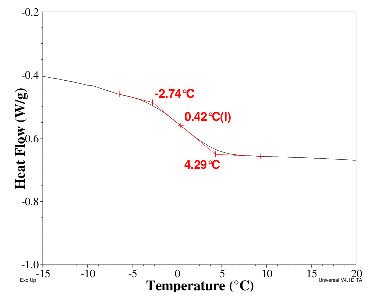
| ritonavir, isolated phase | ultracentrifuge, 10–50 °C; DSC | 94.85–95.97% drug, 4.03–5.15% water; Tg 0.4 °C | ilevbare2013a R5, R6 |
| six fast crystallizers, 25 °C | extinction plus probe | both change at about the same concentration; turbidity lasts minutes | almeidaesousa2015 R1, R3, R5 |
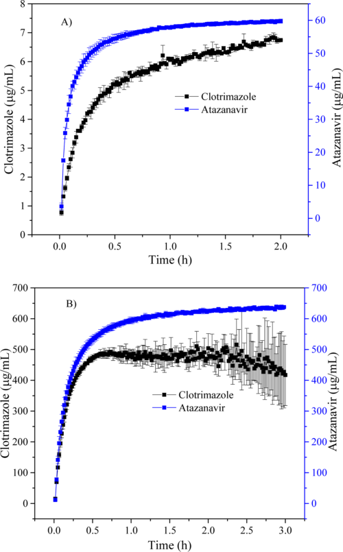
| clotrimazole, atazanavir, nicardipine | XRPD, PLM, DLS, Nile red | halo-only XRPD, no birefringence; 116–387 nm droplets | indulkar2015 R14, R16, R19, R20, R21 |
| pazopanib HCl, pH 1.2 to 6.5 | eye, DLS, NTA, PLM, DSC | milky at once; 496 nm (145 nm with HPMC); dried precipitate Tg about 83 °C | sugihara2018 R11, R14, R15, R16, R17 |
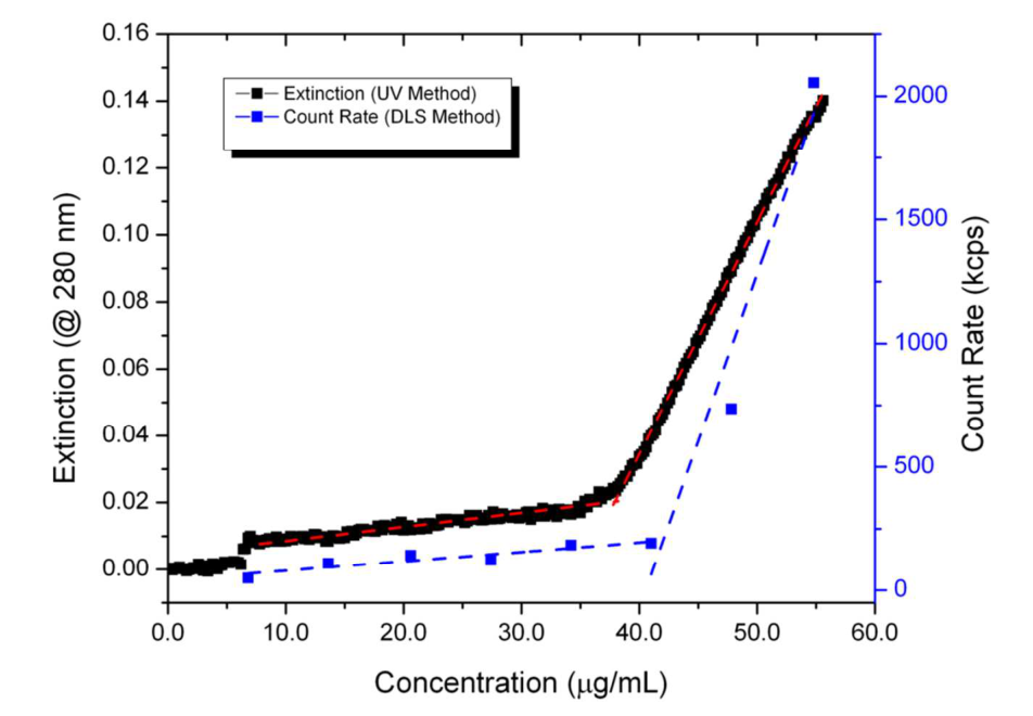
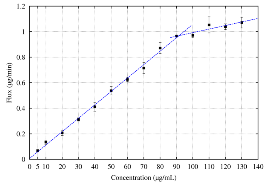
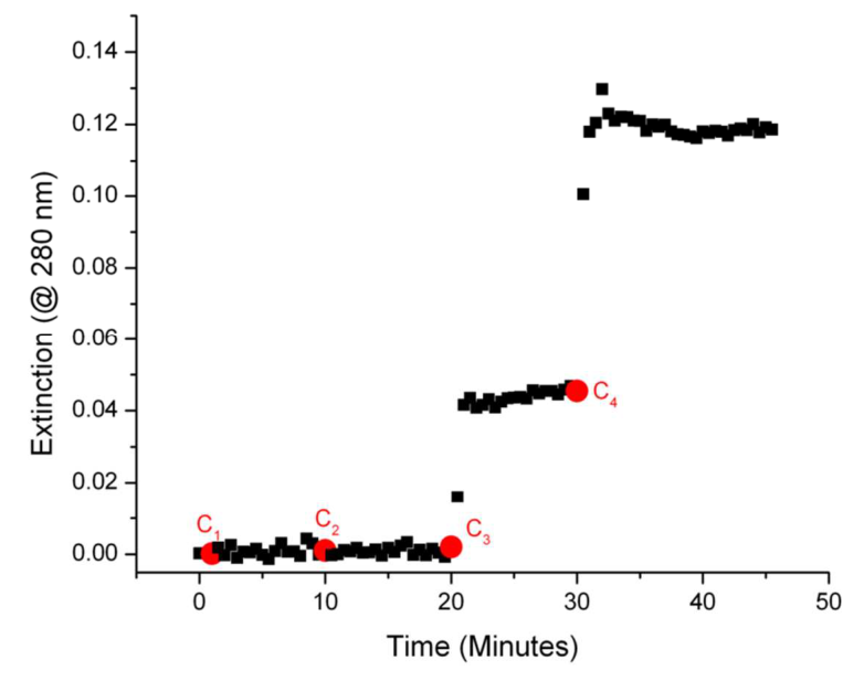
Both signals are flat at first and then rise steeply; the dashed lines cross near 38 µg/mL (UV) and 41 µg/mL (DLS) ilevbare2013a R2.
Why
Ilevbare and Taylor read the colloids as liquid–liquid phase separation: solution and droplets are in equilibrium, so the drug has the same activity in both ilevbare2013a C29, C36. A polarity dye enters a disordered drug-rich phase and changes its emission, whereas crystallization leaves it in solution, so extinction alone cannot tell the two apart almeidaesousa2015 C12, C16.
Limits and transfer
The phase was isolated and examined for five drugs only ilevbare2013a X8, and all 21 compounds are lipophilic (synthesis), so hydrophilic drugs were not tested. A marker band can appear far below the amorphous solubility in SDS, so check it in the medium of use indulkar2017a R18, C27. Raise the concentration slowly while recording extinction and a probe, then isolate the pellet for birefringence, XRPD and a DSC Tg almeidaesousa2015 P1, P2; indulkar2015 P14, P15, P16.
How it was shown
Ilevbare and Taylor (2013) pumped a 4 mg/mL methanol stock into stirred medium at 37 °C and followed extinction at non-absorbing wavelengths (280–450 nm) for eight drugs, each in a medium where it is un-ionized; the drugs had Log P 2.2–5.6 and melting points 121–187 °C ilevbare2013a P5, M1, M2, M3, M5, M6, M7, M8, M9, R8. For ritonavir in water they added DLS count rate in a flow cell and a pyrene I1/I3 titration, and they isolated the phase by ultracentrifugation at 274,356 × g from 0.5 mL of a 60 mg/mL stock in 20 mL water at 10, 37 and 50 °C, then measured pellet water by drying and Tg by DSC at 20 °C/min ilevbare2013a P7, P8, P9, P10, P11. Almeida e Sousa and co-workers (2015) used six fast-crystallizing drugs at 25 °C in 50 mM phosphate buffer, pH 2 for the three acids and pH 6.8 for the others: extinction at 400 or 450 nm during continuous pumping, and, in stepwise additions, a polarity-sensitive probe (1-pyrenecarboxaldehyde, pyrene, 4-Di-2-ASP) or phenylbutazone's own fluorescence almeidaesousa2015 P1, P2. Indulkar and co-workers (2015) used antisolvent addition, DLS, nanoparticle tracking with Nile red, XRPD, polarized microscopy and DSC for three weak bases at 37 °C indulkar2015 P8, P11, P12, P14, P15, P16. Mosquera-Giraldo and Taylor (2015) combined extinction at 370 nm, 4-Di-2-ASP fluorescence, ¹H NMR of dissolved drug and DSC of an ultracentrifuged pellet for telaprevir in pH 6.8 phosphate buffer at 37 °C mosqueragiraldo2015 P4, P7, P9, P10. Trasi and Taylor (2015) located the onset of ritonavir, lopinavir and paclitaxel at 25 °C in 100 mM phosphate buffer by extinction, pyrene, ultracentrifugation and flux trasi2015a P1, P2, P3, P4, P6, and Almeida e Sousa and co-workers (2016) that of the CRH1 free base by extinction at 450 nm and pyrene almeidaesousa2016 P3, P4. Sugihara and Taylor (2018) shifted pazopanib HCl from simulated gastric fluid (pH 1.2) to pH 6.5 phosphate buffer at 37 °C and looked at the solution by eye, by DLS (backscatter at 173°), by nanoparticle tracking, by polarized light microscopy at 20× and by DSC of the air-dried precipitate sugihara2018 P6, P7, P8, P9, P10. Ueda and Taylor (2020) formed ibuprofen droplets by adding a methanol-d4 stock (2% v/v) to pH 2.0 phosphate buffer in D2O at 37 °C, with or without 300 µg/mL polymer, and sized them fresh by DLS ueda2020b P6.
What the data show
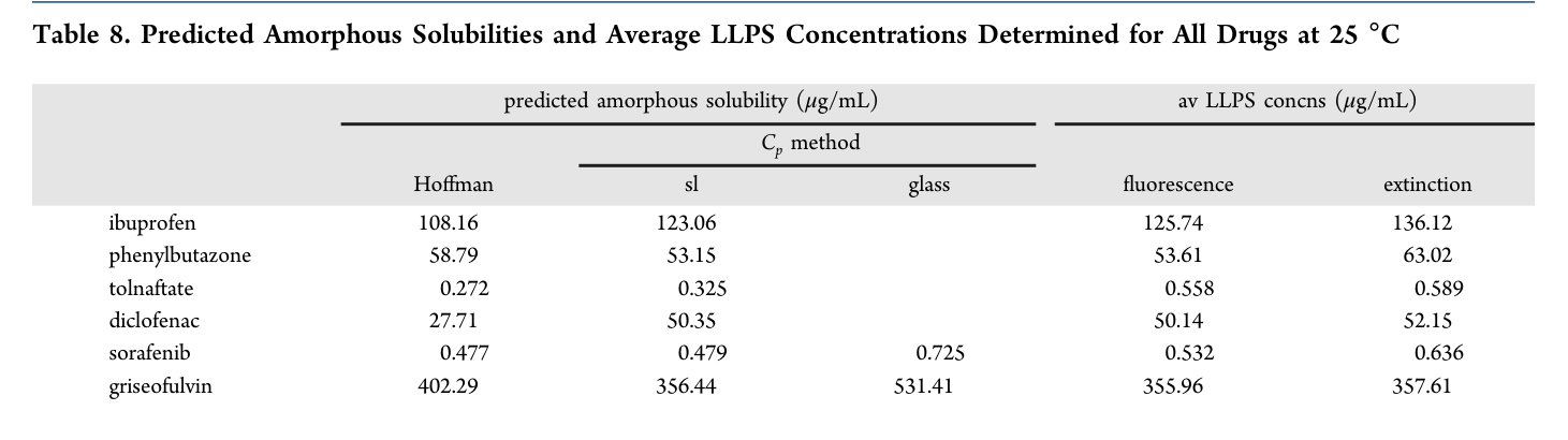
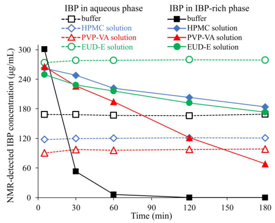
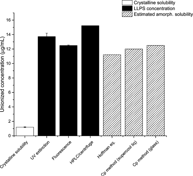
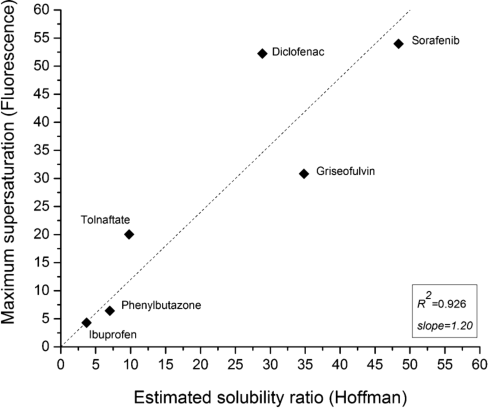
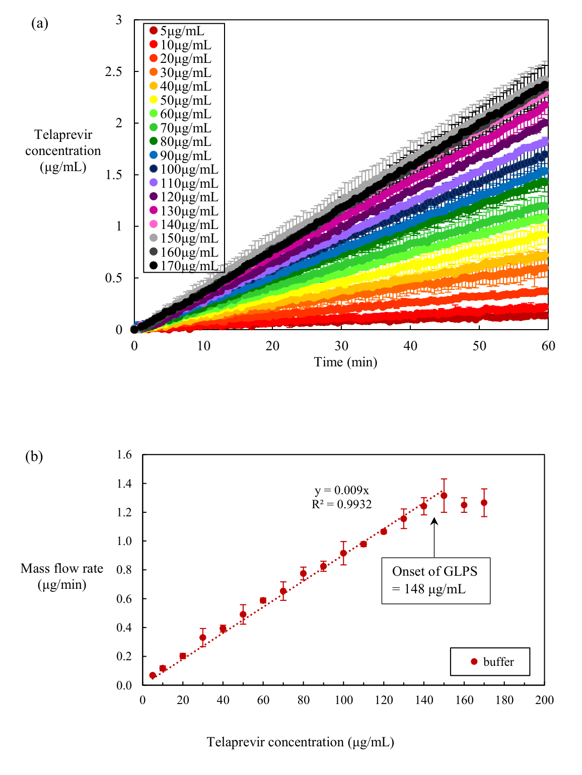
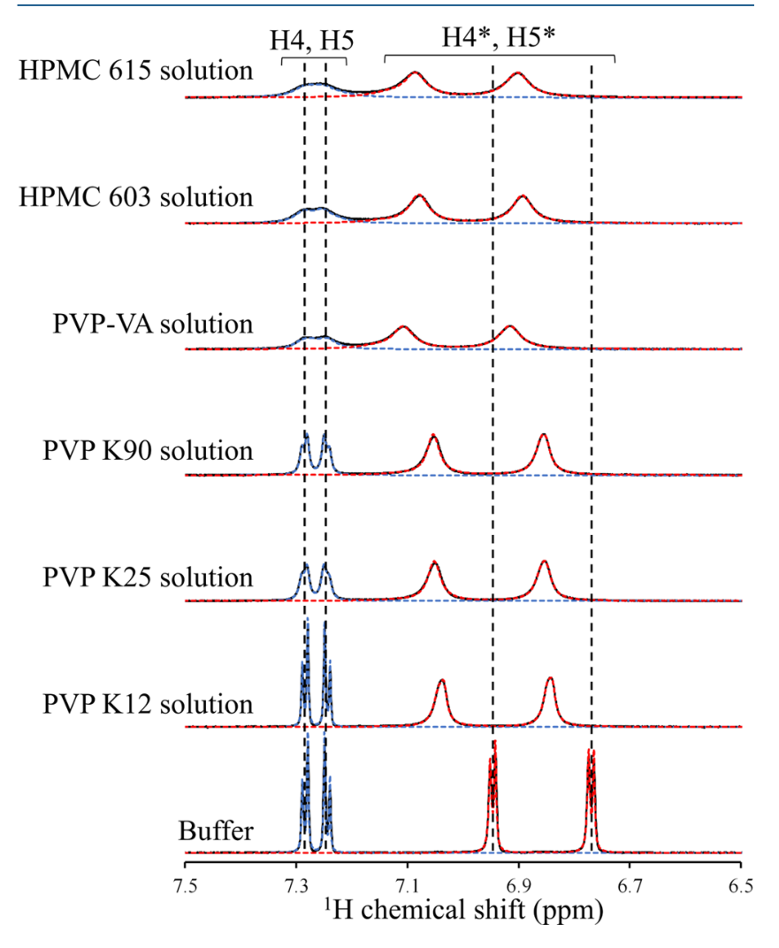
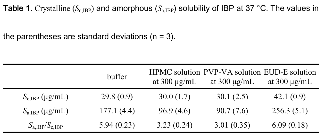
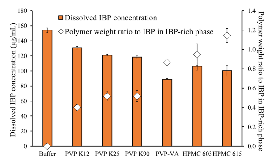
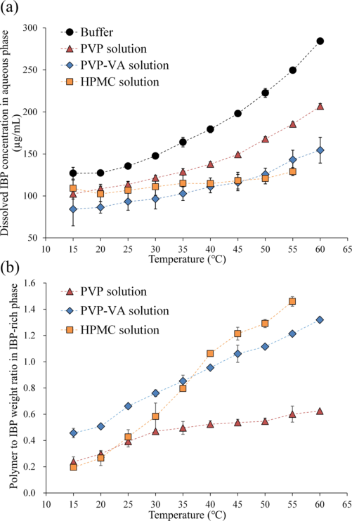
Ritonavir. In water the solution turned cloudy with a bluish tint, and polarized light showed small, non-birefringent, spherical droplets ilevbare2013a R1. Extinction and DLS count rate rose above 37.2 ± 0.9 and 39.8 ± 0.3 µg/mL ilevbare2013a R8. The pyrene I1/I3 ratio stayed at about 1.67–1.69 up to 39.0 µg/mL and fell to about 1.39 at 120 µg/mL ilevbare2013a R4. The droplets grew from about 345 nm at 1 min to about 842 nm at 64 min ilevbare2013a R3. The isolated phase held 94.85–95.97% drug and 4.03–5.15% water between 10 and 50 °C, with a Tg of 0.4 °C against 50.4 °C for dry amorphous ritonavir ilevbare2013a R6, R5, C23. DLS in the second paper gave about 360 nm at 30 µg/mL ritonavir trasi2015a R15. Eight drugs. UV onsets ran from 5.2 µg/mL (clotrimazole) to 135.8 µg/mL (clozapine), and DLS onsets lay close to them (synthesis: the largest UV–DLS difference is 39.8 − 37.2 = 2.6 µg/mL) ilevbare2013a R8. Six fast crystallizers. Extinction and probe signal changed at about the same concentration, and the turbidity lasted only a few minutes before visible particles formed almeidaesousa2015 R1, R3, C17. The probe read-outs were a shift of the 1-pyrenecarboxaldehyde peak from about 468.5–470.5 to about 446–448 nm (ibuprofen), a rise of phenylbutazone's autofluorescence from about 0 to about 980, a rise of the diclofenac pyrene I3/I1 from about 0.52 to about 0.75, and 4–5 nm shifts of the 4-Di-2-ASP peak, to shorter wavelength for tolnaftate and to longer wavelength for sorafenib almeidaesousa2015 R3, X3. Fluorescence onsets were 125.74, 53.61, 0.558, 50.14, 0.532 and 355.96 µg/mL for ibuprofen, phenylbutazone, tolnaftate, diclofenac, sorafenib and griseofulvin almeidaesousa2015 R5, R6. Griseofulvin at pH 6.8 without polymer showed extinction rising near 150 µg/mL with no probe change and small solid particles; with 100 µg/mL HPMCAS-HF in the buffer, extinction and probe intensity rose together near 350 µg/mL and the solution stayed uniformly turbid without visible particles almeidaesousa2015 R4. Weak bases. All six precipitates (clotrimazole from pH 4 and 8, atazanavir from pH 3.5 and 9, nicardipine from pH 6 and 9) showed only broad halos by XRPD and no birefringence indulkar2015 R19, R20, R21; DLS Z-averages were about 116–387 nm indulkar2015 R14; with Nile red in the buffer a population of lipophilic droplets (about 100–450 nm) appeared only above the onset indulkar2015 R16; the DLS polydispersity index of clotrimazole at pH 8 fell from about 0.76 at 6.1 µg/mL to about 0.27 at 6.65 µg/mL at the onset indulkar2015 R11. Telaprevir. Solutions were clear up to about 90 µg/mL and turbid above mosqueragiraldo2015 R1; extinction at 370 nm stayed near zero up to about 93 µg/mL and rose to about 0.048 at about 129 µg/mL mosqueragiraldo2015 R4; the species measured about 300 nm mosqueragiraldo2015 R5; the pelleted material had a Tg of 52 °C mosqueragiraldo2015 R2. CRH1. Solvent shift gave turbidity without visible particles, later clearing as visible particles appeared, with onsets of 14.15 ± 0.87 (extinction) and 11.31 ± 0.56 µg/mL (pyrene) at pH 6.8 and 13.73 ± 0.47 and 12.49 ± 0.08 µg/mL at pH 4.6 with 20 µg/mL HPMC almeidaesousa2016 R5, R3, R4. Pazopanib. Above the amorphous solubility the solution turned milky with a bluish tinge at once sugihara2018 R14; DLS gave Z-averages of 495.9 nm (PDI 0.027) without and 145.4 nm (PDI 0.140) with 50 µg/mL HPMC, NTA found particles of about 50–650 nm sugihara2018 R15, R16, and at the dose below the amorphous solubility no scattering species appeared sugihara2018 R20. Polarized microscopy showed non-birefringent aggregates beside salt crystals at 2 min, and free-base crystals taking over by 10 min without HPMC and by 60 min with it sugihara2018 R11. The dried precipitate had a Tg of about 83 °C sugihara2018 R17. Ibuprofen. In buffer the drug-rich phase was micron-sized (3182–5313 nm at a PDI of 1.00, the top of the scale), with 300 µg/mL HPMC or PVP-VA it was 268–345 nm and with EUD-E 472–808 nm ueda2020b R5, X10; at 400 µg/mL ibuprofen the buffer and all six polymer solutions of the 2021 paper were milky ueda2021 R15.
| paper | system | method and conditions | result | ids |
|---|---|---|---|---|
| ilevbare2013a | ritonavir, water, 37 °C | solvent shift from methanol; extinction, DLS, pyrene | non-birefringent droplets; onset 37.2 (UV), 39.8 (DLS), 39.0 (pyrene) µg/mL | R1, R2, R4, R8 |
| ilevbare2013a | ritonavir, ultracentrifuged phase | 10, 37, 50 °C; gravimetry; DSC | 94.85–95.97% drug, 4.03–5.15% water; Tg 0.4 °C | R5, R6 |
| ilevbare2013a | eight drugs, 37 °C | extinction and DLS in un-ionizing media | onsets 5.2 (clotrimazole) to 135.8 µg/mL (clozapine) | R8 |
| almeidaesousa2015 | six fast crystallizers, 25 °C | extinction plus polarity probe | both change at about the same concentration; turbidity lasts minutes | R1, R3, R5 |
| almeidaesousa2015 | griseofulvin, pH 6.8, with and without 100 µg/mL HPMCAS-HF | same | without polymer: extinction near 150 µg/mL, no probe change, particles; with polymer: both near 350 µg/mL | R4 |
| indulkar2015 | clotrimazole, atazanavir, nicardipine, 37 °C | antisolvent; XRPD; PLM; DLS; Nile red | halo-only XRPD, no birefringence; 116–387 nm droplets | R14, R16, R19, R20, R21 |
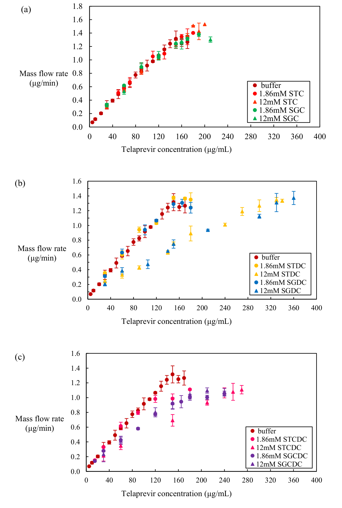
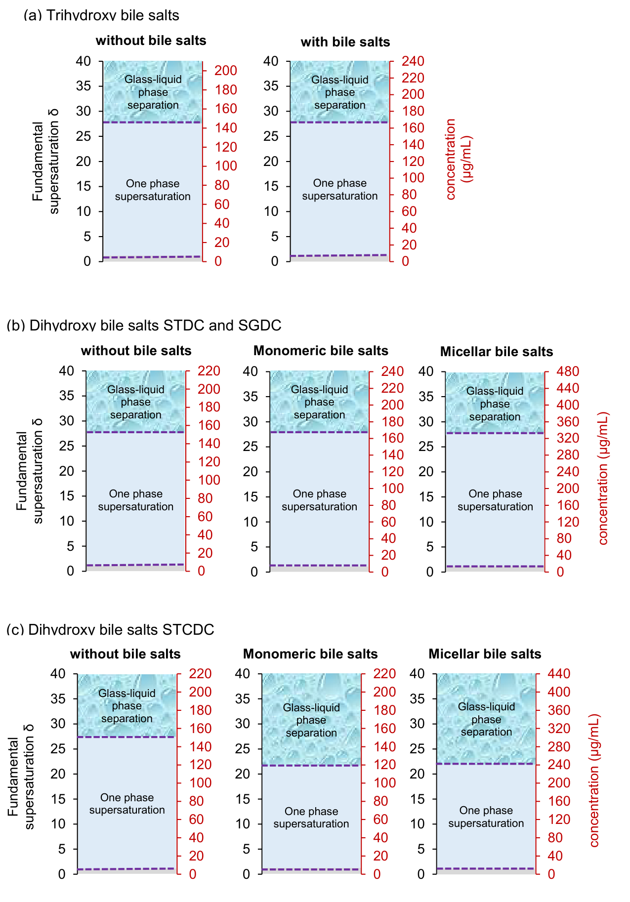
| mosqueragiraldo2015 | telaprevir, pH 6.8, 37 °C | extinction, fluorescence, NMR, DSC of pellet | boundary 90–92 µg/mL; pellet Tg 52 °C; about 300 nm species | R2, R5, R13 |
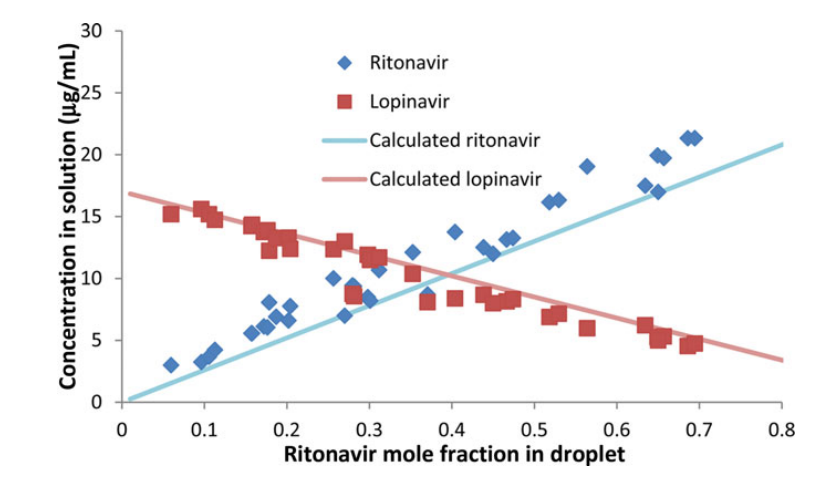
| trasi2015a | ritonavir, lopinavir, paclitaxel, 25 °C | four methods | onsets 26–28, 16–17.3, 27.2–30.1 µg/mL; about 360 nm droplets (ritonavir) | R1, R15 |
| almeidaesousa2016 | CRH1 free base, 25 °C | extinction and pyrene, pH 6.8 and pH 4.6 + HPMC | turbidity, then particles; onsets 11.31–14.15 µg/mL | R3, R4, R5 |
| sugihara2018 | pazopanib HCl, pH 1.2 → 6.5, 37 °C | shift; eye, DLS, NTA, PLM, DSC | milky at once; 496 nm (145 nm with HPMC); non-birefringent aggregates, then free-base crystals; dried precipitate Tg about 83 °C | R11, R14, R15, R16, R17 |
| ueda2020b | ibuprofen, pH 2.0 (D2O), 37 °C, 2% methanol-d4 | solvent shift; DLS | droplets 3182–5313 nm in buffer; 268–345 nm with HPMC or PVP-VA; 472–808 nm with EUD-E | R5 |
Figure
Both signals are flat at first and then rise steeply; the dashed lines cross near 38 µg/mL (UV) and 41 µg/mL (DLS) ilevbare2013a R2.
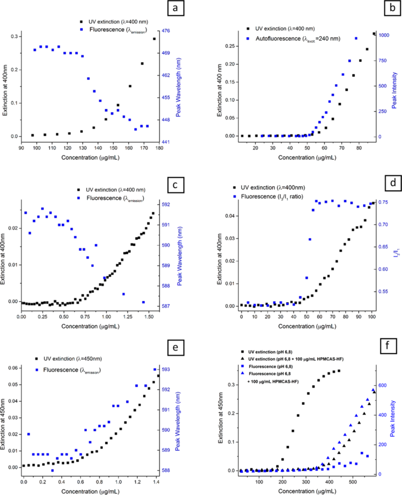
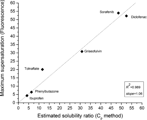
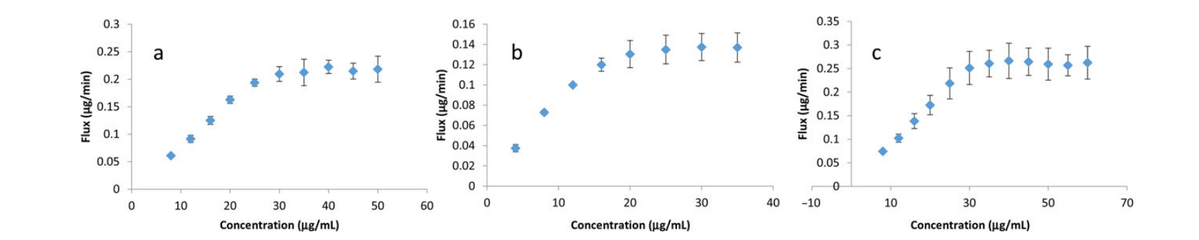
Panels a–f are ibuprofen, phenylbutazone, tolnaftate, diclofenac, sorafenib and griseofulvin; the concentration axes differ about a thousandfold. In panel f the squares (no polymer) show extinction without a probe change, the triangles (HPMCAS-HF) both signals near 350 µg/mL almeidaesousa2015 R3, R4.
Why
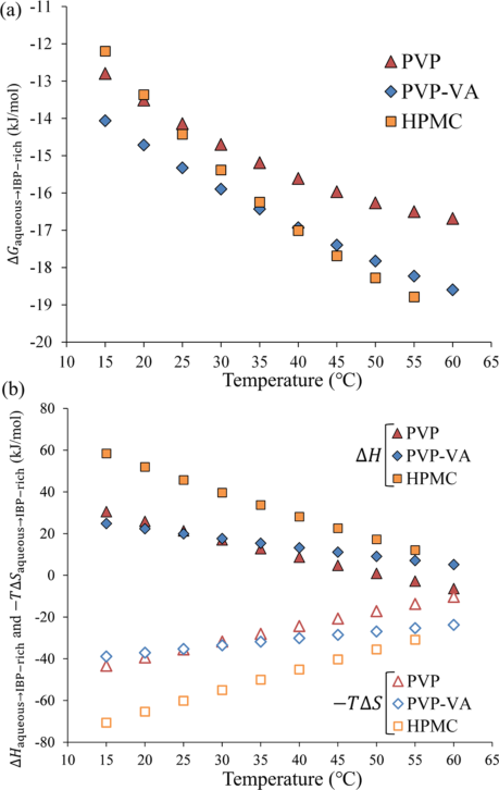
Ilevbare and Taylor read the colloids as liquid–liquid phase separation: the four features reported for drug "aggregation" (no second phase below a threshold, non-crystalline aggregates, constant monomer concentration above the threshold, a reproducible threshold in a given buffer) all appeared in their data, and the solution and the droplets are in equilibrium, so the drug has the same activity in both ilevbare2013a C29, C36. They call a hydrophobic-interaction driving force "intuitive" from the Log P range but did not test it ilevbare2013a C37. The probe logic is that a polarity-sensitive dye partitions into a disordered drug-rich phase and changes its emission, whereas crystallization leaves the dye in solution and the emission unchanged, which is why extinction alone cannot tell the two apart almeidaesousa2015 C12, C16; Indulkar and co-workers add that dyes cannot partition into a crystalline phase and confirm lipophilic droplets with Nile red indulkar2015 C29, C37. NMR sees only dissolved telaprevir because molecules in large, viscous glassy aggregates relax too fast to give a resolved signal ("anticipated") mosqueragiraldo2015 C18. (synthesis: the scattering onset, the probe change, the isolated amorphous pellet and the composition of the pellet are four independent observations, and they agree wherever more than one was made, as for ritonavir and telaprevir.) Sugihara and Taylor read the immediate turbidity, the particle sizes and the Tg together as glass–liquid phase separation into a water-saturated colloidal drug-rich phase ("indicate") sugihara2018 C27, C28, and Ueda and Taylor treat the separated ibuprofen phase as a supercooled-liquid emulsion because its Tg is −45 °C ueda2020b C38, M1. Indulkar and co-workers (2017) show the limit of a spectral marker: the 369 nm emission band that marks the atazanavir drug-rich phase in buffer also appeared in 3.6 mg/mL SDS from about 200 µg/mL, far below the amorphous solubility of about 1450 µg/mL, where they read it as drug–drug contact inside micelles and not as a drug-rich phase indulkar2017a R17, R18, C27. (synthesis: a spectral signature of the drug-rich phase must be checked against the amorphous solubility measured in the same micellar medium.)
Conditions and limits
Temperatures were 25 °C almeidaesousa2015 P1; trasi2015a P1; almeidaesousa2016 P3 or 37 °C ilevbare2013a P5; indulkar2015 P8; mosqueragiraldo2015 P7. Supersaturation came mainly from a solvent shift that left about 1–2% v/v organic solvent almeidaesousa2015 X9; mosqueragiraldo2015 X7. The onset did not depend on the stock solvent or addition rate for ritonavir (methanol, ethanol, THF, or a drug dissolved in 0.1 M HCl) ilevbare2013a C28, R9, R10, and the solvent-free pH titration of the weak bases agreed with the antisolvent runs above the lowest pH indulkar2015 C33, X7. Every compound was poorly soluble and lipophilic, with Log P 2.17–5.75 and melting points 75.9–238 °C almeidaesousa2015 M1, M3, M6; mosqueragiraldo2015 M1. The phase was isolated and examined only for ritonavir, clotrimazole, atazanavir, nicardipine and telaprevir, and the ritonavir DSC trace shown stops at 20 °C, so the absence of melting is stated, not shown ilevbare2013a X8. (synthesis: all 21 compounds are lipophilic and poorly soluble, so hydrophilic drugs were not tested.) The 2017–2022 papers add a pH 1.2 to 6.5 transfer (pazopanib) and a dissolved polymer at 300 µg/mL as ways to generate the phase; the ibuprofen work is in D2O buffer with 2% methanol-d4 sugihara2018 P6; ueda2020b M7, P6.
Counterexamples and conflicts
Griseofulvin without polymer crystallized before it could phase separate, or its probe could not enter a glassy phase; the authors cannot tell which almeidaesousa2015 C18. Crystalline nicardipine lost its birefringence in water within 2 h, so it has no crystal–water equilibrium to compare with indulkar2015 R1. The library finds the same split between slow and fast crystallizers in the fluorescence-probe work: see Solution methods F8. Pazopanib at the dose below its amorphous solubility formed no drug-rich phase at all, as expected sugihara2018 R20, C31.
Transfer check
Raise the concentration slowly in the medium of interest while recording extinction at a wavelength where the drug does not absorb, and repeat with a polarity probe or the drug's own fluorescence almeidaesousa2015 P1, P2. Isolate the pellet by ultracentrifugation and check birefringence, XRPD and a DSC Tg indulkar2015 P14, P15, P16; ilevbare2013a P7, P9. A rise of extinction with a probe change and a birefringence-free, halo-only pellet puts the drug inside this evidence; a rise of extinction without a probe change and with birefringent particles, as for griseofulvin without polymer, means crystallization almeidaesousa2015 R4. In a micellar medium also locate the amorphous solubility in that medium, since a spectral band can appear well before it indulkar2017a R18.
History
Before this subcategory the library's background was "promiscuous aggregation": close to 20% of nearly 300 screened compounds formed aggregates of 50 to more than 1000 nm on dilution into water, and LLPS was proposed as the cause ilevbare2013a C7, C9. Ilevbare and Taylor (2013) were the first in the library to isolate and characterize the phase (ritonavir) and to show the onset for eight drugs ilevbare2013a C2, C56. Almeida e Sousa and co-workers (2015) extended it to six fast crystallizers and argued that a fluorescence check is "clearly essential" for them almeidaesousa2015 C21. Indulkar and co-workers (2015) added XRPD and microscopy for three weak bases, Mosquera-Giraldo and Taylor (2015) a glassy telaprevir phase, and the 2015 and 2016 papers by Trasi and Taylor and by Almeida e Sousa and co-workers used the same onset for ritonavir, lopinavir, paclitaxel and CRH1 indulkar2015 C6; mosqueragiraldo2015 C21; trasi2015a C12; almeidaesousa2016 C17. Sugihara and Taylor (2018) extended the phase to a weak base shifted from stomach to intestinal pH, Ueda and Taylor (2020) to a low-Tg drug whose droplets are stabilized by dissolved polymers, and Indulkar and co-workers (2017) showed where the 369 nm marker fails sugihara2018 C27; ueda2020b C13; indulkar2017a C27.