Topic 1Hydrogen bond between drug and polymer and the ways a dispersion is shown to be mixed1 of 8
In a PVP dispersion the indomethacin acid OH bonds to PVP instead of forming acid dimers; a physical mix shows no such bond.
Infrared spectra of dispersions with 5–70 wt% PVP differ from those of physical mixes of the same composition: the band of the carboxylic acid dimers of the drug falls, and new bands appear at 1726 and 1636 cm⁻¹ taylor1997 R1, R10, R11, R12. The authors assign them to the acid of a PVP-bonded drug molecule and to a hydrogen-bonded PVP carbonyl taylor1997 C33, C35.
moderateModerate: dispersion and mix differ in three bands and two liquid models agree, but every reading is a band position, with no crystallization data taylor1997 K1, K4, X5, X8.
Only the dispersions show the three changes. taylor1997 R10, R11, R12
The bonded PVP carbonyl lies 43 cm⁻¹ below the free one. taylor1997 R5.h, R5.l, R11, R12
For your drug
- For an acid drug in PVP, record the melt-quenched drug, a solvent-evaporated dispersion and a physical mix of the same composition by IR.
- Look for loss of the dimer band, a free acid C=O band about 16 cm⁻¹ higher and a PVP carbonyl far below its free position; do not expect all drug to be bonded at the stoichiometric composition.
taylor1997 R5.h, R11, R12, C37
How it works and key numbers
- Indomethacin has one donor, its acid OH, and PVP can only accept a proton, so any new bond must show in the acid C=O and in the PVP carbonyl taylor1997 C25.
- In the dispersion the drug's acid bonds to PVP at the expense of the dimers; in the physical mix no bond forms, so molecular mixing is needed taylor1997 C35, C26.
- The spectra keep changing above the 24 wt% PVP at which PVP monomer equals drug in moles, so the authors think the equilibrium constant is low and/or the stoichiometry is below 1:1, and dimers, bonded drug and free PVP coexist ("this suggests") taylor1997 R18, C37, C38.
- The authors propose that a γ nucleus, built of dimers, "would require" breaking the PVP–drug bond, which may explain why PVP inhibits crystallization even at low levels (crystallization was not measured here) taylor1997 C43, C45, X8.
Connections to other chapters
Both read the indomethacin carboxylic acid by IR: in solution a dimer band grows with concentration even below saturation towler2007 R13, R17, and in the PVP dispersions the dimer band falls as the acid OH bonds to the PVP carbonyl taylor1997 R10, C35.
Sub-additive water uptake (indomethacin–PVP with 25% PVP, 3.6 measured against 14.0 g/100 g predicted) is explained by drug donors binding the single acceptor of the PVP repeat unit so that those sites are lost to water rumondor2010a R12.h, C11, C12, the bond first shown by IR for indomethacin–PVP taylor1997 R10, C35.
same mechanism PEG dispersions F3: Different measures of drug–PEG interaction agree on which drugs interact, but not on its magnitude.
Both read a drug–polymer bond from a spectrum compared with the physical or additive mixture: loss of the indomethacin dimer band and a 1636 cm⁻¹ band absent from physical mixes taylor1997 R10, R12, and chlorpropamide carbonyl at 1717 against 1663 cm⁻¹ in the additive spectrum baird2011 R10.a.
1 more connection
The PVP carbonyl red shift as a measure of drug–PVP bond strength rumondor2009c R29, C48 follows the band assignments for indomethacin–PVP taylor1997 R11, R12.
Evidence: data, conditions and papers
Finding F1. In PVP dispersions the indomethacin acid OH bonds to the PVP carbonyl at the expense of the acid dimers, physical mixes show no such bond, and some drug stays unbound even with excess polymer
In short. Indomethacin–PVP-K90 solid dispersions with 5–70 wt% PVP were amorphous by XRPD, and their infrared spectra differ from those of physical mixes of amorphous indomethacin and PVP of the same composition: the dimer band falls between 5 and 20 wt% PVP, a single band at 1726 cm⁻¹ appears from 20 wt%, and a band at 1636 cm⁻¹, absent in the physical mixes, appears at 5–50 wt% taylor1997 R1, R10, R11, R12. The authors assign these to the free acid C=O of a PVP-bonded drug molecule and to a hydrogen-bonded PVP carbonyl, and read them as bonding "at the expense of dimer formation" taylor1997 C33, C35. The spectra keep changing above the 24 wt% PVP at which PVP monomer equals drug in moles, so binding is incomplete taylor1997 R18, C37.
Type. single-system observation (indomethacin–PVP), with two liquid models Strength. dispersion and physical mix of the same composition differ in three bands, and two liquid models reproduce the pattern, but all readings are band positions, with no intensities, fits or crystallization data (taylor1997 K1: moderate to strong; K4: weak to moderate; X5, X8).
How it was shown
Taylor and Zografi recorded γ, α and amorphous indomethacin by FT-Raman and transmission IR, because earlier dispersion spectra had been compared with crystalline drug taylor1997 C7, P7, P8. Dispersions with 5–70 wt% PVP-K90 were made from methanol, and physical mixes of amorphous drug and PVP were prepared at the same compositions taylor1997 P3, P4. Acetic acid and indomethacin in methylpyrrolidone served as liquid models taylor1997 P5, M5.
What the data show
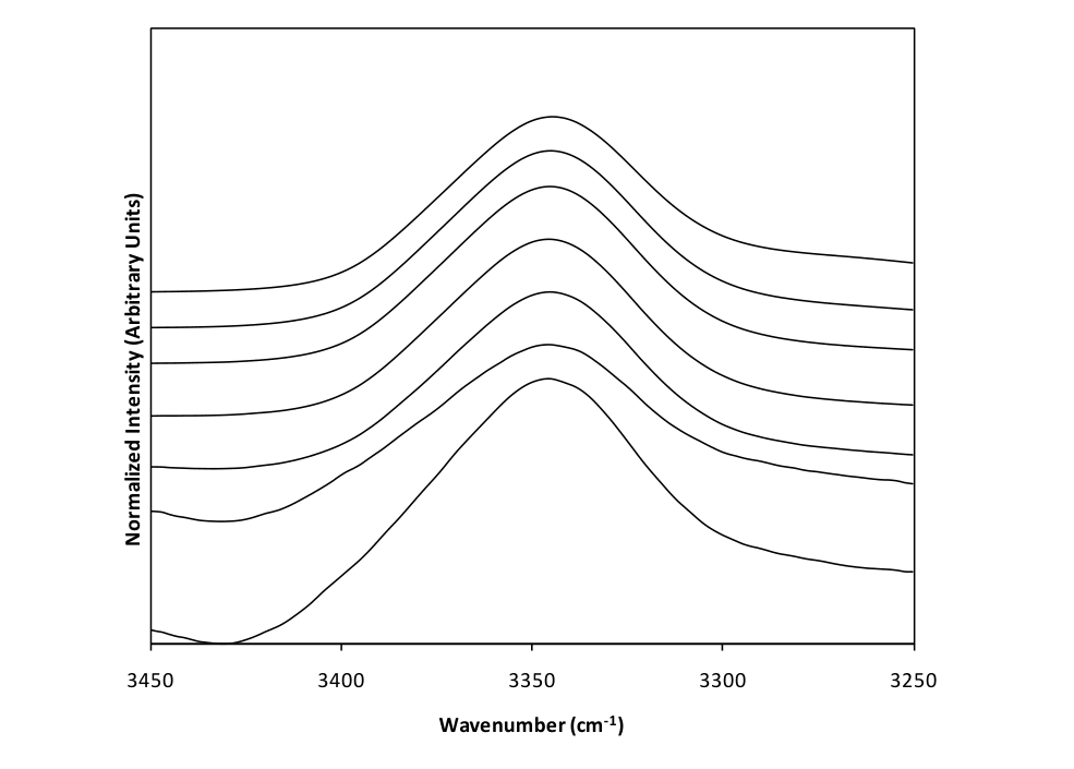
Amorphous indomethacin has an IR acid band at 1710 cm⁻¹ with no Raman counterpart, read as cyclic dimers taylor1997 R5.h, R6, C19. In the physical mixes the amorphous doublet persisted at every composition; in the dispersions the dimer band fell between 5 and 20 wt% PVP, a band at 1726 cm⁻¹ appeared from 20 wt% and one at 1636 cm⁻¹ at 5–50 wt% taylor1997 R10, R11, R12.
| sample | method and conditions | result | ids |
|---|---|---|---|
| amorphous indomethacin | IR (KBr) and FT-Raman, quench-cooled melt | acid band 1710 cm⁻¹ in IR, none in Raman; shoulder 1735 cm⁻¹ | taylor1997 R5.g, R5.h, R6 |
| physical mixes, 5–70 wt% PVP-K90 | IR, KBr | amorphous doublet retained at every composition; no 1636 cm⁻¹ band | taylor1997 R10, R12 |
| solid dispersions, 5–70 wt% PVP-K90 | IR, KBr; XRPD | dimer band falls at 5–20 wt%; 1726 cm⁻¹ from 20 wt%; 1636 cm⁻¹ at 5–50 wt%; no XRPD peaks | taylor1997 R1, R11, R12 |
| acetic acid + methylpyrrolidone | IR, NaCl windows | amide C=O 1688 → 1652 cm⁻¹; indomethacin 1710 cm⁻¹ band gone in methylpyrrolidone | taylor1997 R15, R16 |
| stoichiometry | mole ratio with monomer weight 111 | 1:1 at 24 wt% PVP; changes continue above it | taylor1997 E1, R18 |
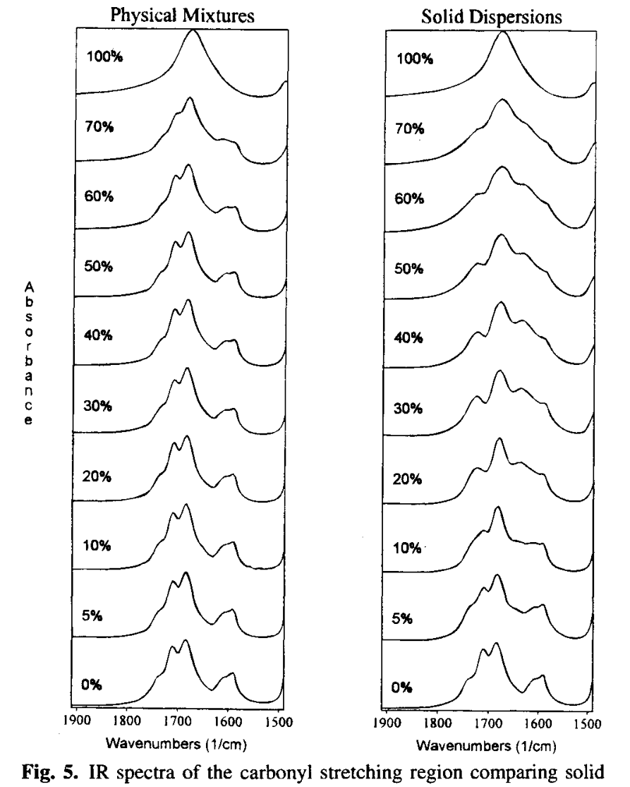
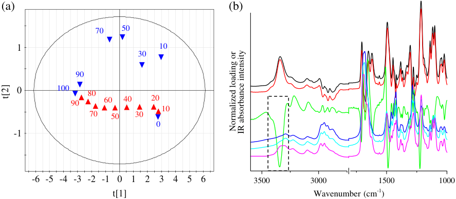
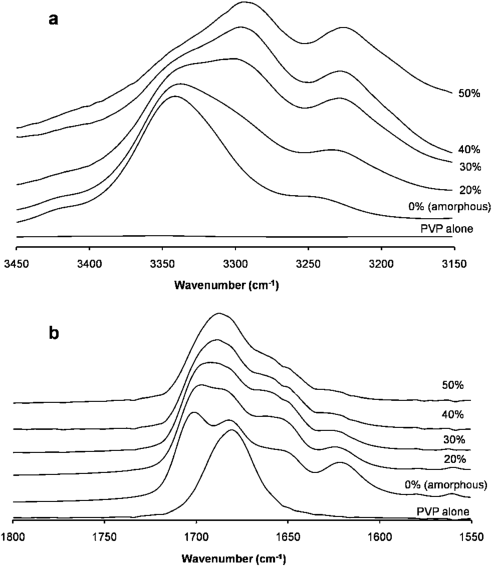
The physical mixes (left) keep the amorphous doublet; the dispersions (right) lose the dimer band and gain bands near 1726 and 1636 cm⁻¹ taylor1997 R10, R11, R12.
Why
Indomethacin has one donor, its acid OH, and PVP can only accept, so a new bond must show in the acid C=O and the PVP carbonyl taylor1997 C25. The authors read the bands as bonding "at the expense of dimer formation"; since the spectra keep changing above 24 wt% PVP, "this suggests" a low equilibrium constant and/or a stoichiometry below 1:1 taylor1997 C35, C37.
Limits and transfer
One acid drug and one PVP grade; crystallization was not measured taylor1997 M4, X8. For an acid drug in PVP, compare melt-quenched drug, a dispersion and a physical mix by IR and look for loss of the dimer band, a free acid C=O 16 cm⁻¹ higher (synthesis: 1726 − 1710) and a PVP carbonyl far below its free position taylor1997 R5.h, R11, R12.
How it was shown
Taylor and Zografi first fixed the reference state, because earlier dispersion spectra had been compared with crystalline drug and crystallinity alone changes vibrational spectra taylor1997 C7. They recorded γ, α and amorphous indomethacin by FT-Raman (Bruker RFS 100, 1064 nm, typically 500 mW, 200 scans at 4 cm⁻¹) and by transmission IR (Mattson Galaxy 5020, solids at 0.8–1% in KBr, 500 scans at 4 cm⁻¹) taylor1997 P7, P8. Amorphous drug was made by melting γ at 165 °C for 5 min and quench cooling in liquid nitrogen, α by precipitation from methanol with water taylor1997 P1, P2. A cyclic carboxylic acid dimer has a centre of symmetry, so by the mutual exclusion rule its asymmetric C=O stretch is seen only in the IR and the symmetric stretch only in the Raman, which the authors checked on liquid acetic acid (IR 1716, Raman 1669 cm⁻¹) taylor1997 C14, R5.j. Dispersions were made by dissolving 5 g of drug and PVP-K90 in 100 ml of anhydrous methanol at 65 °C, rotary evaporation at 50 °C and 24 h under vacuum, at 5, 10, 20, 30, 40, 50, 60 and 70 wt% PVP; physical mixes of amorphous drug and PVP were prepared at the same compositions, by a procedure that is not printed taylor1997 P3, P4, X7. Acetic acid with 10–90 wt% 1-methyl-2-pyrrolidone and indomethacin in 30–70 wt% methylpyrrolidone served as liquid models for the acid–lactam contact taylor1997 P5, M5.
What the data show
Amorphous indomethacin has an IR acid band at 1710 cm⁻¹ with no Raman counterpart, like the 1717 cm⁻¹ band of γ, which is known from its crystal structure to hold cyclic dimers; the authors conclude it is "considered to be composed mainly" of cyclic dimers, with a shoulder at 1735 cm⁻¹ read as free chain ends taylor1997 R5.a, R5.g, R5.h, R6, C19, C20. α-indomethacin has acid bands at 1681 (shoulder) and 1649 cm⁻¹ in the IR that appear at 1680 and 1649 cm⁻¹ in the Raman, which they read as chains, not dimers taylor1997 R5.e, R5.f, C16, C17. In the physical mixes the amorphous doublet near 1710 and 1684 cm⁻¹ persisted at every composition, with the PVP band (1679 cm⁻¹) growing on top taylor1997 R10, R5.l. In the dispersions the dimer peak lost intensity between 5 and 20 wt% PVP while the non-bonded carbonyl peak gained; from 20 wt% there was one broad peak at 1726 cm⁻¹, and a band at 1636 cm⁻¹ was present at 5–50 wt% PVP and merged into a shoulder at 60–70 wt% taylor1997 R11, R12. The shift of the PVP carbonyl from 1679 to 1636 cm⁻¹ is 43 cm⁻¹ (synthesis: 1679 − 1636) taylor1997 R5.l, R12. The PVP C–N stretch did not change in IR or Raman, which points to the carbonyl as acceptor taylor1997 R13, C25. In acetic acid with methylpyrrolidone the amide C=O first appeared at 1652 cm⁻¹, 36 cm⁻¹ below its pure-liquid position, and the free-acid shoulder at 1759 cm⁻¹ grew; indomethacin in methylpyrrolidone lost its 1710 cm⁻¹ band and showed one band at 1723 cm⁻¹ and a bonded amide band at 1649 cm⁻¹ taylor1997 R15, R16. With a monomer molecular weight of 111, a 1:1 mole ratio of PVP monomer to drug is reached at 24 wt% PVP, and the paper states 12 wt% for two hydrogen bonds per carbonyl, which the card recomputes as 13.6 wt% (synthesis: half a monomer per drug molecule gives 0.5/(0.5 + 3.17)) taylor1997 E1, R18, X2. The changes continue above both values taylor1997 R11, R12.
| paper | system | method and conditions | result | ids |
|---|---|---|---|---|
| taylor1997 | amorphous indomethacin | IR (KBr) and FT-Raman, quench-cooled melt | acid band 1710 cm⁻¹ in IR, none in Raman; shoulder 1735 cm⁻¹ | R5.g, R5.h, R6 |
| taylor1997 | α-indomethacin | IR and Raman | acid bands 1681 (sh) and 1649 cm⁻¹ in IR, 1680 and 1649 cm⁻¹ in Raman | R5.e, R5.f, R6 |
| taylor1997 | physical mixes, amorphous indomethacin + PVP-K90, 5–70 wt% PVP | IR, KBr | amorphous doublet retained at every composition; no 1636 cm⁻¹ band | R10, R12 |
| taylor1997 | solid dispersions, 5–70 wt% PVP-K90, methanol, rotary evaporation | IR, KBr; XRPD | dimer band falls at 5–20 wt%; 1726 cm⁻¹ from 20 wt%; 1636 cm⁻¹ at 5–50 wt%; no XRPD peaks | R1, R11, R12 |
| taylor1997 | acetic acid + methylpyrrolidone; indomethacin in methylpyrrolidone | IR, NaCl windows | amide C=O 1688 → 1652 cm⁻¹; indomethacin 1710 cm⁻¹ band gone, 1723 and 1649 cm⁻¹ present | R15, R16 |
| taylor1997 | stoichiometry | mole ratio with monomer weight 111 | 1:1 at 24 wt% PVP; changes continue above it | E1, R18 |
| rumondor2009a | physical mixtures of IND–PVP, from a cited study | cited | physical mixtures did not inhibit crystallization | C3 |
Figure
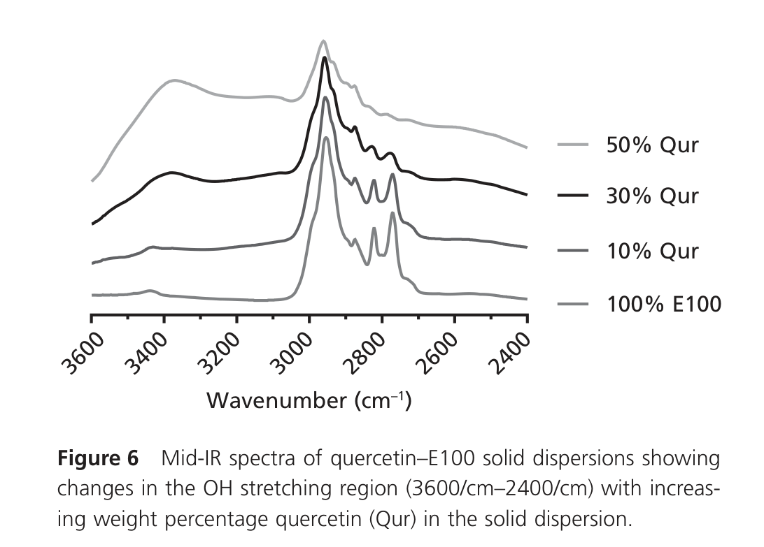
Left, the physical mixes keep the amorphous doublet at every PVP content; right, the dispersions lose the dimer band and gain the bands near 1726 and 1636 cm⁻¹. Percentages are wt% PVP; 0% is amorphous indomethacin and 100% pure PVP taylor1997 R10, R11, R12.
Why
The authors' reasoning, marked as their interpretation: indomethacin has a single donor, its acid OH, and PVP can only accept a proton, so a new bond must show in the acid C=O and in the PVP carbonyl or C–N bands taylor1997 C25. The more basic PVP amide "would explain" why the acid OH bonds to the PVP carbonyl in preference to forming the acid dimer taylor1997 C36. The spectra "support the fact" that the bond forms at the expense of dimers, and the physical mixes show none, so the bond needs molecular mixing taylor1997 C35, C26. The 1726 cm⁻¹ band is assigned to the non-bonded acid C=O of a PVP-bonded molecule and the 1636 cm⁻¹ band to hydrogen-bonded PVP carbonyl; the liquid models reproduce the changes and are used for the assignment taylor1997 C32, C33. Since spectral change continues above the stoichiometric composition, "this suggests" a low equilibrium constant and/or a stoichiometry below 1:1, so dimers, PVP-bonded drug and free PVP coexist, and these species are not taken to be separate phases, on the strength of a single Tg reported earlier taylor1997 C37, C38, C39. Non-ideal mixing or frozen-in concentration fluctuations are not excluded taylor1997 C40. The authors then propose that a γ nucleus, built of dimers, "would require" breaking the PVP–drug bond, so that the bond "offers an explanation" of why PVP inhibits crystallization at levels where its antiplasticizing effect is minimal taylor1997 C43, C45. What was not measured: crystallization, nucleation or stability in these dispersions taylor1997 X8, band intensities, areas or fits taylor1997 X5, and the phase state (the single Tg is cited) taylor1997 X9. The 1735 and 1726 cm⁻¹ bands are both called non-hydrogen-bonded acid carbonyl, and how one turns into the other between 5 and 20 wt% is not shown taylor1997 X4.
Conditions and limits
One acid drug with one donor, one PVP grade (PVP-K90, Tg 177 °C cited), methanol-cast dispersions dried under vacuum; IR solids were ground with KBr, the physical-mix preparation, sample temperature and water content are not printed taylor1997 M4, P3, X7. The comparison with methylpyrrolidone is qualitative and starts at 30 wt% methylpyrrolidone, whereas the dispersion changes begin at 5 wt% PVP taylor1997 X6.
Counterexamples and conflicts
None found in the library that contradicts the bond. The only discrepancy is internal: the two-bond threshold of 12 wt% PVP is a halved weight fraction, not a halved mole ratio, and should be about 13.6 wt% taylor1997 X2.
Transfer check
For an acid drug in PVP, record amorphous drug (melt-quenched), a solvent-evaporated dispersion and a physical mix of the same composition by IR, and look for three markers without any crystal reference: loss of the dimer band (1710 cm⁻¹ here), a free acid C=O band 16 cm⁻¹ higher (1726 cm⁻¹; synthesis: 1726 − 1710) and a PVP carbonyl band far below its free position (1636 against 1679 cm⁻¹) taylor1997 R5.h, R11, R12. Use Raman to test for dimers by the mutual exclusion rule taylor1997 C14. Do not expect all drug to be bonded at the stoichiometric composition taylor1997 C37. For a drug with several donors, intramolecular bonds or no donor, see F4 and F14.
History
Yoshioka and co-workers found that PVP inhibits indomethacin crystallization at low levels beyond what antiplasticization explains; this paper (1997) supplied the first spectroscopic evidence in the library of the molecular contact that could be responsible taylor1997 C5, C45. Kestur and Taylor (2010) took the bond as a proposed stabilization mechanism and tested it across polymers for felodipine (see F12) kestur2010a C3. Marsac and co-workers (2009) later measured a 45 cm⁻¹ PVP carbonyl shift for indomethacin, the largest of five compounds (see F2) marsac2009 R12.c. Rumondor and co-workers (2010) cited the larger carbonyl red shift of indomethacin against felodipine, quinidine and pimozide as evidence of stronger interactions with PVPVA rumondor2010c C28. Wegiel and co-workers (2013) read polyphenol–polymer OH and carbonyl shifts the same way (see F25) wegiel2013a C8, C36, and Que and co-workers (2020) took the 20–30 cm⁻¹ red shifts of drug–PVP hydrogen bonds as the comparator for the 2–6 cm⁻¹ shifts of halogen bonds (see F20) que2020 R17.q, C25.
Related: PEG dispersions F3, Crystallization tendency F8.