Topic 1Light-scattering and absorption read-outs in particle-containing solutions1 of 3
An in-situ UV probe overestimates the dissolved drug concentration when submicron drug particles are suspended in the medium.
A felodipine suspension of 300 nm particles was read as 3.4 to 8.95 µg/mL "dissolved" by a UV probe, although only about 0.81 µg/mL can dissolve. Coarse particles (10–380 µm) read only about 0.9–2.1 µg/mL vaneerdenbrugh2011b R1, R2, R6.a, R7, M1.
moderateModerate: the effect is large, rises in a straight line with the amount of solid (R² 0.995) and is absent for coarse particles, but the cause is inferred by ruling out other explanations, not measured directly vaneerdenbrugh2011b K1, K3, K4, X4.
Felodipine in 1 mg/mL PVP at 25 °C: the reading for 300 nm particles keeps rising with the amount of solid, while the solubility stays at 0.81. vaneerdenbrugh2011b R6.a, R7, R6.b–e, M1
For your drug
- Compare an in-situ UV reading with a separation-based value (ultracentrifugation then HPLC, or flux) on the same suspension, and check that it does not keep rising with added solid above the known solubility.
- Measure the particle size: the evidence is for particles below about 1 µm, and a flat baseline correction may suffice above about 2 µm.
vaneerdenbrugh2011b C33, C36, C38, X5
How it works and key numbers
- Light taken out of the beam by a particle is either absorbed or scattered; for small particles absorption dominates vaneerdenbrugh2011b C3, C26.
- The usual correction (a second derivative) removes sloping scattering baselines, but not a particle absorption band that lies inside the drug's own band vaneerdenbrugh2011b C2, C23, C25.
- So the probe reports an apparent concentration that rises with the amount of solid vaneerdenbrugh2011b C25.
- In support, NMR showed no solubility gain (89 ± 10% of the coarse value), no amorphous form was found, and a Mie calculation matches the size of the signal (see F2); the authors word the claim "might absorb" in the abstract and "demonstrate" in the conclusions vaneerdenbrugh2011b R8, R10, R13, C24.
Evidence: data, conditions and papers
Finding F1. In-situ UV concentration readings overstate the dissolved drug when submicron drug particles are present, even after scattering correction
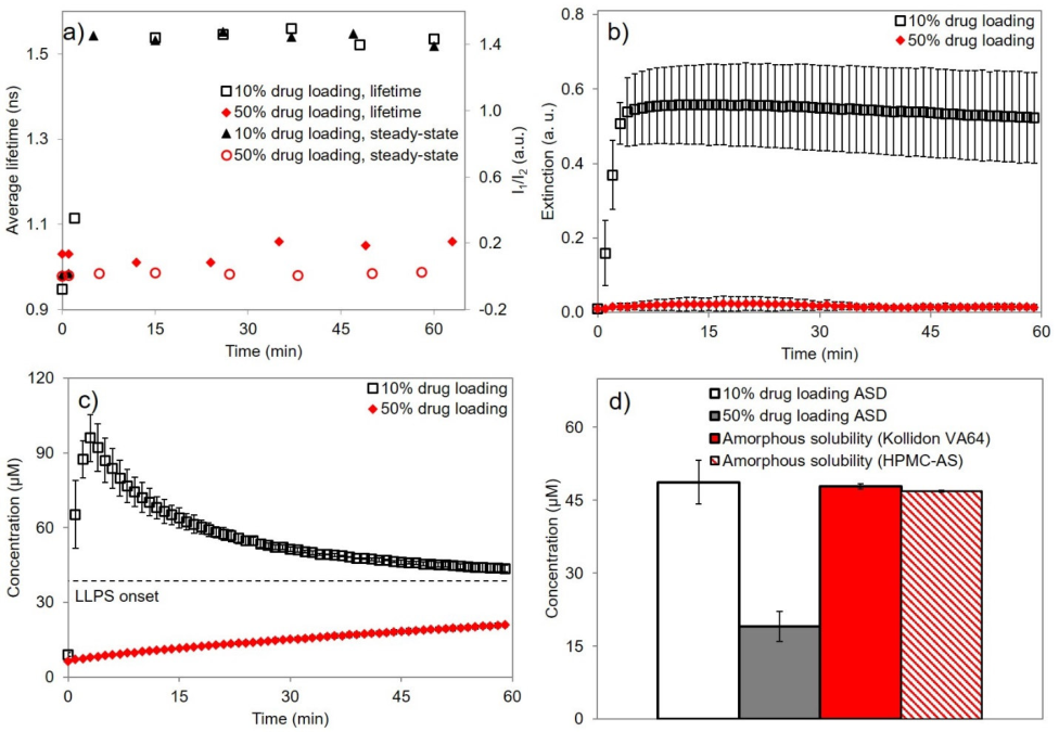
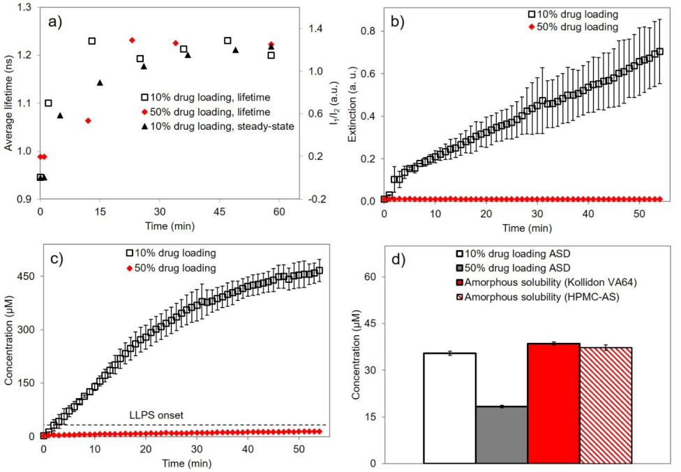
In short. In a 300 nm felodipine nanosuspension in 1 mg/mL PVP, a fiber-optic UV probe with second-derivative correction read 3.4, 5.75, 7.85 and 8.95 µg/mL of "dissolved" drug at 10, 25, 40 and 50 µg/mL total, a slope of 0.14, against a solubility of about 0.81 µg/mL; suspensions with median sizes of 10–380 µm read about 0.9–2.1 µg/mL vaneerdenbrugh2011b R1, R2, R6, R7, M1. The excess was not a solubility gain (NMR gave 89 ± 10% of the coarse-material value, no amorphous form was found), and the authors conclude that the submicron particles absorb light like dissolved drug vaneerdenbrugh2011b R8, R10, C24. The same overreading was reported when drug-rich nanoparticles or droplets formed during dissolution of felodipine, danazol, ritonavir and lopinavir dispersions alonzo2011 C27; jackson2015 R2.c, R9.i; tres2018 R16, R17.
Type. observation in one drug (five particle sizes), with later confirmations in dispersions of felodipine, danazol, ritonavir and lopinavir Strength. the artifact is large, linear over four concentrations (R² 0.995) and absent for coarse particles, but its attribution to particle absorption rests on excluding a solubility gain in a different solvent and on a Mie match vaneerdenbrugh2011b K1, K3, K4.
How it was shown
Van Eerdenbrugh, Alonzo and Taylor asked whether undissolved drug particles disturb in-situ UV readings only by scattering light or also by absorbing it vaneerdenbrugh2011b C2, C4. A felodipine nanosuspension (300 nm) and four coarser suspensions (median 10–380 µm) were diluted to 1 mg/mL PVP and 10–50 µg/mL total drug at 25 °C and read with a fiber-optic UV probe using second derivatives vaneerdenbrugh2011b R1, R2, P4, A1, P9. A correct reading would stay at the solubility, about 0.81 µg/mL vaneerdenbrugh2011b M1, A2.
What the data show
The nanosuspension read 3.4, 5.75, 7.85 and 8.95 µg/mL at 10, 25, 40 and 50 µg/mL total (slope 0.14), the coarse suspensions only about 0.9–2.1 µg/mL vaneerdenbrugh2011b R6.a, R7, R6.b–e. NMR gave 89 ± 10% of the coarse solubility and no amorphous form was found vaneerdenbrugh2011b R8, R10. Later ASD dissolution studies report the same overreading (table) alonzo2011 R1; jackson2015 R2.c; tres2018 R8.
| paper | system | result | ids |
|---|---|---|---|
| vaneerdenbrugh2011b | felodipine nanosuspension, 300 nm, 1 mg/mL PVP, 10–50 µg/mL total | apparent dissolved ≈3.4 to ≈8.95 µg/mL; solubility ≈0.81 | vaneerdenbrugh2011b R6.a, R7, M1 |
| vaneerdenbrugh2011b | jetmilled and sieved suspensions, 10.19–380.21 µm | ≈0.9–2.1 µg/mL, slight upward trend | vaneerdenbrugh2011b R6.b–e, R2, C20 |
| alonzo2011 | 90:10 and 50:50 HPMC–felodipine ASDs | UV peak ≈25–30 against ≈9 µg/mL; diffusion and NMR nearly equal | alonzo2011 R1, R14, R15 |
| jackson2015 | danazol 90:10 HPMCAS film | UV ≈16.2 against flux 8.2 ± 1.2 µg/mL | jackson2015 R2.c, R9.i |
| tres2018 | ritonavir 10% dispersion | UV peak 96 then 44 µM; supernatant 49 µM | tres2018 R8, R9 |
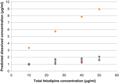
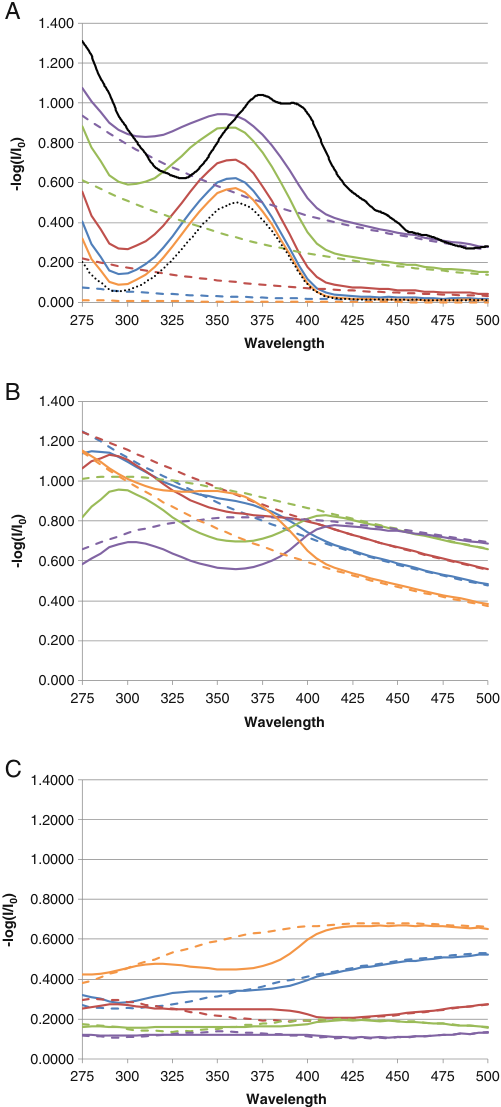
The orange nanosuspension series climbs to about 9 µg/mL, while the coarse series stay near 1–2 µg/mL vaneerdenbrugh2011b R6, R7.
Why
Light removed by a particle is absorbed or scattered, and for small particles absorption dominates vaneerdenbrugh2011b C3, C26. A second derivative removes sloping scattering baselines but not a particle absorption band inside the drug's own band vaneerdenbrugh2011b C2, C23, C25. The attribution rests on excluding a solubility gain and on a Mie match; the aqueous dissolved concentration was not measured vaneerdenbrugh2011b C24, X4.
Limits and transfer
One drug, crystalline particles, 1 mg/mL PVP, 25 °C; nothing was measured between 0.38 and 10 µm vaneerdenbrugh2011b X5, X7. Coarse suspensions read up to about 2.6 times the solubility (synthesis: 2.1/0.81), which the authors ascribe to noise vaneerdenbrugh2011b C22. Compare any in-situ UV reading with ultracentrifugation plus HPLC or flux, and measure the particle size vaneerdenbrugh2011b C36, C38.
How it was shown
Van Eerdenbrugh, Alonzo and Taylor asked whether undissolved drug particles disturb in-situ UV concentration readings only by scattering light, as the dissolution literature assumed, or also by absorbing it vaneerdenbrugh2011b C2, C4. A crystalline felodipine nanosuspension was made by wet media milling: 0.5 g felodipine and 0.5 g PVP in 5 mL water per vial, eight vials, 5 g of 0.5 mm zirconia beads, stirring at 100 rpm until dynamic light scattering showed a mean size near 300 nm; the batches were pooled, left overnight so that large particles and agglomerates settled, and the supernatant was kept vaneerdenbrugh2011b P1. Four coarser suspensions came from jet milling (sieved at 53 µm) and from hand grinding and sieving (three fractions) vaneerdenbrugh2011b P2, P3. All suspensions were diluted in aqueous PVP to 1 mg/mL PVP and 10, 25, 40 or 50 µg/mL total felodipine at 25 °C vaneerdenbrugh2011b P4, A1, A2. A six-channel Ocean Optics system with 1 cm transflectance dip probes scanned 200–700 nm; spectra were smoothed (Savitzky–Golay, which changed neither peaks nor intensities), and the apparent dissolved concentration came from second derivatives over 270–440 nm calibrated with 0.5–20 µg/mL methanolic standards vaneerdenbrugh2011b P9, R11. A correct reading would stay at the solubility whatever the solids content, because total drug is 12 to 62 times above 0.81 µg/mL (synthesis: 10/0.81 = 12.3; 50/0.81 = 61.7) vaneerdenbrugh2011b M1, A2. Three explanations other than particle absorption were checked: higher solubility of small crystals (cited theory and an earlier study put the gain at 100–115% for crystals of this size), tested by ¹H NMR relative solubility in ethanol/water 25/75 for 24 h at 25 °C in triplicate; an amorphous fraction, tested by DSC, XRPD, Raman and mid-IR; and solubilisation by PVP vaneerdenbrugh2011b R16, P8, P5, R9. Particle size came from DLS (nanosuspension) and laser diffraction (all five) vaneerdenbrugh2011b P6, P7.
What the data show
The five suspensions covered three orders of magnitude: the nanosuspension had a Z-average of 300 ± 3 nm (PDI 0.141 ± 0.006) and d(v,0.5) 0.38 µm with a tail to d(v,0.9) 1.90 µm; the others had d(v,0.5) of 10.19 (jetmilled), 38.39, 105.03 and 380.21 µm vaneerdenbrugh2011b R1, R2. Nanosuspension spectra rose at every wavelength with total felodipine: at 276 nm from about 0.24 to 1.28 and at the band maximum near 375 nm from about 0.20 to 1.04, on top of a Tyndall-shaped scattering baseline, against a band of only about 0.015 for 0.81 µg/mL in methanol vaneerdenbrugh2011b R3. The jetmilled suspension peaked at about 0.073 (band maximum at 50 µg/mL); its baseline was almost flat and rose from about 0.011 to 0.046 at 425 nm between 10 and 50 µg/mL vaneerdenbrugh2011b R4. After second-derivative correction the nanosuspension read about 3.4, 5.75, 7.85 and 8.95 µg/mL, and the regression slope was 0.14 (R² = 0.995), that is 1.4 µg/mL of apparent increase per 10 µg/mL of particles vaneerdenbrugh2011b R6.a, R7. A straight line through the four points has an intercept of about 2.1 µg/mL (the extractor's fit), so the reading at 10 µg/mL is about 4.2 times the solubility (synthesis: 3.4/0.81), more than the slope alone accounts for; the slope should not be used alone to correct readings vaneerdenbrugh2011b X9. The coarse suspensions stayed nearly flat with a slight upward trend: jetmilled about 1.0 to 1.6, fraction 1 (38.39 µm) about 0.94 to 1.25 (no marker at 50 µg/mL), fraction 2 about 1.07 to 1.9 at 40 µg/mL (1.75 at 50) and fraction 3 about 1.0 to 2.1 µg/mL vaneerdenbrugh2011b R2, R6.b–e, X11. The highest value is about 2.6 times the solubility (synthesis: 2.1/0.81); the authors put it "most likely" down to the low absolute absorbance of a saturated solution, which turns small absolute errors into large relative ones, and did not pursue it vaneerdenbrugh2011b C22. The three alternative explanations did not hold: the nanosuspension solubility was 89 ± 10% of that of the coarse powder, no amorphous felodipine was detected in any powder or in the freeze-dried nanosuspension, and PVP raised solubility only above 5 mg/mL, five times the 1 mg/mL used vaneerdenbrugh2011b R8, R10, R9. The authors write that the NMR result "independently confirms" that solubility is not the cause and that the excess "strongly suggests" absorption by submicron particles vaneerdenbrugh2011b C15, C24.
| paper | system | method and conditions | result | ids |
|---|---|---|---|---|
| vaneerdenbrugh2011b | felodipine nanosuspension (Z-average 300 nm), 1 mg/mL PVP | fiber-optic UV, second derivative, 25 °C, 10–50 µg/mL total | apparent dissolved ≈3.4 to ≈8.95 µg/mL; slope 0.14, R² 0.995; solubility ≈0.81 µg/mL | R6.a, R7, M1 |
| vaneerdenbrugh2011b | jetmilled and sieved suspensions, d(v,0.5) 10.19–380.21 µm | same | ≈0.9–2.1 µg/mL, slight upward trend | R6.b–e, R2, C20 |
| vaneerdenbrugh2011b | nanosuspension against coarse felodipine | ¹H NMR, ethanol/water 25/75, 24 h, 25 °C | 89 ± 10% of the coarse solubility | R8 |
| alonzo2011 | 90:10 and 50:50 HPMC–felodipine ASDs, 37 °C | in-situ UV against diffusion-cell slope and NMR peak area | UV peak ≈25–30 µg/mL (90:10) against ≈9 µg/mL (50:50); slope ratios 10.5 and 9.5; NMR areas 1370 and 1014 | R1, R14, R15 |
| alonzo2012 | felodipine with HPMC, 25 °C | second-derivative PLS UV, check for a red shift of the 361 nm peak | no red shift seen, so no nanoparticle signature | P3, R20 |
| jackson2015 | danazol 90:10 HPMCAS ASD film, 25 °C | in-situ UV against diffusion-cell flux | UV maximum ≈16.2 µg/mL; flux-derived free drug 8.2 ± 1.2 µg/mL | R2.c, R9.i |
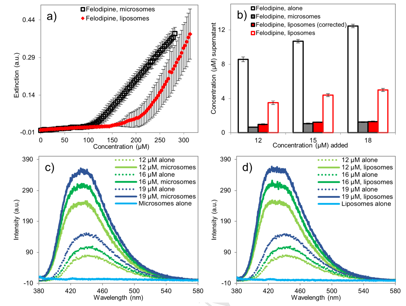
| almeidaesousa2016 | weak base CRH1, forms in pH 4.6 buffer with HPMC | in-situ UV | apparent concentration; nanometre LLPS species absorb like dissolved drug | C20 |
| tres2018 | ritonavir and lopinavir, 10% drug-loading ASDs | in-situ UV (240 or 260 nm minus 450 nm) against ultracentrifuged HPLC | ritonavir UV peak 96 µM then 44 µM, supernatant 49 µM; lopinavir UV ≈466 µM, supernatant 36 µM | R8, R16, R9, R17 |
Figure
Why
The authors' reasoning, step by step (their interpretation): light removed from the beam by a particle is absorbed or scattered, and Mie theory gives both from size, wavelength and refractive indices; for small particles absorption dominates vaneerdenbrugh2011b C3, C26. A second derivative removes constant and smoothly sloping baselines, which is how Tyndall scattering is usually corrected, but it cannot remove a particle absorption band that lies inside the drug's own band vaneerdenbrugh2011b C2, C23, C25. So the probe reports an apparent concentration that rises with the amount of solid vaneerdenbrugh2011b C25. They word it "might absorb" in the abstract, "strongly suggests" in the results and "demonstrate" in the conclusions vaneerdenbrugh2011b C24. What was measured in support: no solubility gain by NMR, no amorphous form, and a Mie calculation with the drug's own absorptivity that reproduces the size of the signal for 300 nm particles (see F2) vaneerdenbrugh2011b R8, R10, R13. What was not measured: the dissolved concentration in the aqueous nanosuspension itself (the NMR test used ethanol/water) and a particle-free aqueous reference spectrum, so absorption by the particles was inferred by exclusion plus modelling, not isolated vaneerdenbrugh2011b X4. A practical sign is the band position: the nanosuspension band peaked near 375 nm, against about 355–365 nm for the methanolic standard and the jetmilled suspensions, and the nanoparticle spectrum shows a red shift of the 361 nm peak vaneerdenbrugh2011b R3, R4; alonzo2012 R20. In the 90:10 HPMC–felodipine dispersion the shift appeared within 5 min of the start of dissolution and was absent for the 50:50 dispersion, pure amorphous felodipine and the dissolved controls alonzo2011 R20. Alonzo and co-workers explain their UV overreading for the 90:10 dispersions by submicron particles of 200–400 nm that absorb like the solution and are not removed by second derivatives alonzo2011 C27. Jackson and co-workers (2015) and Almeida e Sousa and co-workers (2016) say the same of drug-rich nanospecies above the amorphous solubility; Tres and co-workers (2018) add that apparent concentrations are then "not quantitative" jackson2015 C12; almeidaesousa2016 C20; tres2018 C37.
Conditions and limits
The controlled experiment covers one drug (felodipine), crystalline milled or ground particles, 1 mg/mL PVP, water at 25 °C and a 1 cm probe vaneerdenbrugh2011b A1, P9. No suspension between 0.38 and 10 µm was measured, no precipitated or amorphous particle was measured, and the expectation that lower solubility, more solids and smaller particles raise the error was tested only for solids and size vaneerdenbrugh2011b X5, X7, X6, C44. The derivative feature used for quantification is not stated and calibration was in methanol, whose band lies at shorter wavelength than the aqueous nanosuspension band vaneerdenbrugh2011b X8. The later dispersion studies did not test particle absorption directly; they compared UV with flux, NMR or ultracentrifugation (table above).
Counterexamples and conflicts
None found in the library for the existence of the overreading. Open: the coarse suspensions also read above the solubility, up to about 2.6 times (synthesis: 2.1/0.81), which the authors ascribe to noise on a weak band vaneerdenbrugh2011b C22, and the text value of 0.034 a.u. for a saturated solution disagrees with the plotted peak of about 0.016 vaneerdenbrugh2011b R5, X2.
Transfer check
Before trusting an in-situ UV reading of another drug: (1) compare it with a separation-based value (ultracentrifugation followed by HPLC, or flux) on the same suspension; (2) test whether the reading keeps rising in proportion to added solid above the known solubility, which is the signature of this artifact; (3) compare the band position with the solution standard, since a red shift points to particles; (4) measure the particle size (DLS or NTA): the evidence is for particles below about 1 µm, and a flat baseline correction may suffice above about 2 µm, a statement based on calculation only vaneerdenbrugh2011b C33, C36, C38, X5; (5) a drug with lower solubility, higher solids or smaller particles is outside the tested range and the error should be expected to be larger, not smaller vaneerdenbrugh2011b C44.
History
2011: Van Eerdenbrugh and co-workers first show the artifact for a felodipine nanosuspension and explain it by particle absorption vaneerdenbrugh2011b C23, C24. Alonzo and co-workers (2011) use it to read the discrepancy between UV, diffusion and NMR for 90:10 dispersions alonzo2011 C27. Alonzo and co-workers (2012) adopt the second-derivative method and check the 361 nm peak for the red shift alonzo2012 P3, R20. Jackson and co-workers (2015) measure the size of the overreading for a danazol dispersion against flux (16.2 against 8.2 ± 1.2 µg/mL) jackson2015 R2.c, R9.i. Almeida e Sousa and co-workers (2016) and Tres and co-workers (2018) carry the caveat to a weak base and to ritonavir and lopinavir almeidaesousa2016 C20; tres2018 C37.