Category

Characterization methods

The methods that show what a drug sample contains and what misleads, grouped by the state of the sample. The solid-state chapter covers powders, films, extrudates and spray-dried dispersions.

What this page covers

crystallinity by FT-Raman, PXRD with chemometrics and second harmonic generation (SHG), trace crystals and the onset of crystallization, nanoscale infrared mapping of phases, and fluorescent probes, confocal imaging, DSC and solid-state NMR as tests of phase separation. The solution chapter covers supersaturated solutions and dissolving dispersions: in-situ UV concentration probes, extinction as a marker of a second phase, environment-sensitive fluorescent probes, fluorescence lifetime, and their use to follow liquid-liquid phase separation (LLPS), crystallization of the drug-rich phase and ASD dissolution at two drug loadings. The category is a toolbox for the other categories, so its findings say which method answers which formulation question and what each method cannot see. It has two chapters, Solid-state methods and Solution methods, and 14 papers from 1998 to 2018. The category findings are in findings.md, the links between the chapters in links.md and the citation keys in sources.md.

2 chapters14 papers1998–20187 category findings

01How the parts fit together

  • Solution methods → Solid-state methods polarity probes calibrated in solution are carried to films and dispersions (F1)
  • Solid-state methods → Solution methods birefringence, XRD and crystal-selective SHG are the checks that solution probes lack (F3, F4)
  • Solid-state methods → Solution methods matrix separation on wetting versus a drug-rich phase in the medium (F2)

The two chapters read the same events, a drug-rich region and its crystals, in the sample before and after water arrives. purohit2015b R6, R11; tres2018 R6, R9; raina2015a R16

The two chapters ask the same few questions of a drug sample in two states: has a drug-rich region formed in the polymer matrix or in water, how big is it, and has it crystallized. The solid-state chapter asks them of a powder, film or extrudate as made, stored and wetted; the solution chapter asks them of the medium after water has arrived and drug has been released. Their questions meet where an ASD is wetted: the matrix can separate in the first seconds, and the medium can reach the amorphous solubility and form a drug-rich phase or not.

The bridge between the chapters is the fluorescent probe. Pyrene, prodan and the styryl dyes were calibrated in solvents and in supersaturated solutions, then carried to films and dispersions, and in both states they report that a less polar, drug-like region exists, as a rank within one drug, probe and medium and not as a polarity (F1). Where the two states meet in ritonavir, a PVP matrix that separates when wetted did not reach the amorphous solubility at high loading, while an HPMCAS matrix that stayed mixed did, so separation in the matrix and a drug-rich phase in the medium are different events (F2). When the drug-rich region crystallizes the probe signal returns in both states, but only the film was confirmed by a second method (F3).

The other three findings come from reading what the methods can and cannot say. Every method has a blind spot or a look-alike signal that appeared only against a second method (F4); detection limits, onsets and induction times belong to the read-out and its protocol (F5); and an optical signal becomes an amount only when the standard has the same physical form as the sample (F6). Together they give the map of F7: for each question of the other categories, a method set and the second read-out it needs.

02Chapters

chapterpapersyearswhat it coversits key findings
Solid-state methods101998–2017Crystallinity of powders and dispersions by Raman, PXRD with PLS and SHG; trace crystals and early crystallization; AFM-IR of phases and drug particles; probes, imaging, DSC and ssNMR for phase separation under humidity and by drying routeF7 detection limits, F10 SHG earlier than XRPD, F11 what a dark SHG image proves, F17 humidity and the probe signal
Solution methods42011–2018UV probes and submicron particles, Mie theory, extinction, environment-sensitive probes and lifetime for LLPS, crystallization of the drug-rich phase, ASD dissolution at two loadings, vesicle mediaF3 extinction onset, F5 four onsets agree, F10 10% against 50% loading, F11 UV and probe can mislead

03Category findings

Conclusions that appear only when the chapters are read together. Click a card to read it.

04Which chapter each finding draws on

Show the table of chapters and findings
Probe read-outs across solid and solutionWhat a reading can and cannot saySignal to amountChoosing the method for each formulation question of the other categories
chapterF1F2F3F4F5F6F7
Solid-state methods
Solution methods

Dot size = number of chapter findings each category finding draws on.

05Connections to other categories

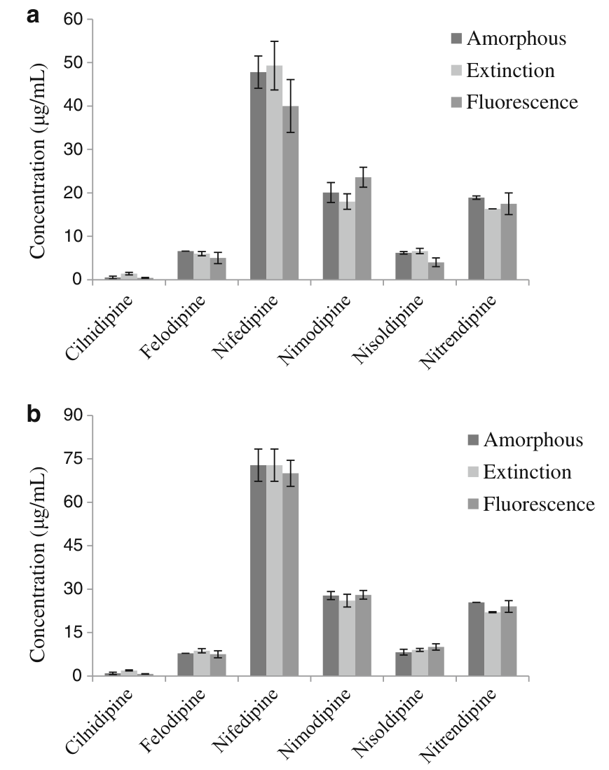
Dissolution / C. Amorphous solubility and LLPS. Question D. The extinction, probe and lifetime onsets are the methods used there: pyrene and 450 nm extinction gave 11.31 ± 0.56 and 14.15 ± 0.87 µg/mL for the weak base CRH1, and a dye blue shift marked the UV LLPS concentration of nicardipine almeidaesousa2016 R3; indulkar2015 R12. The reference onsets of felodipine (19 µM), clotrimazole (22), efavirenz (58), ritonavir (39) and lopinavir (28) were measured there tres2017 M1, M2, M3; tres2018 M1, M2. See F5 and F7.

Dissolution / B. Supersaturation from ASD dissolution. Question E. The particle-absorption explanation of UV overreading was used for felodipine and danazol dispersions, and the 10%-against-50% loading contrast reappears for posaconazole in FaSSIF alonzo2011 C27; jackson2015 C12; elkhabaz2019a C15. See F2, F6 and F7.

Dissolution / A. Release mechanism and LoC. Question C. The polymer-controlled and drug-controlled readings of the 10% and 50% dispersions match the loading dependence found for ritonavir films, and the probe evidence that PVP films separate on hydration is the starting point of the race between dissolution and separation in those films tres2018 C23, C24; purohit2017b C34, C54, C5, C3. See F2.

Dissolution / D. Crystallization from solution. Question F. The pyrene return and the UV fall define induction times for danazol, the solution-state ranking of the six dihydropyridines was made there, and SONICC separated crystals from undissolved amorphous material in biorelevant-media dissolution jackson2014 C30, C35; raina2015b C13, C28; elkhabaz2017 C8. See F3, F5.

Solid state stability / Miscibility and drug-polymer interactions. Question B. The chapters take the partial miscibility of felodipine–PAA and the non-additive halo of a molecular-level dispersion, and give AFM-nanoIR, probe and ssNMR evidence of phase separation in itraconazole–HPMC despite one Tg, and the spectral-mixing caution that applies to telaprevir films vaneerdenbrugh2012b R1; rumondor2010b C8; purohit2015a C36, C23; li2016c C14, C21, C46. See F1, F4.

Solid state stability / Physical stability. Questions A and C. The probe read-out of water-induced separation and the SHG detection of crystallization in storage tests were used there for miconazole–PVPVA, celecoxib dispersions and indomethacin films, and the felodipine crystallization kinetics against humidity motivated the PXRD quantification saboo2017 R12, R13, C41; xie2016b C5, C32; tian2016 C16, C17; rumondor2010b C4; rumondor2009b C7. See F3, F5.

Solid state stability / Crystallization tendency. Question F. The low crystallization tendency of felodipine is why it was assumed to stay amorphous in the AFM-IR films, SHG imaged seeds in a quenched film, and the observation that fast crystallizers crystallize before they phase separate is a solution-state test of the neat-drug classification vaneerdenbrugh2012b C4; wanapun2010 R4, C8; raina2015a R17, C37, C11. See F3, F4.

ASD process. Questions A and B. SHG and TEM find residual crystals in extrudates below the XRPD limit, the ladder of method sensitivity comes from extrudate studies, and water in the solvent or air can cause phase separation of itraconazole–HPMC and evacetrapib films shah2024 R10; moseson2018a R9, C10; moseson2021 R6; mugheirbi2017 C1, C37; li2017 C50. See F4, F5, F7.

Bioperformance. Questions A and G. The rise of the felodipine aggregation onset with microsomes and liposomes extends the micelle and FaSSIF results of the membrane-transport work, and the tacrolimus capsule papers report an apparent lag before crystals are seen with XRPD limits of 1–10% (synthesis: part of such a lag is set by the limit) tres2017 R5; raina2015c R11, C30; hate2019 R2; purohit2018 R1, R2. See F5, F7.

How to read this page. Each finding is one conclusion: a headline, a plain statement, one picture and what it means for your drug. Open How it works for the steps and numbers, and Evidence for the data and conditions, at medium (B) or full (C) length. The coloured tag grades the evidence as strong, moderate or weak. Dotted words show a definition. An id such as hate2017 R15 names a paper and an item in its card. Text marked (synthesis: …) joins several papers and is not one paper's claim.

Evidence length

Topic 1 of 4 · 3 findings · F1–F3

Probe read-outs across solid and solution

F1

Topic 1Probe read-outs across solid and solution1 of 3

A fluorescent probe whose emission shifts in less polar surroundings shows that a drug-rich region exists, but not its polarity.

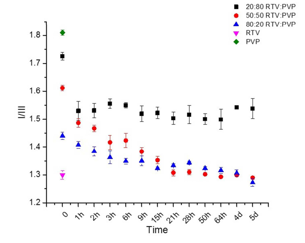
Fluorescent probes shift their emission when they are in less polar surroundings. In films and dispersions, and in supersaturated solutions, the probe signal moved toward the value it gives in the pure drug once a drug-rich region formed. For example, the ratio of the pyrene probe, which falls as the surroundings become less polar, went from 1.60 to 1.30 in ritonavir–PVP films at 97% RH purohit2015b R6, R15; purohit2017a R7; raina2015a R1, R6.

moderateModerate: the direction is the same in both states and was checked by other methods in the films, but the location of the probe was not measured purohit2015b K2, K3, X8; raina2015a K1, X17.

beyond the benzene valuedrug-rich phase ≈ 0.76benzene 1.07buffer 1.60.60.91.21.51.8pyrene I1/I3 ratio (lower = less polar surroundings)

Nifedipine with HPMC reached about 0.76, past the benzene value; buffer and benzene are the solvent references of the same paper. raina2015a R1, R6, R9, R16

dispersion powderspure drugpure polymeritraconazole–HPMCthree drying routes1.38–1.39itraconazole alone1.39HPMC alone1.541.31.351.41.451.51.551.6pyrene I1/I3 ratio of the powder

The powders read like the pure drug, not somewhere between drug and polymer. purohit2017a R7

For your drug

  1. Record the probe in films of the pure drug and the pure polymer prepared the way you prepare the dispersion; a miscible dispersion should lie between them and a separated one near the drug value.
  2. In solution, record the probe in a solvent series on your own instrument, read a result beyond the benzene value as "non-aqueous environment" and not as a polarity, and confirm the phase state by one probe-free method.

purohit2017a R24, R7; raina2015a R1, X6

How it works and key numbers
  1. A probe emits at a shorter wavelength (or, for pyrene, a lower ratio) in a less polar surrounding purohit2015b C4, C5, R4; raina2015a C25, C26.
  2. A lipophilic (poorly water-soluble) probe moves from the polar phase into a drug-rich region as soon as one forms purohit2015b C4, C5; raina2015a C25, C26.
  3. In the films the region is made when sorbed water replaces drug–polymer contacts in PVP, and pyrene "will preferentially partition" into it "given sufficient mobility" purohit2015b C24, C27; purohit2017a C7.
  4. In solution the region forms above the amorphous solubility (the drug concentration above which a drug-rich liquid phase forms), and more drug adds more phase without raising the free concentration, so more probe moves in raina2015a C26, C36; tres2017 C34.
  5. The location of the probe and its partition coefficient were not measured in either state purohit2015b C3; raina2015a X17. (synthesis: so a shift shows that a non-aqueous, drug-like region exists and, inside one drug–probe pair, at most a rank.)
Analogy. A thermometer calibrated in only one cup can say that this cup is warmer than that one, but not what the temperature is.
1.60 → 1.30pyrene ratio of ritonavir–PVP 50:50 films at 97% RH, the value of amorphous ritonavirpurohit2015b R6, R15
1.38–1.39itraconazole–HPMC powders, equal to the drug alone (1.39) and not to HPMC (1.54)purohit2017a R7
442.5 vs ≈ 451 nmthe same HPMC 606 read by the same probe in two papers, so each pair needs its own referencespurohit2015b R3; purohit2017a R13, X9
Evidence: data, conditions and papers

Finding F1. The same lipophilic probes move toward the pure-drug value when a drug-rich region forms, in amorphous films and dispersions as in supersaturated solutions, but the value is a rank within one drug, probe and medium and not a polarity

In short. In spin-coated films of amorphous probucol and procaine pyrene read 1.2 against 1.4 and prodan 418 against 448 nm, and prodan across ten polymers ran from 460.0 nm in PAA to 427.0 nm in PVAc in the order of their solubility parameters purohit2015b R1, R3 (Solid-state methods F16). In separated dispersions the pyrene ratio sat at the value of the drug: ritonavir–PVP at 97% RH fell from 1.60 to 1.30, and itraconazole–HPMC powders read 1.38–1.39, equal to itraconazole alone (1.39) and not between itraconazole and HPMC (1.54) purohit2015b R6; purohit2017a R7 (Solid-state methods F17, Solid-state methods F19). In supersaturated solutions the same direction holds from buffer to benzene (pyrene 1.6 to 1.07), and in a felodipine or nifedipine drug-rich phase three probes went past their benzene values raina2015a R1, R6, R9, R16 (Solution methods F4). (synthesis: a scale built in one state cannot be carried to the other.)

Type. observation in two states, films and dispersions of 4 drugs and 10 polymers, and solutions of 10 drugs Strength. the direction is the same in both states and was checked against probe-free methods in the films (IR, DSC, imaging, XRD) and against extinction and a thermodynamic estimate in solution, but probe partitioning was inferred and not measured in either purohit2015b K2, K3, X8 (Solid-state methods F17); raina2015a K1, X17 (Solution methods F5).

How it was shown

Purohit and Taylor spin coated amorphous probucol and procaine with probes, ten polymers with prodan, and drug–polymer films with 0.1% probe stored at 97% RH purohit2015b P2, P4, P1, P5. Raina and co-workers diluted six probes in water, buffer and organic solvents, then read each within 1 min after stepwise drug additions raina2015a P8, P9.

What the data show

The pyrene ratio of ritonavir–PVP 50:50 at 97% RH fell from 1.60 to 1.30, whereas ritonavir–HPMCAS went from 1.42 to 1.47 purohit2015b R6, R15. Itraconazole–HPMC powders read 1.38–1.39, equal to the drug alone and not between drug and HPMC (1.54) purohit2017a R7. In solution, three probes in a felodipine or nifedipine drug-rich phase went past their benzene values raina2015a R1, R6, R9, R16.

systemmethod and conditionsresultids
amorphous probucol / procaine filmsfour probes in spin-coated filmspyrene I1/I3 1.2 / 1.4; prodan 418 / 448 nmpurohit2015b R1
ten polymer filmsprodan emission maximum460.0 nm (PAA) to 427.0 nm (PVAc), in the order of the solubility parameterpurohit2015b R3
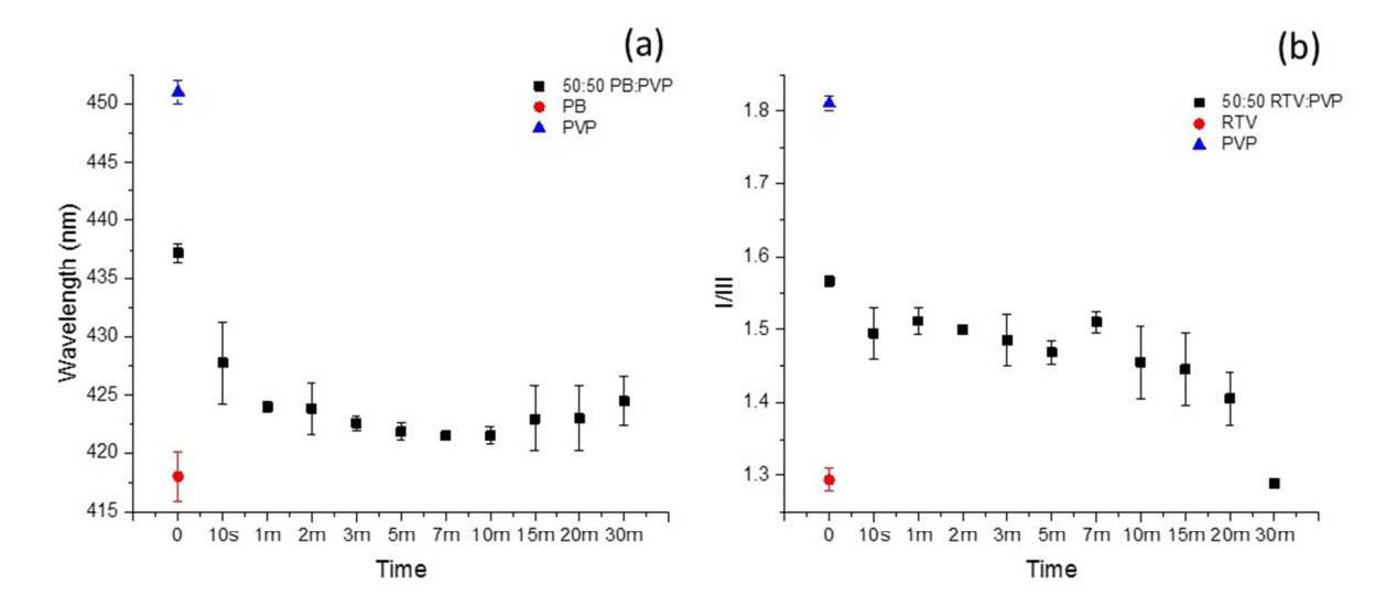
ritonavir–PVP 50:50, 97% RHpyrene I/III on the film1.60 to 1.30, the value of amorphous ritonavirpurohit2015b R6, R15
itraconazole–HPMC 40:60, three drying routespyrene I1/I31.38 / 1.39 / 1.39 against itraconazole 1.39 and HPMC 1.54purohit2017a R7
six probes, buffer to benzeneemission maximum in solventspyrene 1.6 / 1.2 / 1.07 (buffer / methanol / benzene); PRODAN 520 / 498 / 463 nmraina2015a R1
felodipine, 37 °C; nifedipine with HPMCDASPEI, 4-Di-2-ASP and pyrene, stepwise additionspyrene about 1.8 to 0.76 (benzene 1.07)raina2015a R6, R9, R16, R1
Fig. 5 of purohit2015b: pyrene I/III against time at 97% RH for ritonavir–PVP at three ratios
Fig. 5 · purohit2015bpyrene I/III against time at 97% RH for ritonavir–PVP at three ratios

A lower pyrene ratio means less polar surroundings, and the 50:50 and 80:20 curves approach the value of pure ritonavir purohit2015b R4, R6.

Why

Both groups of authors read a shorter wavelength or a lower ratio as a lipophilic probe moving into a drug-rich, less polar region purohit2015b C4, C5; raina2015a C25, C26. Neither the probe's location nor a partition coefficient was measured purohit2015b C3; raina2015a X17. (synthesis: a probe shift is a presence test for a non-aqueous, drug-like region and, inside one drug–probe pair, at most a rank purohit2015b R3; raina2015a R6, R9, R16.)

Limits and transfer

The same HPMC 606 gave 442.5 nm in one paper and about 451 nm in another, so each drug–probe pair needs its own references purohit2015b R3; purohit2017a R13, X9. Mugheirbi and co-workers found itraconazole–HPMC films spun at 7% RH homogeneous by TEM mugheirbi2017 R10, C1. Record the probe in pure-drug and pure-polymer films prepared as the dispersion is, and confirm the phase state by one probe-free method.

How it was shown

Purohit and Taylor spin coated amorphous probucol and procaine with a trace of probe, ten polymers with prodan, and drug–polymer films with 0.1% probe (prodan for probucol–PVP, pyrene for the ritonavir films), and stored the dispersion films at 97% RH purohit2015b P2, P4, P1, P5 (Solid-state methods F16, Solid-state methods F17). Purohit and co-workers dried one 40:60 itraconazole–HPMC solution by four routes and read pyrene on films and powders purohit2017a P1, P2, P3, P4, P5 (Solid-state methods F19). In solution, Raina and co-workers diluted each of six probes to 1 µg/mL in water, buffer and organic solvents to record the emission maximum, then read each probe within 1 min after stepwise additions of drug from methanol raina2015a P8, P9 (Solution methods F4). Tres and co-workers used PRODAN at 0.9 µM to read lifetimes tres2017 M4, P5 (Solution methods F4).

What the data show

In films probucol was the less polar host with all four probes, and the prodan maxima of ten polymers fell in exactly the order of their solubility parameters purohit2015b R1, R3 (Solid-state methods F16). At 97% RH the pyrene ratio of ritonavir–PVP fell from 1.60 to 1.30 at 50:50 and from 1.44 to 1.29 at 80:20, but only from 1.72 to 1.48 at 20:80, whereas ritonavir–HPMCAS went from 1.42 to 1.47 purohit2015b R6, R15 (Solid-state methods F17). The itraconazole powders of three drying routes all read like the pure drug purohit2017a R7 (Solid-state methods F19). In solution the emission moved from buffer to benzene by 16 nm (DASPEI) to 103 nm (dapoxyl) raina2015a R1 (Solution methods F4), and the probe onsets for five of six dihydropyridines lay within a factor of 1.0–1.7 of the extinction onset and of the estimated amorphous solubility raina2015a R11, X9 (Solution methods F5).

chapter findingsystemmethod and conditionsresultids
Solid-state methods F16amorphous probucol / procaine filmsfour probes in spin-coated filmspyrene I1/I3 1.2 / 1.4; prodan 418 / 448 nm; 4-Di-2-ASP 548 / 570 nm; dansyl glycine 473 / 501 nmpurohit2015b R1
Solid-state methods F16ten polymer filmsprodan emission maximum460.0 nm (PAA) to 427.0 nm (PVAc), in the order of the solubility parameterpurohit2015b R3
Solid-state methods F17ritonavir–PVP 50:50, 97% RHpyrene I/III on the film1.60 to 1.30, the value of amorphous ritonavirpurohit2015b R6, R15
Solid-state methods F17ritonavir–HPMCAS 50:50, 97% RHpyrene I/III; IR1.42 to 1.47; IR spectrum unchangedpurohit2015b R13, R15, R18
Solid-state methods F19itraconazole–HPMC 40:60, three drying routespyrene I1/I31.38 / 1.39 / 1.39 against itraconazole 1.39 and HPMC 1.54purohit2017a R7
Solution methods F4six probes, buffer to benzeneemission maximum in solventspyrene 1.6 / 1.2 / 1.07 (buffer / methanol / benzene); PRODAN 520 / 498 / 463 nmraina2015a R1
Solution methods F4felodipine, 37 °C; nifedipine with HPMC, 37 and 25 °CDASPEI, 4-Di-2-ASP and pyrene, stepwise additionsDASPEI about 578 to 558 nm (benzene 564); 4-Di-2-ASP about 610.5 to 579 nm (585); pyrene about 1.8 to 0.76 (1.07)raina2015a R6, R9, R16, R1
Solution methods F5six dihydropyridines, 25 and 37 °CDASPEI emission, extinction at 450 nm, thermodynamic estimatehighest over lowest of the three onsets 1.0–1.7 for five drugs, about 3 for cilnidipineraina2015a R11, X9

Figure

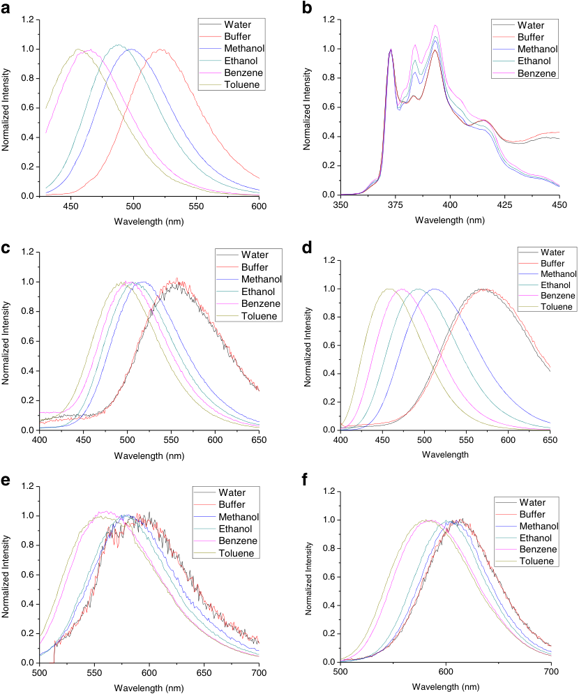
Fig. 3 of raina2015a: probe emission spectra in six solvents
Fig. 3 · raina2015aprobe emission spectra in six solvents
Fig. 5 of purohit2015b: pyrene I/III against time at 97% RH for ritonavir–PVP at three ratios
Fig. 5 · purohit2015bpyrene I/III against time at 97% RH for ritonavir–PVP at three ratios
In the solvent series each normalised spectrum moves to shorter wavelength from water and buffer towards toluene raina2015a R2 (Solution methods F4); in the film series a lower pyrene ratio means less polar surroundings, and the 50:50 and 80:20 curves approach the value of pure ritonavir purohit2015b R4, R6 (Solid-state methods F17).

Why

The authors of both groups of papers give the same mechanism as their interpretation: an environment-sensitive probe emits at shorter wavelength in a less polar surrounding, and a lipophilic probe moves from the polar phase into a drug-rich region purohit2015b C4, C5 (Solid-state methods F16); raina2015a C25, C26 (Solution methods F4). In the films the drug-rich region is made when sorbed water replaces drug–polymer contacts in PVP (a "classic case of solvent-induced immiscibility"), and pyrene "will preferentially partition" into it "given sufficient mobility" purohit2015b C24, C27; purohit2017a C7 (Solid-state methods F17, Solid-state methods F19). In solution the region forms above the amorphous solubility, and further drug adds phase without raising the free concentration, so more probe moves in raina2015a C26, C36; tres2017 C34 (Solution methods F5). Neither the location of the probe nor a partition coefficient was measured in either state purohit2015b C3 (Solid-state methods F16); raina2015a X17 (Solution methods F5). (synthesis: the same unmeasured step stands under both read-outs, so a probe shift is a presence test for a non-aqueous, drug-like region and, inside one drug–probe pair, at most a rank. The film values lie between those of pure-component films, while a solution drug-rich phase reads beyond the benzene value purohit2015b R3 (Solid-state methods F16); raina2015a R6, R9, R16 (Solution methods F4).)

Conditions and limits

In the films the extent of separation compares probe shifts only within a drug–probe pair, because prodan was used for probucol and pyrene for ritonavir, and only one HPMCAS dispersion was run at 97% RH purohit2015b P5, X7, X12 (Solid-state methods F17). The same probe gave 442.5 nm in one paper and about 451 nm in another for nominally the same HPMC 606, so each drug–probe pair needs its own references purohit2015b R3; purohit2017a R13, X9 (Solid-state methods F16). Water alone moves the probe the other way in single-component films, for example prodan in PVP from about 450.5 to 455.3 nm purohit2015b R9 (Solid-state methods F17). In solution the pyrene baseline differs between experiments (about 1.8 in the nifedipine series, 1.6 in the solvent table), so ratios are compared only within one experiment raina2015a X6, R1, R16 (Solution methods F4), and the drug's own fluorescence can mask a probe signal: with felodipine four of six probes gave an onset of 5.00–6.00 µg/mL, while the emission of dansyl glycine and dapoxyl was masked by the felodipine band raina2015a R12, R13 (Solution methods F6).

Counterexamples and conflicts

The itraconazole result is contested: Mugheirbi and co-workers found itraconazole–HPMC films spun at 7% RH homogeneous by TEM and films at 20–40% RH phase separated, so a pure-drug probe value shows that a drug-rich region exists in that sample, not that the pair is inherently immiscible mugheirbi2017 R10, C1 (Solid-state methods F19). The authors of the solution paper rank the styryl dyes as most polarity-sensitive, but these two dyes show the smallest solvent shifts in their own table raina2015a C29, R1, X4 (Solution methods F4). None found that contradicts the direction of the shift in either state.

Transfer check

(1) For a dispersion, record the probe in films of the pure drug and the pure polymer prepared as the dispersion is prepared; a miscible dispersion should lie between them and a separated one near the drug value. (2) For a solution, record the probe in a solvent series on your own instrument and take the aqueous value from a probe-only solution measured the same day; read a result beyond the benzene value as "non-aqueous environment" and not as a polarity. (3) Include pure-component films as the control for water alone in humidity tests. (4) Check that the drug does not emit in the probe window. (5) Confirm the phase state by one probe-free method before quoting it.

History

2014: pyrene I1/I3 had been used for danazol solutions jackson2014 R6, R8 (Solution methods F4). 2015: Purohit and Taylor calibrated prodan and pyrene in solid films and then applied them to dispersions, and Raina and co-workers published the solution-state calibration of six probes in the same year raina2015a C8 (Solid-state methods F16). 2017: Tres and co-workers extended PRODAN from spectra to lifetime tres2017 C33, C37 (Solution methods F4), and Purohit and co-workers used the pyrene ratio of drug and polymer references as the test for a miscible dispersion and carried it to powders purohit2017a R24, R7 (Solid-state methods F16, Solid-state methods F19). 2018: Tres and co-workers kept PRODAN for ASD dissolution tres2018 M5 (Solution methods F4).
F2

Topic 1Probe read-outs across solid and solution2 of 3

At high loading, the ritonavir matrix that phase separates on wetting gave a medium concentration below the amorphous solubility.

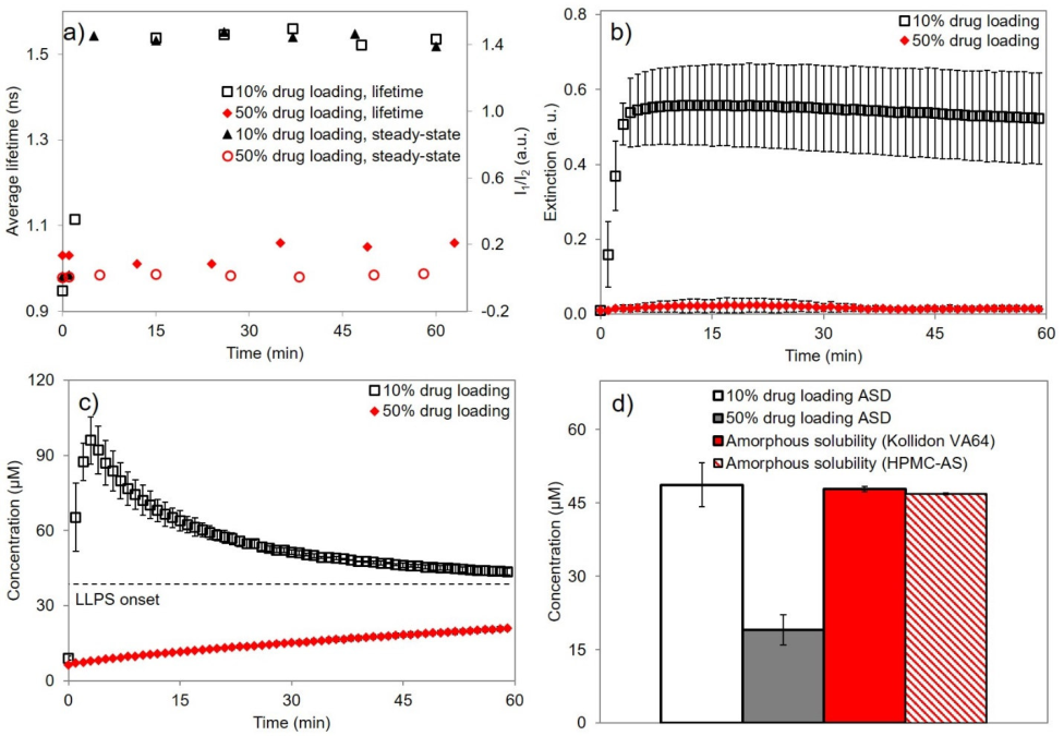
Ritonavir was mixed with PVP-type polymers or with HPMCAS. When wetted, the PVP films gave the pure-drug probe value, which the authors interpret as phase separation into a drug-rich phase. In the dissolution medium the 50% powder gave only 19 µM of drug in solution against an amorphous solubility of 48 µM, while the 10% powder reached 49 µM purohit2015b R6, R15; tres2018 R6, R9. HPMCAS films formed a drug-rich phase at every loading purohit2017b C37.

moderateModerate for the contrast but weak for its explanation: the probe readings were taken on leftover films, films and powders differ, and release rates were not measured purohit2015b K4, X5; tres2018 K2, K3, X10.

Ritonavir in solution after dissolution from Kollidon VA64 powders, µM10% drug loadingultracentrifuged supernatant4950% drug loadingultracentrifuged supernatant19amorphous solubilitysame medium48

Only the 10% powder reached the amorphous solubility. tres2018 R6, R9

ritonavir–PVPritonavir–HPMCASritonavir–PVPseparated when wetamorphous ritonavir 1.301.60 → 1.30ritonavir–HPMCASstayed mixed1.42 → 1.471.21.31.41.51.61.7pyrene I/III of the 50:50 ritonavir film at 97% RH

In the PVP film the probe ends at the value of amorphous ritonavir; in the HPMCAS film it stays near its start. purohit2015b R6, R13, R15

For your drug

  1. Read the wetted matrix with a probe and pair it with a probe-free check (residue composition, wet-film imaging or release profile); read the medium by ultracentrifuged supernatant (the clear liquid left after spinning out all particles) with a filtration control.
  2. Compare the supernatant with the amorphous solubility measured in the same medium, at three or more drug loadings and with your polymer; do not infer one result from the other.

purohit2015b X5; tres2018 X10, K4

How it works and key numbers
  1. PVP interacts favourably with water and ritonavir unfavourably: sorbed water replaces drug–polymer contacts and a drug-rich phase forms (the authors' interpretation) purohit2015b C24, C27.
  2. At 10% loading dissolution follows the soluble polymer ("clearly polymer-controlled") and carries the drug beyond the amorphous solubility tres2018 C23, C24.
  3. At 50% loading it follows the poorly soluble drug tres2018 C23, C24, and the medium stayed below the amorphous solubility tres2018 R9.
  4. (synthesis: in a matrix that phase separates into a drug-rich domain, that domain controls the release, while in a matrix that stays mixed the polymer carries the drug into solution, where the drug-rich phase forms tres2018 C23, C24.)
  5. An alternative reading of the leftover films is that PVP dissolves from the film faster than the drug and the residue becomes drug-rich without separation purohit2015b X5.
To feel the numbers. At 50% loading the medium reached 19 ÷ 48 = 0.40 of the amorphous solubility; at 10% it reached 49 ÷ 48 = 1.02.
49 vs 19 µMritonavir in solution from the 10% and 50% powders; amorphous solubility 48 µMtres2018 R6, R9
1.60 → 1.30 vs 1.42 → 1.47pyrene ratio of PVP and HPMCAS films at 97% RHpurohit2015b R6, R13, R15
10–20% vs 30–50%PVP loadings at which ritonavir films did and did not form a drug-rich liquid phase (LLPS) in dissolutionpurohit2017b C30, C33, C34, C37
Evidence: data, conditions and papers

Finding F2. For ritonavir at high drug loading, the PVP-type matrices that separated when wetted did not reach the amorphous solubility in the medium, while the HPMCAS matrix that stayed mixed under humidity did

In short. At 97% RH the pyrene ratio of 50:50 ritonavir–PVP films fell from 1.60 to 1.30, the value of amorphous ritonavir, while ritonavir–HPMCAS went from 1.42 to 1.47; in pH 6.8 buffer the leftover PVP film fell from about 1.565 to about 1.29 at 30 min and the HPMCAS film rose from 1.425 to 1.50–1.53 purohit2015b R6, R13, R15, R11, R14 (Solid-state methods F17, Solid-state methods F18). In the dissolution medium the 10% ritonavir–Kollidon VA64 dispersion left 49 µM in the ultracentrifuged supernatant against an amorphous solubility of 48 µM and the 50% dispersion only 19 µM, and in ritonavir films LLPS occurred at 10–20% PVP but not at 30–50%, while HPMCAS films reached it at every loading tres2018 R6, R9; purohit2017b C30, C33, C34, C37 (Solution methods F10). The authors tie the high-loading case to drug-controlled release, which no paper measured by release rates tres2018 C23, C24, X10 (Solution methods F10). (synthesis: separation in the wetted matrix and a drug-rich phase in the medium are different events.)

Type. method comparison within one drug (ritonavir), 3 polymers and loadings of 10–50%, across 3 papers and 2 sample forms Strength. for the contrast, weak for its explanation — the probe readings were taken on leftover films (weak to moderate on their own), films and powders differ, and release rates were not measured purohit2015b K4, X5 (Solid-state methods F18); tres2018 K2, K3, X10 (Solution methods F10).

How it was shown

Purohit and Taylor stored spin-coated 50:50 films at 97% RH or immersed them in pH 6.8 buffer for 10 s to 30 min and read the leftover film purohit2015b P1, P5, P6. Tres and co-workers added ritonavir–Kollidon VA64 powders (10 and 50% drug) to pH 7.4 buffer with PRODAN and ultracentrifuged the medium for HPLC tres2018 P1, P4, P6, P9.

What the data show

At 97% RH the pyrene ratio of ritonavir–PVP fell from 1.60 to 1.30, the value of amorphous ritonavir, while ritonavir–HPMCAS went from 1.42 to 1.47 purohit2015b R6, R13, R15. In the medium the 10% powder left 49 µM against an amorphous solubility of 48 µM, the 50% powder only 19 µM tres2018 R6, R9. LLPS occurred in ritonavir films at 10–20% PVP but not at 30–50%, and in HPMCAS films at every loading purohit2017b C30, C33, C34, C37.

systemmethod and conditionsresultids
ritonavir–PVP 50:50 film, 97% RHpyrene I/III on the film1.60 to 1.30purohit2015b R6, R15
ritonavir–HPMCAS 50:50 film, 97% RHpyrene I/III; IR1.42 to 1.47; IR unchangedpurohit2015b R13, R15, R18
ritonavir–PVP 50:50 film, pH 6.8 bufferpyrene on the leftover film1.565 to about 1.49 (10 s) to about 1.29 (30 min)purohit2015b R11
ritonavir–HPMCAS 50:50 film, pH 6.8 bufferpyrene on the leftover film1.425 to 1.50–1.53purohit2015b R14
ritonavir–Kollidon VA64 10%lifetime, extinction, ultracentrifuged supernatant, pH 7.4, 24 °C1.54 ns, about 0.51, 49 µM (amorphous solubility 48 µM)tres2018 R6, R7, R9
ritonavir–Kollidon VA64 50%sameabout 1.01–1.06 ns, about 0.02, 19 µMtres2018 R6, R7, R9
Fig. 4 of tres2018: ritonavir dispersions, 10% against 50% drug loading
Fig. 4 · tres2018ritonavir dispersions, 10% against 50% drug loading

Lifetime rises only for the 10% dispersion, and the supernatant matches the amorphous solubility only at 10% tres2018 R6, R7, R9.

Why

The authors think sorbed water replaces drug–polymer contacts in PVP, so a drug-rich phase forms purohit2015b C24, C27; at 10% loading dissolution follows the soluble polymer ("clearly polymer-controlled"), at 50% the poorly soluble drug tres2018 C23, C24. Leftover films may instead lose PVP faster than drug, without separation purohit2015b X5. (synthesis: when the matrix phase separates into a drug-rich domain, that domain controls the release, so the medium stays below the amorphous solubility tres2018 C23, C24.)

Limits and transfer

Films and powders differ in buffer, polymer and probe purohit2015b P6; tres2018 P1, P4, X10, and for posaconazole the 50% HPMC-AS dispersion never reached the amorphous solubility in FaSSIF elkhabaz2019a C15. For another dispersion, pair the wetted-matrix probe with a probe-free check, read the medium by ultracentrifuged supernatant, and test three or more loadings.

How it was shown

Purohit and Taylor spin coated 50:50 films with 0.1% probe and either stored them at 97% RH over saturated K₂SO₄ (1 h to 5 days) or immersed them in 100 mM pH 6.8 phosphate buffer for 10 s to 30 min, removed them and read the leftover film; pure PVP, ritonavir and HPMCAS films were the controls purohit2015b P1, P5, P6 (Solid-state methods F17, Solid-state methods F18). Tres and co-workers dissolved ritonavir (and lopinavir) with Kollidon VA64 at 10:90 and 50:50, rotary-evaporated, dried and sieved the products to amorphous powders, added powder to 50 mL of pH 7.4 buffer at 24 °C with 0.9 µM PRODAN, followed lifetime, I1/I2, in-situ UV and extinction at 450 nm for about 60 min, and ultracentrifuged the medium for HPLC against the amorphous solubility measured by antisolvent addition tres2018 P1, P2, R10, P4, P6, P7, P8, P9 (Solution methods F10). The ritonavir films with PVP, PVPVA and HPMCAS at 10–50% drug that both chapters cite were studied by Purohit and Taylor in non-sink dissolution with pyrene purohit2017b C30, C32 (Solution methods F10).

What the data show

In the humid film the 80:20 ritonavir–PVP film also reached 1.29, and in PVP dispersions the 1654 cm⁻¹ hydrogen-bond band disappeared and one Tg became two, while the HPMCAS spectrum did not change purohit2015b R6, R17, R19, R20, R18 (Solid-state methods F17). In buffer the largest ritonavir–PVP step lay between 20 and 30 min, not within seconds purohit2015b R11, X4 (Solid-state methods F18). Outside the two chapters, ritonavir films with 50% drug in PVP and PVPVA phase separated within 10 s of water contact and released drug under drug control at or below the amorphous solubility, dispersions that stayed miscible during early dissolution formed nanodroplets, and HPMCAS dispersions did so at every loading, fresh or after humidity purohit2017b C5, C3, C53, C54, C6, C55, C30, R4, R9 (Solid-state methods F18). In the medium, the 10% ritonavir powder raised the PRODAN lifetime from about 0.95 to 1.54 ns and the extinction to about 0.51 by 3 min, whereas the 50% powder stayed at about 1.01–1.06 ns and about 0.02 tres2018 R6, R7, R9 (Solution methods F10). One probe read-out in the medium needs care: the 50% lopinavir lifetime rose to about 1.23 ns because of undissolved particles and fell to 1.00 ns, the probe-alone value being 0.99 ns, after 1 µm filtration tres2018 R11, R13, C33 (Solution methods F11).

chapter findingsystemmethod and conditionsresultids
Solid-state methods F17ritonavir–PVP 50:50 film, 97% RHpyrene I/III on the film1.60 to 1.30purohit2015b R6, R15
Solid-state methods F17ritonavir–HPMCAS 50:50 film, 97% RHpyrene I/III; IR1.42 to 1.47; IR unchangedpurohit2015b R13, R15, R18
Solid-state methods F18ritonavir–PVP 50:50 film, pH 6.8 bufferpyrene on the leftover film1.565 to about 1.49 (10 s) to about 1.29 (30 min)purohit2015b R11
Solid-state methods F18ritonavir–HPMCAS 50:50 film, pH 6.8 bufferpyrene on the leftover film1.425 to 1.50–1.53purohit2015b R14
Solid-state methods F18ritonavir films, 50% drug in PVP and PVPVAfilms in pH 6.8 buffersplit within 10 s; drug-controlled releasepurohit2017b C5, C53, C54, R4, R9
Solution methods F10ritonavir–Kollidon VA64 10%lifetime, extinction, ultracentrifuged supernatant, pH 7.4, 24 °C1.54 ns, about 0.51, 49 µM (amorphous solubility 48 µM)tres2018 R6, R7, R9
Solution methods F10ritonavir–Kollidon VA64 50%sameabout 1.01–1.06 ns, about 0.02, 19 µMtres2018 R6, R7, R9
Solution methods F10ritonavir films, PVP, PVPVA, HPMCAS, 10–50%non-sink film dissolution, pyrene I/IIILLPS at 10–20% PVP, none at 30–50%; HPMCAS at every loadingpurohit2017b C30, C33, C34, C37, C38
Solution methods F11lopinavir–Kollidon VA64 50%lifetime and 1 µm filtrationabout 1.23 ns; filtrate 1.00 ns; probe alone 0.99 nstres2018 R11, R13

Figure

Fig. 8 of purohit2015b: probe signal of 50:50 probucol–PVP (a) and ritonavir–PVP (b) films against immersion time
Fig. 8 · purohit2015bprobe signal of 50:50 probucol–PVP (a) and ritonavir–PVP (b) films against immersion time
Fig. 4 of tres2018: ritonavir dispersions, 10% against 50% drug loading
Fig. 4 · tres2018ritonavir dispersions, 10% against 50% drug loading
Left: each point is measured on the undissolved film, and ritonavir–PVP drifts down to the ritonavir value at 30 min purohit2015b R10, R11 (Solid-state methods F18). Right: lifetime and I1/I2 rise only for the 10% dispersion (panel a) and the supernatant matches the amorphous solubility only at 10% (panel d) tres2018 R6, R7, R9 (Solution methods F10).

Why

The authors' interpretation of the film results is that PVP interacts favourably with water while ritonavir interacts unfavourably with it, so sorbed water replaces drug–polymer contacts and a drug-rich phase forms into which the probe moves purohit2015b C24, C27 (Solid-state methods F17); they add that separation during dissolution "might be anticipated" to lead to crystallization in the matrix and a lower supersaturation, none of which was measured purohit2015b C25, C31, C33 (Solid-state methods F18). For the medium they say that at 10% loading dissolution follows the soluble polymer ("clearly polymer-controlled") and carries the drug beyond the amorphous solubility, whereas at 50% it follows the poorly soluble drug tres2018 C23, C24 (Solution methods F10). The alternative reading of the leftover films is that PVP dissolves from the film faster than the drug and the residue becomes drug-rich without separation inside a hydrated matrix purohit2015b X5 (Solid-state methods F18). (synthesis: the two sets of results are consistent if, in a high-loading matrix that phase separates into a drug-rich domain, that domain controls the release, so the medium stays below the amorphous solubility, while in a matrix that stays mixed the polymer carries the drug into solution, where the drug-rich phase forms. The probe lifetime rise caused by undissolved ASD particles in the 50% lopinavir run tres2018 C33 (Solution methods F11) is the same kind of confound that leftover-film readings cannot exclude.)

Conditions and limits

Films and powders differ in preparation, buffer (pH 6.8 phosphate against pH 7.4 phosphate), polymer (PVP K-90 against Kollidon VA64) and probe, and the film immersion readings carry no dissolution profile purohit2015b P6, C33 (Solid-state methods F18); tres2018 P1, P4, X10 (Solution methods F10). The loading at which dissolution stops reaching LLPS lies between 10% and 50% for the powders and was not located tres2018 X10 (Solution methods F10). The filtration control for the lifetime was done once, with a 1 µm filter, and probe with undissolved solid alone was not measured tres2018 K4 (Solution methods F11).

Counterexamples and conflicts

The polymer order is not general. For posaconazole the 50% HPMC-AS dispersion never reached the amorphous solubility in FaSSIF, and for danazol the 50:50 PVP film did reach LLPS elkhabaz2019a C15; jackson2015 R4 (Solution methods F10); the chapter's own reading is that loading alone is not the switch purohit2017b C37, C38 (Solution methods F10). The link from separation to supersaturation stays an inference in the papers of the solid-state chapter purohit2015b C33 (Solid-state methods F18).

Transfer check

For another dispersion: (1) read the wetted matrix with a probe, pair it with a probe-free check such as the composition of the residue, wet-film imaging or the release profile, and make sure a drug-rich residue is not produced by selective polymer loss; (2) read the medium with extinction at a wavelength where the drug does not absorb, an ultracentrifuged supernatant and a filtration control, and compare the supernatant with the amorphous solubility measured in the same medium; (3) do both at three or more drug loadings and with the polymer of interest; (4) do not infer one result from the other, and measure drug and polymer release if the mechanism matters.

History

2015: Purohit and Taylor gave the first probe read-out of separation in a hydrating film, in humid air and in buffer purohit2015b R4, R10 (Solid-state methods F17, Solid-state methods F18). 2017: Purohit and co-workers added dissolution profiles, nanodroplets and PVPVA and found the loading and polymer dependence for ritonavir films purohit2017b C5, C33, C34, C37 (Solid-state methods F18, Solution methods F10). 2018: Tres and co-workers added the PRODAN lifetime and the ultracentrifuged supernatant for ritonavir and lopinavir at two loadings tres2018 R6, R9, R11, R17 (Solution methods F10) and showed both directions of error in the in-situ read-outs tres2018 R8, R16, R13 (Solution methods F11). 2019: Elkhabaz and co-workers saw the loading effect for posaconazole in FaSSIF elkhabaz2019a C15 (Solution methods F10).

F3

Topic 1Probe read-outs across solid and solution3 of 3

The probe signal returns toward its aqueous value when the drug-rich region crystallizes, but only one film was confirmed.

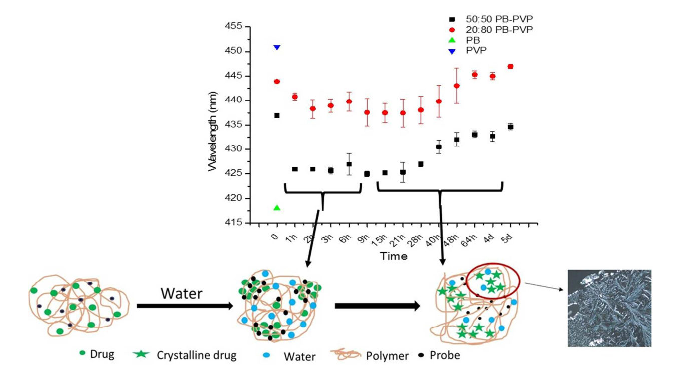
In a humid probucol–PVP film the probe signal first moved to the drug-rich value and then returned toward its initial value once birefringent crystals appeared at 21 h; X-ray diffraction showed probucol peaks at 30 h purohit2015b R4, R5, R21, R22. In solutions the signal also returned toward its aqueous value, but no solution sample was checked by diffraction or microscopy raina2015a R16; tres2018 R20, R21.

weakWeak to moderate: the confirmation by a second method exists for one crystallizing film, and each solution paper shows one sample purohit2015b K2; raina2015a K6; tres2017 K2; tres2018 K5, X4.

1 h ≈ 42640 h 430.55 days 434.5start 437returns as crystals form420425430435440prodan emission maximum, nm (probucol–PVP film, 97% RH)

Birefringent crystals were seen at 21 h and probucol XRD peaks at 30 h. purohit2015b R4, R5, R21, R22

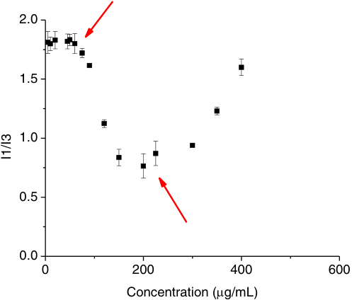
200 µg/mL ≈ 0.76400 µg/mL ≈ 1.60up to 60 µg/mL ≈ 1.80.60.951.31.652pyrene I1/I3 ratio, nifedipine with HPMC at 25 °C

The ratio drops when the drug-rich phase forms and rises again as particles appear. raina2015a R16, C22

For your drug

  1. Run a polymer-free sample at a stated supersaturation and confirm the solid by polarized-light microscopy or XRPD, or by a fall of the ultracentrifuged supernatant below the amorphous solubility.
  2. Record the probe-alone and probe-plus-crystals values on your instrument so that "returned to aqueous" is defined, and read the lifetime or the ratio, not the intensity.

purohit2015b R21, R22; raina2015a R16; tres2018 R21

How it works and key numbers
  1. While the drug-rich region exists, the probe partitions into it and the signal stays at the drug-like value purohit2015b R4; raina2015a R16.
  2. Crystal growth depletes the drug-rich phase and, the authors think, expels the probe into water raina2015a C23; tres2018 C43, C45; purohit2015b C9.
  3. The signal therefore returns toward its aqueous value: the pyrene ratio rises, the PRODAN lifetime (the mean time the probe spends in the excited state) falls and the prodan emission returns toward the polar value raina2015a C23; tres2018 C43, C45.
  4. The film result has its own uncertainty: water alone raises the prodan value in PVP by about 5 nm over 5 days, so part of the late shift may be hydration purohit2015b X9, C9.
  5. (synthesis: an external probe can show that the drug-rich liquid has gone, not what replaced it; only polarized light, XRD or, for SHG-active crystals, SHG shows the crystal purohit2015b C9; raina2015a R16.)
To feel the numbers. The film lost 437 − 426 = 11 nm on forming the drug-rich region and regained 434.5 − 426 = 8.5 nm, 8.5 ÷ 11 = 0.77 of the shift.
437 → 426 → 434.5 nmprodan emission of the probucol–PVP film at the start, at 1 h and at 5 dayspurohit2015b R4, R5
0.76 → 1.60pyrene ratio of nifedipine with HPMC at 200 and 400 µg/mLraina2015a R16
1.31 → 1.06 nsPRODAN lifetime of lopinavir at 94 and 213 min, near the probe-alone 1.00 nstres2018 R21
Evidence: data, conditions and papers

Finding F3. The probe signal returns toward its aqueous value when a drug-rich region crystallizes, in a humid film and in supersaturated solutions, but only the film was confirmed by a second method

In short. In 50:50 probucol–PVP films at 97% RH the prodan emission fell from 437 to about 426 nm and then rose to 434.5 nm after birefringent crystals appeared at 21 h, and XRD showed probucol peaks at 30 h purohit2015b R4, R5, R21, R22 (Solid-state methods F17). In solution the pyrene ratio of nifedipine with HPMC fell to about 0.76 at 200 µg/mL and rose to about 1.60 at 400 µg/mL as particles appeared, and the PRODAN lifetime of lopinavir fell from 1.31 ns at 94 min to about 1.06 ns at 213 min, near the probe-alone 1.00 ns, while ritonavir stayed at about 1.31–1.37 ns raina2015a R16; tres2018 R20, R21 (Solution methods F9). None of the solution samples was confirmed by diffraction or microscopy in the papers of the chapter, and the probes show no change when crystals form before any liquid phase raina2015a X15, R8; tres2017 X6; tres2018 X5 (Solution methods F9, Solution methods F8). (synthesis: an external probe can show that the drug-rich liquid has gone, not what replaced it.)

Type. observation, one confirmed film system and four unconfirmed solution samples Strength. to moderate — the direction agrees in the two states, but the confirmation by a second method exists for one crystallizing film, and each solution paper shows one sample, without polymer in the lopinavir and ritonavir tests purohit2015b K2 (Solid-state methods F17); raina2015a K6; tres2017 K2; tres2018 K5, X4 (Solution methods F9).

How it was shown

Purohit and Taylor followed the prodan emission of 50:50 probucol–PVP films at 97% RH for 5 days, with polarized-light microscopy and XRD as checks purohit2015b P1, P5, P8, P9, P10, P11, P12. Raina and co-workers raised nifedipine with HPMC to 400 µg/mL while reading pyrene raina2015a P11, R16, and Tres and co-workers followed the PRODAN lifetime of lopinavir and ritonavir tres2018 P5, R20.

What the data show

The film read 437 nm at the start, about 426 nm at 1 h and 434.5 nm at 5 days, with birefringent crystals at 21 h and probucol peaks by XRD at 30 h purohit2015b R4, R5, R21, R22. The nifedipine ratio fell to about 0.76 at 200 µg/mL and rose to about 1.60 at 400 µg/mL raina2015a R16, C22. The lopinavir lifetime fell from about 1.31 ns at 94 min to about 1.06 ns tres2018 R21. The probes showed no change when nifedipine and nimodipine crystallized in buffer raina2015a R4, R5, R8.

systemmethod and conditionsresultids
probucol–PVP 50:50 film, 97% RHprodan; polarized-light microscopy; XRD437 to 425–427 nm (1–28 h) to 434.5 nm; birefringent crystals at 21 h; probucol peaks at 30 hpurohit2015b R4, R5, R21, R22
nifedipine with HPMC, 25 °Cpyrene I1/I3 up to 400 µg/mLabout 1.8 to about 0.76 (200 µg/mL) to about 1.60 (400 µg/mL); particles appearraina2015a R16, C22
felodipine 108 µM, no HPMC, 15 hlifetime, bi-exponentialτ2 9.04 ns (17%) against 1.87–2.04 ns with aggregatestres2017 R1.g, R1.d–f
lopinavir and ritonavir without polymer, 24 °CPRODAN lifetime, 213 minlopinavir 1.31 ns (94 min) to about 1.06 ns; ritonavir about 1.31–1.37 nstres2018 R21, R20
nifedipine (25, 37 °C) and nimodipine (37 °C), bufferextinction, visual, polarized-light microscopy; probescrystals at about 38, 58 and 21.5 µg/mL; probes silent (data not shown)raina2015a R5, R4, R8
Fig. 3 of purohit2015b: prodan wavelength against time at 97% RH for probucol–PVP
Fig. 3 · purohit2015bprodan wavelength against time at 97% RH for probucol–PVP

The film signal falls, plateaus and rises again once crystals form purohit2015b R4, R5.

Why

The authors think crystal growth depletes the drug-rich phase and expels the probe into water raina2015a C23; tres2018 C43, C45; purohit2015b C9; in the film, water alone raises the prodan value by about 5 nm, so part of the late shift may be hydration purohit2015b X9, C9. (synthesis: an external probe can show that the drug-rich liquid has gone, not what replaced it purohit2015b C9; raina2015a R16.)

Limits and transfer

Only one crystallizing film carries a second-method confirmation; the lopinavir and ritonavir tests had no polymer and the felodipine sample was measured once purohit2015b R6, C12; tres2018 X4; tres2017 X6. To follow crystallization, run a polymer-free sample at a stated supersaturation and confirm the solid by polarized-light microscopy or XRPD.

How it was shown

Purohit and Taylor followed the prodan emission of 50:50 probucol–PVP films stored at 97% RH from 1 h to 5 days and checked the same system with polarized-light microscopy, DSC and XRD of powders purohit2015b P1, P5, P8, P9, P10, P11, P12 (Solid-state methods F17). Raina and co-workers raised nifedipine with pre-dissolved HPMC to 400 µg/mL at 25 °C while reading pyrene I1/I3 and noted particles by eye raina2015a P11, R16 (Solution methods F9). Tres and co-workers left 108 µM felodipine in pH 7.4 buffer without HPMC for 15 h and recorded the decay, and followed the PRODAN lifetime of ritonavir and lopinavir solutions without polymer for about 213 min at 24 °C tres2017 P2, P1; tres2018 P5, R20, R22, X4 (Solution methods F9). The extinction method that cannot separate liquid from crystal, and the visual and polarized-light classification used with it, are in the same paper as the nifedipine series raina2015a P7, C9 (Solution methods F3).

What the data show

The probucol–PVP film read 437 nm at the start, about 426 nm at 1 h, 425–427 nm from 2 to 28 h, 430.5 nm at 40 h and 434.5 nm at 5 days, with branched birefringent crystals at 21 h and probucol peaks by XRD at 30 h, while ritonavir, a very slow crystallizer, did not crystallize and its probe signal did not return purohit2015b R4, R5, R6, C12, R19, R20, R21, R22 (Solid-state methods F17). In the nifedipine series the ratio was about 1.8 up to 60 µg/mL, about 0.76 at 200 µg/mL and about 1.60 at 400 µg/mL, but the arrow marking the LLPS onset sits near 80 µg/mL in the figure against 40 µg/mL in the text raina2015a R16, C22, X8 (Solution methods F9). The felodipine sample gave a 9.04 ns component (17%) against 1.87–2.04 ns in solutions with aggregates tres2017 R1.g, R1.d–f (Solution methods F9). The lopinavir lifetime was about 1.31 ns at 94 min and about 1.05–1.06 ns at 193–213 min, and the ritonavir lifetime stayed at about 1.31–1.37 ns tres2018 R21, R20 (Solution methods F9). In buffer, nifedipine at 25 and 37 °C and nimodipine at 37 °C gave an extinction rise that went with visible crystals, and the probes showed "no distinct change" (data not shown) raina2015a R4, R5, R8 (Solution methods F8). Extinction alone cannot tell the two cases apart, and the authors separated them by haze, absence of visible particles and absence of birefringence raina2015a R4, C9, P7 (Solution methods F3). A solid-state method that is crystal-selective did detect crystals earlier than XRPD in dispersions: SHG saw flutamide crystals at 11 days against 53 days by XRPD, but only for SHG-active forms above about 100 nm correasoto2017 R1 (Solid-state methods F10); kestur2012 R17, C34 (Solid-state methods F11).

chapter findingsystemmethod and conditionsresultids
Solid-state methods F17probucol–PVP 50:50 film, 97% RHprodan; polarized-light microscopy; XRD437 to 425–427 nm (1–28 h) to 434.5 nm; birefringent crystals at 21 h; probucol peaks at 30 hpurohit2015b R4, R5, R21, R22
Solution methods F9nifedipine with HPMC, 25 °Cpyrene I1/I3 up to 400 µg/mLabout 1.8 to about 0.76 (200 µg/mL) to about 1.60 (400 µg/mL); particles appearraina2015a R16, C22
Solution methods F9felodipine 108 µM, no HPMC, 15 hlifetime, bi-exponentialτ2 9.04 ns (17%) against 1.87–2.04 ns with aggregatestres2017 R1.g, R1.d–f
Solution methods F9lopinavir and ritonavir without polymer, 24 °CPRODAN lifetime, 213 minlopinavir 1.31 ns (94 min) to about 1.06 ns; ritonavir about 1.31–1.37 nstres2018 R21, R20
Solution methods F8nifedipine (25, 37 °C) and nimodipine (37 °C), bufferextinction, visual, polarized-light microscopy; probescrystals at about 38, 58 and 21.5 µg/mL; probes silent (data not shown)raina2015a R5, R4, R8
Solid-state methods F10flutamide 15%, 40 °C, 75% RHSHG against XRPD, first detectioncrystals at 11 against 53 dayscorreasoto2017 R1

Figure

Fig. 3 of purohit2015b: prodan wavelength against time at 97% RH for probucol–PVP
Fig. 3 · purohit2015bprodan wavelength against time at 97% RH for probucol–PVP
Fig. 12 of raina2015a: pyrene I1/I3 against nifedipine concentration with HPMC at 25 °C
Fig. 12 · raina2015apyrene I1/I3 against nifedipine concentration with HPMC at 25 °C
Left: the film signal falls, plateaus and rises again once crystals form purohit2015b R4, R5 (Solid-state methods F17). Right: the solution ratio falls to about 0.76 and then rises towards about 1.6; the first arrow points near 80 µg/mL against the 40 µg/mL in the text raina2015a R16, X8 (Solution methods F9).

Why

The authors' interpretation in both states is that crystal growth depletes the drug-rich phase and expels the probe into water, so pyrene I1/I3 rises, the PRODAN lifetime falls and the prodan emission returns towards the polar value raina2015a C23; tres2018 C43, C45 (Solution methods F9); purohit2015b C9 (Solid-state methods F17). For felodipine, which is its own reporter, the opposite direction fits a rigid crystal lattice, "most likely" tres2017 C22, C41 (Solution methods F9). The film result has its own uncertainty: water alone raises the prodan value in PVP by about 5 nm over 5 days, so part of the late red shift may be hydration, and some probucol "might be" left amorphous purohit2015b X9, C9 (Solid-state methods F17). Not measured in solution: the location of the probe, the lifetime of the expelled probe and the identity of the solid raina2015a X17; tres2018 C45; tres2017 C22 (Solution methods F9). (synthesis: the signal reflects the location of the reporter, so a return of an external probe to its aqueous value shows loss of the non-aqueous region, and only a method that responds to the crystal itself, such as polarized light, XRD or, for SHG-active forms, SHG, shows what took its place.)

Conditions and limits

The lopinavir and ritonavir tests had no polymer and an unstated supersaturation, so the 94 and 213 min figures cannot be compared with other induction times tres2018 X4 (Solution methods F9). The felodipine sample was measured once, without microscopy, diffraction or a concentration check tres2017 X6 (Solution methods F9). The film result is one system that crystallizes; the other ritonavir film did not purohit2015b R6, C12 (Solid-state methods F17). SHG sees only non-centrosymmetric crystals larger than about 100 nm, and a dark image does not prove that no crystals are present wanapun2010 X11; kestur2012 C34 (Solid-state methods F11).

Counterexamples and conflicts

None found for the direction of the change in either state; only the nifedipine onset in the figure differs from the text raina2015a X8 (Solution methods F9). An outside study of weak bases takes the probe response as proof that the second phase is non-crystalline, because the dyes cannot partition into a crystalline phase and behave there as in water, which agrees with the absence of a probe response for nifedipine and nimodipine indulkar2015 C28, C29; raina2015a R8 (Solution methods F8).

Transfer check

To follow crystallization of a drug-rich phase: (1) run a polymer-free sample at a stated supersaturation and confirm the solid by polarized-light microscopy or XRPD, or by a fall of the ultracentrifuged supernatant below the amorphous solubility; (2) if the crystals are expected to be SHG-active and larger than about 100 nm, add SHG on the collected solid, after establishing the space group of the expected form; (3) record the probe-alone and probe-plus-crystals values on your instrument, so that "returned to aqueous" is defined; (4) read the lifetime or the ratio, not the intensity; (5) if the drug is its own reporter, expect the lifetime to rise in crystals.

History

2010: Wanapun and co-workers showed that the lag before crystals are seen depends on the sensitivity of the method wanapun2010 C2 (Solid-state methods F10). 2014: Jackson and co-workers coupled the fall of the UV signal with the rise of I1/I3 to define the induction time of danazol jackson2014 C30, C35 (Solution methods F9). 2015: Purohit and Taylor showed the red shift of the probe in a crystallizing film, and Raina and co-workers the pyrene return for nifedipine purohit2015b R4, C9 (Solid-state methods F17); raina2015a R16 (Solution methods F9). 2017: Correa-Soto and co-workers showed crystals by SHG days to weeks before XRPD correasoto2017 R1 (Solid-state methods F10), and Tres and co-workers a 9 ns crystal component for felodipine tres2017 R1.g (Solution methods F9). 2018: Tres and co-workers used the PRODAN lifetime to separate a persistent drug-rich phase (ritonavir) from a crystallizing one (lopinavir) tres2018 R20, R21 (Solution methods F9).

Topic 2 of 4 · 2 findings · F4–F5

What a reading can and cannot say

F4

Topic 2What a reading can and cannot say1 of 2

Every method has a detection limitation, and each was found only by comparison with a second method.

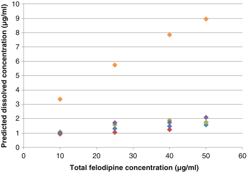
A dark image, a single glass transition, a rising turbidity or a UV reading can each give a misleading result. For example, TEM found crystals below 100 nm in an extrudate whose X-ray pattern showed only a halo, and a UV probe read 300 nm drug particles as dissolved drug moseson2018a R9, C10; vaneerdenbrugh2011b R6.a, M1.

moderateModerate: each limitation was shown in at least one paper, but in different drugs, and no paper put one sample through all the methods kestur2012 K3, X10; correasoto2017 K2, X7.

UV probe reading ÷ true amount (our arithmetic from the paper's numbers)felodipine, 300 nm particles8.95 vs 0.81 µg/mL≈ 11×lopinavir, 10% dissolution466 vs 36 µM≈ 13×

In both cases the in-situ UV probe over-read the dissolved drug. vaneerdenbrugh2011b R6.a, M1; tres2018 R16, R17

TEM onlyXRPD limit ≈ 1polarized light 3.5DSC and SEM 30110100crystal content of the extrudate, % (log scale)

In the same extrudate series, each method first detected crystals at a different content. moseson2021 P2, R6

For your drug

  1. Before trusting a method, find the property it needs and test it: the space group of the expected form for SHG, the drug's absorbance at the extinction wavelength, its own emission at the probe's excitation, the particle size for the UV probe.
  2. Add a second method with a different limitation (TEM or polarized light beside XRPD, a probe or imaging beside DSC, an ultracentrifuged supernatant beside UV) and read a negative result as bounded by the method's limits, never as absence.

wanapun2010 R13; kestur2012 R17, C34; vaneerdenbrugh2011b C23; purohit2017a R3, R7

How it works and key numbers
  1. SHG (second harmonic generation, a laser method for crystals) needs order without an inversion centre, so glass, liquid, solution and centrosymmetric crystals give no net signal kestur2012 C4, C37; wanapun2010 C15.
  2. XRPD fails to detect crystals because of dilution by the polymer and peak broadening, and DSC (differential scanning calorimetry) detects crystals with low sensitivity because they can dissolve into the molten matrix kestur2012 C8; moseson2018a C26, C27; moseson2018 C31.
  3. A single Tg (glass transition temperature) is a bulk criterion: DSC resolves domains only above about 15–30 nm purohit2017a R28, R29, C25.
  4. Extinction (the loss of transmitted light by absorption and scattering) records any scattering centre, and the probes respond to a non-crystalline drug-rich phase, so they showed no change when crystals formed first raina2015a C6, C40.
  5. The second-derivative UV correction removes sloping scattering baselines but not a particle absorption band inside the drug's own band vaneerdenbrugh2011b C2, C23, C25.
  6. (synthesis: the size windows overlap between about 100 nm and 1 µm, where an optical read-out of a second phase is least secure kestur2012 C34; vaneerdenbrugh2011b R13, R14, R15.)
0.1% vs 5 wt%naproxen–HPMCAS: SONICC (SHG imaging) imaged 0.1% while PXRD showed no peaks below 5 wt%kestur2012 R8, R6
below 100 nmcrystals found by TEM in an extrudate whose XRPD showed only a halomoseson2018a R9, C10
8.95 vs 0.81 µg/mLUV reading of 300 nm felodipine particles against the solubilityvaneerdenbrugh2011b R6.a, M1
Evidence: data, conditions and papers

Finding F4. Each method has a detection limitation or a non-specific signal, and in the papers of both chapters it was revealed only when the method was compared with a second one

In short. A dark SHG image says only that no SHG-active crystal above about 100 nm is present: chlorpropamide has two SHG-inactive polymorphs, and TEM found crystals below 100 nm in an extrudate whose XRPD showed only a halo wanapun2010 R13; kestur2012 R17, C34; moseson2018a R9, C10 (Solid-state methods F11). One Tg is not one phase: itraconazole–HPMC with a single Tg of 87 °C read like the pure drug with pyrene and showed drug-rich domains by confocal imaging purohit2017a R3, R7, R16, R17 (Solid-state methods F19). In solution, extinction rises for a liquid drug-rich phase and for crystals alike, the probes show no change when crystals form first, and a UV probe read 300 nm felodipine particles as 3.4–8.95 µg/mL dissolved drug against a solubility of 0.81 µg/mL raina2015a R4, C9, R8; vaneerdenbrugh2011b R6.a, R7, M1 (Solution methods F3, Solution methods F8, Solution methods F1).

Type. method comparison, with one or more observations for each of 7 methods Strength. every limitation is shown in at least one paper, mostly by a second method on the same or a similar sample, but they were found in different drugs, and no paper put one sample through all the methods kestur2012 K3, X10; correasoto2017 K2, X7 (Solid-state methods F7); raina2015a K1, X12, X16 (Solution methods F3).

How it was shown

Reading across the chapters for what each method cannot see, Moseson and co-workers compared XRPD, DSC, SEM, polarized light and TEM on the same extrudates moseson2021 P2, R6; Purohit and co-workers added pyrene, confocal imaging and ssNMR to DSC of itraconazole–HPMC powders purohit2017a P6, P9, P10, P11; Van Eerdenbrugh and co-workers read a 300 nm felodipine nanosuspension by UV vaneerdenbrugh2011b R1, P4.

What the data show

In extrudates with an XRPD limit of about 1%, crystals below 1% were seen only by TEM moseson2021 P2, R6. Itraconazole–HPMC with one Tg of 87 °C read like the pure drug with pyrene and showed drug-rich domains by confocal imaging purohit2017a R3, R7, R16, R17. The UV probe read the 300 nm suspension as 8.95 against a solubility of 0.81 µg/mL, and a 10% lopinavir dissolution as 466 µM against 36 µM in the supernatant vaneerdenbrugh2011b R6.a, M1; tres2018 R16, R17.

systemmethod and conditionsresultids
BCL–PVPVA extrudatesXRPD (limit about 1%) against DSC, SEM, polarized light, TEMbelow 1%: TEM only; 3.5%: polarized light and TEM; 30%: DSC and SEM toomoseson2021 P2, R6
chlorpropamide films, 30 °CSHG against cited structuresα, γ SHG-active; β, δ SHG-inactivewanapun2010 R13, R7
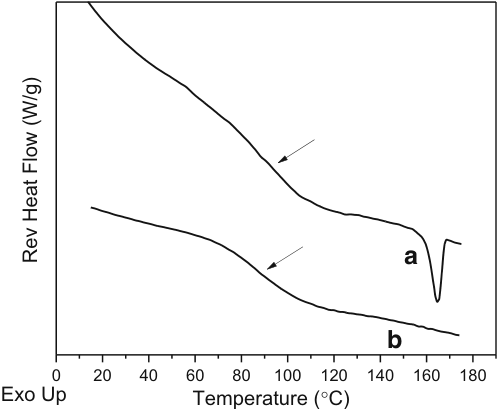
itraconazole–HPMC 40:60 powdersmDSC, XRD; pyrene; confocalone Tg, 87 °C, halo only; pyrene 1.38 / 1.39 / 1.39 (drug 1.39); drug-rich domainspurohit2017a R1, R3, R7, R16, R17
nifedipine, nimodipine, bufferextinction at 450 nm, visual, polarized light; six probesrise went with visible crystals; probes silent (data not shown)raina2015a R4, C9, R8
felodipine 300 nm nanosuspension, 1 mg/mL PVPsecond-derivative UV probe3.4 to 8.95 µg/mL against solubility 0.81 µg/mLvaneerdenbrugh2011b R6.a, R7, M1
lopinavir 10% dispersionin-situ UV against ultracentrifuged supernatantUV about 466 µM; supernatant 36 µMtres2018 R16, R17
Fig. 3 of vaneerdenbrugh2011b: apparent dissolved concentration against total felodipine for five particle sizes
Fig. 3 · vaneerdenbrugh2011bapparent dissolved concentration against total felodipine for five particle sizes

The nanosuspension series climbs to about 9 µg/mL while the coarse series stay near 1–2 µg/mL vaneerdenbrugh2011b R6, R7.

Why

Each method responds to one property and cannot detect a species that lacks it: SHG gives no net signal for glass, liquid, solution or centrosymmetric crystals kestur2012 C4, C37; wanapun2010 C15; XRPD fails to detect crystals because of dilution by the polymer and peak broadening kestur2012 C8; DSC resolves domains only above about 15–30 nm purohit2017a R28, R29, C25; and the UV correction removes sloping baselines but not a particle absorption band in the drug's own band vaneerdenbrugh2011b C2, C23, C25.

Limits and transfer

The Mie result that places the UV problem below about 1 µm rests on homogeneous monodisperse spheres and one measured spectrum vaneerdenbrugh2011b C39, X5. Before trusting a method for a new drug, test the property it needs, add a second method with a different limitation, and read a negative result as bounded by the method's size and form limits, never as absence.

How it was shown

The finding reads across the chapters for what each method cannot see. For SHG, Wanapun and co-workers computed coherence lengths of 4.8 µm forward and 120 nm backward for griseofulvin and read the signal-to-background where forward and backward SHG separate wanapun2010 R9, R10 (Solid-state methods F11). For XRPD, Moseson and co-workers compared XRPD, DSC, SEM, polarized-light microscopy and TEM on the same extrudates moseson2021 P2, R6 (Solid-state methods F7). For DSC, Purohit and co-workers ran modulated DSC at 2 °C/min and XRD on itraconazole–HPMC powders and pyrene, confocal imaging and ssNMR on the same solution dried by four routes purohit2017a P6, P7, P9, P10, P11 (Solid-state methods F19). For extinction and the probes, Raina and co-workers read extinction at 450 nm with visual and polarized-light classification, and probes within 1 min of each addition raina2015a P6, P7, P9 (Solution methods F3, Solution methods F8). For the UV probe, Van Eerdenbrugh and co-workers diluted a 300 nm felodipine nanosuspension and four coarser suspensions and read them with second-derivative UV vaneerdenbrugh2011b R1, R2, P4, P9 (Solution methods F1).

What the data show

In the extrudate series with an XRPD limit of about 1%, crystals below 1% were seen only by TEM, 3.5% by polarized light and TEM, and 30% also by DSC and SEM moseson2021 P2, R6 (Solid-state methods F7). In naproxen–HPMCAS blends PXRD showed no peaks below 5 wt% while SONICC imaged 0.1% kestur2012 R8, R6 (Solid-state methods F7). Chlorpropamide has two SHG-active (α, γ) and two SHG-inactive (β, δ) polymorphs, and flutamide, though achiral, is SHG-active (Pna2₁) wanapun2010 R13; correasoto2017 M1, C8 (Solid-state methods F11). DEX6–PVP90 was miscible by DSC but showed domains of about 0.3–0.6 µm by AFM, and the itraconazole powders of the 2017 paper read as pure drug with pyrene under a single Tg vaneerdenbrugh2012c R19; purohit2017a R3, R7 (Solid-state methods F15, Solid-state methods F19). In AFM-IR the stronger absorber dominates: PVP gave ringdown responses up to ten times those of dextran, and AFM-IR imaging failed for telaprevir–HPMCAS domains 122 nm wide and 13 nm high while stiffness imaging still gave contrast vaneerdenbrugh2012c R6; li2016c R7, R11, C49 (Solid-state methods F13, Solid-state methods F14). In solution, the extinction rise for nifedipine at 25 and 37 °C and nimodipine at 37 °C went with visible crystals, the probes showed "no distinct change", and felodipine's own fluorescence masked the emission of dansyl glycine and dapoxyl raina2015a R4, R8, R12, R13 (Solution methods F3, Solution methods F8, Solution methods F6). The UV probe read the 300 nm suspension as 8.95 against a solubility of 0.81 µg/mL, about 11 times (synthesis: 8.95/0.81 = 11.05), and a 10% lopinavir dissolution as 466 µM against 36 µM in the supernatant, about 13 times (synthesis: 466/36 = 12.9) vaneerdenbrugh2011b R6.a, M1; tres2018 R16, R17 (Solution methods F1, Solution methods F11).

chapter findingsystemmethod and conditionsresultids
Solid-state methods F7BCL–PVPVA extrudatesXRPD (limit about 1%) against DSC, SEM, polarized light, TEMbelow 1%: TEM only; 3.5%: polarized light and TEM; 30%: DSC and SEM toomoseson2021 P2, R6
Solid-state methods F11indomethacin–PVPVA extrudateXRPD, DSC, TEMhalo and one Tg; TEM crystals below 100 nm and 5–10 nm domainsmoseson2018a R9, R5
Solid-state methods F11chlorpropamide films, 30 °CSHG against cited structuresα, γ SHG-active; β, δ SHG-inactive; signal levelled off or dippedwanapun2010 R13, R7
Solid-state methods F19itraconazole–HPMC 40:60 powdersmDSC, XRD; pyrene; confocalone Tg, 87 °C, halo only; pyrene 1.38 / 1.39 / 1.39 (drug 1.39); drug-rich domainspurohit2017a R1, R3, R7, R16, R17
Solid-state methods F15DEX6–PVP90, 50/50cited DSC class against AFMDSC miscible; AFM domains of about 0.3–0.6 µm; spectra at six positions alikevaneerdenbrugh2012c R19, R20
Solid-state methods F13dextran and PVP filmsAFM-IR ringdown amplitudePVP up to ten times dextranvaneerdenbrugh2012c R6
Solid-state methods F14telaprevir–HPMCAS 50:50AFM-IR imaging against stiffness imagingAFM-IR failed; stiffness contrast workedli2016c R7, R11, C49
Solution methods F3nifedipine (25, 37 °C), nimodipine (37 °C), bufferextinction at 450 nm, visual, polarized lightrise went with visible crystals; liquid and crystal told apart only by haze, clarity and birefringenceraina2015a R4, C9, P7
Solution methods F8nifedipine and nimodipine, buffersix probes within 1 min of each additionprobes silent (data not shown)raina2015a R8
Solution methods F6felodipine, six probes, 25 °Cprobe emission with the drug's own emissiononset 5.00–6.00 µg/mL with four probes; dansyl glycine and dapoxyl swampedraina2015a R12, R13
Solution methods F1felodipine 300 nm nanosuspension, 1 mg/mL PVPsecond-derivative UV probe3.4 to 8.95 µg/mL against solubility 0.81 µg/mLvaneerdenbrugh2011b R6.a, R7, M1
Solution methods F11lopinavir 10% dispersionin-situ UV against ultracentrifuged supernatantUV about 466 µM; supernatant 36 µMtres2018 R16, R17

Figure

Fig. 3 of purohit2017a: DSC of spray-dried and rotary-evaporated itraconazole–HPMC dispersions
Fig. 3 · purohit2017aDSC of spray-dried and rotary-evaporated itraconazole–HPMC dispersions
Fig. 3 of vaneerdenbrugh2011b: apparent dissolved concentration against total felodipine for five particle sizes
Fig. 3 · vaneerdenbrugh2011bapparent dissolved concentration against total felodipine for five particle sizes
Left: one broad Tg step in each DSC trace, against prodan-stained discrete domains in the confocal images of the same drug and polymer purohit2017a R3, R16, R17 (Solid-state methods F19). Right: the nanosuspension series climbs to about 9 µg/mL while the coarse series stay near 1–2 µg/mL vaneerdenbrugh2011b R6, R7 (Solution methods F1).

Why

Each method responds to one property, and it cannot detect a species that lacks that property. SHG needs order without an inversion centre, so glass, liquid, solution and centrosymmetric crystals give no net signal, and it adds in phase only over a coherence length kestur2012 C4, C37; wanapun2010 C15 (Solid-state methods F11). XRPD fails to detect crystals because of dilution by the polymer and peak broadening of nanometre crystallites, and DSC detects crystals with low sensitivity because they can dissolve into the molten matrix during the run kestur2012 C8; moseson2018a C26, C27; moseson2018 C31 (Solid-state methods F7). A single Tg is a bulk criterion: DSC resolves domains only above about 15–30 nm, and heating towards Tg can remix the phases purohit2017a R28, R29, C25 (Solid-state methods F19). Extinction records any scattering centre and says little about its structure raina2015a C6, C40 (Solution methods F3, Solution methods F8). The probes respond to a non-crystalline drug-rich phase: they showed no change for the crystallizing drugs, which the authors call "important verification", and an outside study takes the response as proof that the phase is non-crystalline because the dyes cannot partition into a crystalline phase raina2015a C40; indulkar2015 C28, C29 (Solution methods F8). The second-derivative UV correction removes sloping scattering baselines but not a particle absorption band inside the drug's own band vaneerdenbrugh2011b C2, C23, C25 (Solution methods F1). (synthesis: the size windows overlap in the range 100 nm to 1 µm. SHG starts at about 100 nm kestur2012 C34 (Solid-state methods F11), AFM-IR placed a 90 nm particle in an image harrison2013 R8 (Solid-state methods F14), the UV probe overreads below about 1 µm vaneerdenbrugh2011b R13, R14, R15 (Solution methods F2), and the drug-rich phases of 10% ritonavir and lopinavir media measured 696.4 ± 213.6 and 294.3 ± 67.7 nm by NTA tres2018 R18 (Solution methods F11); an optical read-out of a second phase is least secure in that range.)

Conditions and limits

Each limitation was shown for a few drugs. The SHG size estimates of 90 and 125 nm are upper-bound back-calculations through an unprinted scaling wanapun2010 X2, X3 (Solid-state methods F11). The itraconazole phase separation may reflect water in the solvent or the air rather than the drying route mugheirbi2017 C1, C37 (Solid-state methods F19). The extinction classification of nifedipine and nimodipine depends on the infusion time, which is not stated raina2015a X12, X16 (Solution methods F3). The Mie result that places the UV problem below about 1 µm rests on homogeneous monodisperse spheres and one measured spectrum vaneerdenbrugh2011b C39, X5 (Solution methods F2).

Counterexamples and conflicts

None found that a listed limitation does not exist. The size claims for SHG are worded differently across papers: "<150 nm", a smallest detectable crystal of 90 nm, and "on the order of 100 nm" with smaller nuclei "unlikely" to be detected wanapun2010 C22, R10; kestur2012 C34 (Solid-state methods F11).

Transfer check

Before trusting a method for a new drug, find the property it needs and test it: the space group of the expected crystal form for SHG, the drug's absorbance at the extinction wavelength, the drug's own emission at the probe's excitation, a drug-only band and a thin film for AFM-IR, and the size distribution of the particles for the UV probe. Then add a second method with a different limitation (TEM or polarized light beside XRPD, a probe or imaging beside DSC, an ultracentrifuged supernatant beside UV), and read a negative result as bounded by the size and form limits of the method, never as absence.

History

2010: Wanapun and co-workers showed the SHG selectivity rule and size scale wanapun2010 C15, R10 (Solid-state methods F11). 2011: Van Eerdenbrugh and co-workers showed that submicron particles absorb light and are not removed by second-derivative correction vaneerdenbrugh2011b C23, C24 (Solution methods F1). 2012: Kestur and co-workers carried the 100 nm limit and the chiral space group restriction into powders kestur2012 C34, C37 (Solid-state methods F11), and Van Eerdenbrugh and co-workers showed that the stronger absorber dominates AFM-IR spectra vaneerdenbrugh2012c R6, C16 (Solid-state methods F13). 2015: Raina and co-workers used the absence of a probe response for crystallizing drugs as a negative control raina2015a C11, C40 (Solution methods F8). 2016–2017: Li and co-workers reported the failure of AFM-IR imaging for narrow low domains, and Purohit and co-workers the single Tg with phase separation li2016c C49 (Solid-state methods F14); purohit2017a R3, R7 (Solid-state methods F19). 2018–2021: Moseson and co-workers showed crystals below the XRPD limit by TEM and polarized light, and Tres and co-workers showed the UV overreading in both directions during ASD dissolution moseson2018a R9; moseson2021 R6 (Solid-state methods F7, Solid-state methods F11); tres2018 R8, R16, R13 (Solution methods F11).

F5

Topic 2What a reading can and cannot say2 of 2

Detection limits and onsets depend on the method: crystal limits differ about 1,000-fold, LLPS onsets about 1.7-fold.

Crystals in an amorphous powder were detectable at about 10 ppm by SHG (calculated) but only from 1% by XRPD, Raman or DSC. In solution, extinction, a fluorescent probe and a thermodynamic estimate put the onset of the drug-rich phase within 1.0–1.7-fold of each other for five of six drugs kestur2012 R7, R6, R8; raina2015a R11, X9.

moderateModerate: the order of sensitivity is the same in every solid-state paper that compares methods, but the numbers come from different drugs, bases and definitions kestur2012 K3, X10; correasoto2017 K2, X7; raina2015a X12.

SHG calculated 0.001 (10 ppm)SHG flutamide 0.03XRPD ≈ 0.4XRPD, Raman, DSC ≥ 1≈ 1,000×0.0010.010.1110crystal content detectable in an amorphous powder, % (log scale)

Limits come from different drugs and definitions, so only the order of sensitivity is solid. kestur2012 R7, R6, R8; correasoto2017 R8

five dihydropyridines25 °C1.16–1.65cilnidipineall values below 2 µg/mL3.511.522.533.54highest ÷ lowest of three onsets (extinction, probe, thermodynamic estimate)

Three independent read-outs of one solution onset agree closely for most drugs. raina2015a R11

For your drug

  1. Quote a limit, an onset or an induction time only with its read-out, its basis (per drug or per powder) and its definition.
  2. In a powder study, sample a crystal-selective method and XRPD at the same times from day one and read an XRPD-defined induction time as an upper bound; in a solution study repeat the onset at two addition rates and confirm a design limit by an ultracentrifuged supernatant.

kestur2012 C23, C25; correasoto2017 X8; ueda2021 C3; tres2018 X1

How it works and key numbers
  1. In an amorphous powder the event to detect is a trace of crystals diluted in polymer, so PXRD and Raman "most likely" miss crystals that SHG sees kestur2012 C2, C8, C23, C25; correasoto2017 C19.
  2. The limits are defined differently: 3 SD of a pure form contains no sampling error, a regression residual contains the scatter of the standards, and a single-voxel limit is a calculation on a sample without crystals taylor1998a E7; correasoto2017 P9; kestur2012 X10.
  3. (synthesis: in a solvent-shift series all read-outs respond to one bulk transition at a concentration, so with one protocol they should agree raina2015a C9, C26, C36.)
  4. Even so, the extinction onset is kinetic and depends on mixing, addition rate and detection limit, and the cited extinction onsets of ritonavir and lopinavir sat 9–10 µM below the ultracentrifuged values ueda2021 C3; tres2018 X1.
  5. (synthesis: a detection limit and an onset concentration are different kinds of number, so this is a contrast and not a ratio of ratios kestur2012 R7; raina2015a R11.)
To feel the numbers. In 100 g of powder, 1% is 1 g of crystals, while 10 ppm is 100 × 0.00001 = 0.001 g, one milligram.
10 ppm vs 1%calculated SHG limit against the usual XRPD, Raman or DSC limitkestur2012 R7, R6, R8
11 vs 53 daysfirst flutamide crystals by SHG and by XRPDcorreasoto2017 R1
1.0–1.7×spread of the extinction, probe and thermodynamic onsets for five of six dihydropyridinesraina2015a R11, X9
Evidence: data, conditions and papers

Finding F5. Detection limits, onsets and induction times belong to the read-out and its protocol as much as to the sample: solid-state limits span about three orders of magnitude, whereas four read-outs of an LLPS onset agree within about 1.7-fold, but only as same-system checks

In short. Quoted detection limits for crystals in amorphous powders run from about 10 ppm (SHG, calculated) to 1% or more (XRPD, Raman, DSC), a factor of about 1,000 (synthesis: 1%/0.001% = 1,000), and SHG detected the first crystals of spray-dried flutamide at 11 days against 53 days by XRPD, and of ezetimibe at 15 days against none within 60 days kestur2012 R7, R6, R8; correasoto2017 R1, R8; moseson2021 R6 (Solid-state methods F7, Solid-state methods F10). In solution the extinction, probe and thermodynamic onsets of five of six dihydropyridines lay within a factor of 1.0–1.7, and lifetime onsets bracketed the earlier extinction onsets of felodipine, clotrimazole and efavirenz raina2015a R11, X9; tres2017 R14 (Solution methods F5). Those solution onsets are protocol values too: the extinction onset is kinetic and depends on mixing, addition rate and detection limit, and in the dissolution study the cited extinction onsets of ritonavir and lopinavir sat 9–10 µM below the ultracentrifuged values ueda2021 C3 (Solution methods F3); tres2018 X1 (Solution methods F5).

Type. method comparison across the solid-state and solution papers of both chapters and the dissolution and ASD-process papers that apply the same methods Strength. the order of sensitivity is the same in every solid-state paper that compares methods and the solution read-outs agree for most drugs, but the numbers come from different drugs, bases and definitions, and most solution values are plot readings or concentration brackets kestur2012 K3, X10; correasoto2017 K2, X7 (Solid-state methods F7); raina2015a X12; tres2017 X7, X8 (Solution methods F5).

How it was shown

For the solid state the finding collects SHG limits from a size back-calculation, a single-voxel calculation, a regression residual and visual inspection, and XRPD limits from spiked standards wanapun2010 P10; kestur2012 P13; correasoto2017 P9; moseson2021 P2. In solution, Raina and co-workers compared extinction, DASPEI and a thermodynamic estimate for six dihydropyridines raina2015a P6, P9, R11, and Tres and co-workers added lifetime and an ultracentrifuged supernatant tres2017 P1, P4; tres2018 P3, R9.

What the data show

In naproxen–HPMCAS the calculated SHG limit was 10 ppm, the lowest imaged level 0.1% and the lowest PXRD level with peaks 5 wt% kestur2012 R7, R6, R8. SHG detected the first flutamide crystals at 11 days against 53 days by XRPD correasoto2017 R1. In solution the highest over the lowest of three onsets was 1.16–1.65 for five dihydropyridines and 3.5 for cilnidipine raina2015a R11, and danazol induction times were 6 ± 1, 7 ± 1 and 10 ± 2 min by three read-outs jackson2014 R17, C35.

systemmethod and conditionsresultids
naproxen–HPMCAS blendsSHG single voxel at S/B 3; PXRD10 ppm calculated, 0.1% imaged; no PXRD peaks below 5 wt%kestur2012 R7, R6, R8
flutamide 15% / ezetimibe 30% dispersionsSHG and XRPD, residual SD of the calibrationSHG 0.03 / 0.047%; XRPD about 0.4 / 0.7%correasoto2017 R8
flutamide 15%, 40 °C, 75 / 82% RHSHG against XRPD, first detection11 against 53 days; 8 against 29 dayscorreasoto2017 R1
six dihydropyridines, pH 6.8, 25 and 37 °CDASPEI, 450 nm extinction, estimatehighest over lowest 1.0–1.7 for five drugs; 2.7–3.5 for cilnidipineraina2015a R11, X9
felodipine, clotrimazole, efavirenz, pH 7.4, 25 °Clifetime and spectra against extinctionbrackets 19–24, 16–27, 49–69 µM; extinction 19, 22, 58 µMtres2017 R14, M1, M2, M3
danazol 15 µg/mL, no polymer, pH 6.8, 25 °Cinduction time by UV absorbance, UV extinction, fluorescence6 ± 1, 7 ± 1, 10 ± 2 minjackson2014 R17, C35
Fig. 8 of raina2015a: estimated amorphous solubility, extinction onset and fluorescence onset for six drugs at 25 and 37 °C
Fig. 8 · raina2015aestimated amorphous solubility, extinction onset and fluorescence onset for six drugs at 25 and 37 °C

For each drug the three bars should be of similar height raina2015a R11.

Why

In powders the polymer dilutes the crystallizing drug, so PXRD and Raman "most likely" miss crystals that SHG sees kestur2012 C2, C8; correasoto2017 C19, and the limits are defined differently: 3 SD of a pure form, a regression residual, a single-voxel calculation taylor1998a E7; correasoto2017 P9; kestur2012 X10. (synthesis: a detection limit and an onset concentration are different kinds of number raina2015a R11; kestur2012 R7.)

Limits and transfer

The numbers come from different drugs, polymers, instruments and definitions, and detection times are the first positive sampled points, so they are upper bounds kestur2012 R7; correasoto2017 R8, X8. Quote a limit, an onset or an induction time only with its read-out, its basis (per drug or per powder) and its definition; compare solution onsets across papers only as same-system checks raina2015a X12.

How it was shown

For the solid state the finding collects limits that were obtained in four ways for SHG (size back-calculation in films, a single-voxel calculation on a crystal-free sample, the residual standard deviation of a regression, and visual inspection of standards) and from spiked standards for XRPD wanapun2010 P10, E3; kestur2012 P13, E9; correasoto2017 P9, R5; moseson2021 P2 (Solid-state methods F7). The storage tests sampled SHG, XRPD and Raman on the same dispersions at 60 °C (naproxen) or 40 °C and 75 or 82% RH (flutamide, ezetimibe) kestur2012 P3; correasoto2017 P2, R1, X8 (Solid-state methods F10). For solution, Raina and co-workers compared the extinction onset at 450 nm, the DASPEI onset and a thermodynamic estimate for six dihydropyridines at 25 and 37 °C, and Tres and co-workers added the fluorescence lifetime and an ultracentrifuged supernatant raina2015a P6, P9, R11; tres2017 P1, P4, P5; tres2018 P3, R9, R17 (Solution methods F5). The estimate is

\sigma_a = \sigma_c \, e^{-I(\alpha_a)} \, e^{\Delta G / RT}

raina2015a E1 (Solution methods F5), and the basis problem of the solid state is the conversion of a per-drug figure to a per-powder figure,

w_{\text{cryst}} = f_{\text{drug,cryst}} \times \mathrm{DL}

where w_{\text{cryst}} is the crystalline mass fraction of the powder, f_{\text{drug,cryst}} the fraction of the drug that is crystalline and \mathrm{DL} the drug loading correasoto2017 E1 (Solid-state methods F4).

What the data show

In naproxen–HPMCAS the calculated SHG limit was 10 ppm, the lowest imaged level 0.1% and the lowest PXRD level with peaks 5 wt% kestur2012 R7, R6, R8 (Solid-state methods F7). For flutamide and ezetimibe dispersions the computed limits were 0.03 and 0.047% for SHG and about 0.4 and 0.7% for XRPD, a factor of 30–47 below the naproxen figure for SHG (synthesis: 0.03/0.001 and 0.047/0.001) correasoto2017 R8; kestur2012 R7 (Solid-state methods F7). The same crystal content looks 3.3–6.7 times smaller on a powder basis than on a drug basis at drug loadings of 30–15% (synthesis: 1/0.30 to 1/0.15) correasoto2017 E1; rumondor2010b E3 (Solid-state methods F4). In the naproxen dispersion SONICC read about 24% crystalline drug at 0 h while PXRD and Raman could not quantify before 2 h kestur2012 R16, R15, C17 (Solid-state methods F10). In solution the highest over the lowest of the three onsets was 1.16–1.65 for felodipine, nifedipine, nimodipine, nisoldipine and nitrendipine at 25 °C and 3.5 for cilnidipine, whose values are all below 2 µg/mL raina2015a R11 (Solution methods F5). The lifetime brackets were 19–24, 16–27 and 49–69 µM against extinction onsets of 19, 22 and 58 µM tres2017 R14, M1, M2, M3 (Solution methods F5). For danazol without polymer the induction times read from UV absorbance, UV extinction and fluorescence were 6 ± 1, 7 ± 1 and 10 ± 2 min at 15 µg/mL jackson2014 R17, C35 (Solution methods F9) (synthesis: a spread of 10/6 = 1.7-fold).

chapter findingsystemmethod and conditionsresultids
Solid-state methods F7naproxen–HPMCAS blendsSHG single voxel at S/B 3; PXRD10 ppm calculated, 0.1% imaged; no PXRD peaks below 5 wt%kestur2012 R7, R6, R8
Solid-state methods F7flutamide 15% / ezetimibe 30% dispersionsSHG and XRPD, residual SD of the calibrationSHG 0.03 / 0.047%; XRPD about 0.4 / 0.7%correasoto2017 R8
Solid-state methods F7BCL–PVPVA extrudatesXRPD (limit about 1%) with TEM, polarized light, DSCbelow 1%: TEM onlymoseson2021 P2, R6
Solid-state methods F10flutamide 15%, 40 °C, 75 / 82% RHSHG against XRPD, first detection11 against 53 days; 8 against 29 dayscorreasoto2017 R1
Solid-state methods F10ezetimibe 30%, 40 °C, 75 / 82% RHsame15 days against none within 60 days; 15 against 48 dayscorreasoto2017 R1
Solid-state methods F10naproxen–HPMCAS 75/25, 60 °CSONICC against PXRD and Ramanabout 24% at 0 h against not quantifiable before 2 hkestur2012 R15, R16, C17
Solid-state methods F4felodipine–PVP at 75% PVP; ezetimibe at 30% drugbasis of the percentage4.7% of the drug is about 1.2% of the sample; 11.5% of the drug is about 3.5% of the powderrumondor2010b E3, R19; correasoto2017 E1, R21, R22
Solution methods F5six dihydropyridines, pH 6.8, 25 and 37 °CDASPEI, 450 nm extinction, estimatehighest over lowest 1.0–1.7 for five drugs; 2.7–3.5 for cilnidipineraina2015a R11, X9
Solution methods F5felodipine, clotrimazole, efavirenz, pH 7.4, 25 °Clifetime and spectra against extinctionbrackets 19–24, 16–27, 49–69 µM; extinction 19, 22, 58 µMtres2017 R14, M1, M2, M3
Solution methods F5ritonavir, lopinavir, pH 7.4, 24 °Ccited extinction onset against ultracentrifuged39 and 28 µM against 48 and 38 µMtres2018 M1, M2, R9, R17, X1
Solution methods F3any solvent-shift onsetextinction at a non-absorbing wavelengthkinetic: depends on mixing, addition rate and detection limitueda2021 C3
Solution methods F9danazol 15 µg/mL, no polymer, pH 6.8, 25 °Cinduction time by UV absorbance, UV extinction, fluorescence6 ± 1, 7 ± 1, 10 ± 2 minjackson2014 R17, C35

Figure

Fig. 8 of raina2015a: estimated amorphous solubility, extinction onset and fluorescence onset for six drugs at 25 and 37 °C
Fig. 8 · raina2015aestimated amorphous solubility, extinction onset and fluorescence onset for six drugs at 25 and 37 °C
Fig. 12 of correasoto2017: ezetimibe crystallized fraction of the drug by SHG against storage time
Fig. 12 · correasoto2017ezetimibe crystallized fraction of the drug by SHG against storage time
Left: for each drug the three bars should be of similar height raina2015a R11 (Solution methods F5). Right: the y-axis is per cent of the drug, not of the powder, and at 60 days the SHG value (about 11%) sits next to the XRPD value (about 12%) correasoto2017 R21, R22 (Solid-state methods F4).

Why

In the powders the authors' interpretation is that the polymer dilutes the crystallizing drug, so PXRD and Raman "most likely" miss crystals that SHG sees, and the period before XRPD detection "would normally be considered the induction time" although the SHG data show crystallization progressing in it kestur2012 C2, C8, C23, C25; correasoto2017 C19 (Solid-state methods F10). The limits are also defined differently: 3 SD of a pure form contains no sampling error, a regression residual contains the scatter of the standards, and a single-voxel limit is a calculation on a sample without crystals taylor1998a E7; correasoto2017 P9; kestur2012 X10 (Solid-state methods F4). In solution the authors' interpretation is that a new phase scatters light where the drug does not absorb and that the lipophilic probe moves into the drug-rich liquid above the amorphous solubility raina2015a C9, C26, C36 (Solution methods F3, Solution methods F5); the chapter notes that fluorescence onsets lie below extinction onsets for four of six drugs at 25 °C, which fits a small amount of drug-rich phase shifting a probe before it scatters enough to be seen, but this was not tested raina2015a R11 (Solution methods F5). (synthesis: in a solvent-shift series all read-outs respond to one bulk transition at a concentration, so with one protocol they should agree; in an amorphous powder the event is a trace of crystals diluted in polymer, and each read-out meets its own dilution limit. A detection limit and an onset concentration are different kinds of number, so this is a contrast and not a ratio of ratios, and no paper tested it.)

Conditions and limits

The solid-state numbers come from different drugs, polymers, loadings, instruments and definitions, and the SHG limits hold only for SHG-active crystals above about 100 nm kestur2012 R7; correasoto2017 R8 (Solid-state methods F7). Detection times are the first positive sampled points, so they are upper bounds, and the earlier detection in the naproxen dispersion is at about 24% of the drug, not at trace level correasoto2017 X8; kestur2012 X9 (Solid-state methods F10). Almost all solution values are readings of bars or points, the lifetime onsets are brackets set by the tested concentrations without replicate decays, and the buffers, temperatures and polymers differ between papers, so onsets compare across papers only as same-system checks raina2015a X12; tres2017 X7, X8 (Solution methods F5).

Counterexamples and conflicts

In solution, cilnidipine's three values differ about threefold and the two reference values for the amorphous solubility of the dissolution study differ by 9–10 µM raina2015a X9; tres2018 X1 (Solution methods F5). In the powders the SONICC authors' headline of less than 0.01% rests on visual inspection while the computed limits are higher correasoto2017 C36, X7 (Solid-state methods F4). None found that a more sensitive method gives a later detection.

Transfer check

(1) Quote a limit, an onset or an induction time only with its read-out, its basis (mole, mass or volume; per drug or per powder) and its definition. (2) In a powder study, sample a crystal-selective method and XRPD at the same times from the first day and read an XRPD-defined induction time as an upper bound. (3) In a solution study repeat the onset at two addition rates, report it as a bracket, name the polymer and its concentration, and confirm a design limit by an ultracentrifuged supernatant. (4) Do not compare onsets or limits across papers except as same-system checks.

History

1998: Taylor and Zografi set a Raman limit of 0.6% from three standard deviations of a pure form taylor1998a R14, E7 (Solid-state methods F7). 2010: Wanapun and co-workers gave a model-based SHG limit near 10^{-9} % in films and showed that the lag before crystals are seen depends on the method wanapun2010 R11, C2 (Solid-state methods F7, Solid-state methods F10). 2012: Kestur and co-workers gave 10 ppm and no PXRD peaks below 5 wt% kestur2012 R7, R8 (Solid-state methods F7). 2014–2015: Jackson and co-workers compared pyrene fluorescence, UV extinction and an estimate for danazol, and Raina and co-workers extended the comparison to six dihydropyridines jackson2014 R5; raina2015a R11 (Solution methods F5). 2017: Correa-Soto and co-workers computed limits of 0.03–0.047% with visual detection at 0.005%, and Tres and co-workers showed that lifetime brackets the same onsets for a fluorescent and two dark drugs correasoto2017 R8, R5 (Solid-state methods F7); tres2017 R14 (Solution methods F5). 2018: Tres and co-workers added ultracentrifuged values 9–10 µM above the cited onsets tres2018 R1, R2, X1 (Solution methods F5).

Topic 3 of 4 · 1 finding · F6

Signal to amount

F6

Topic 3Signal to amount1 of 1

A signal can be converted into an amount only if the standard is in the same physical state as the sample.

Where the standards were weighed mixtures of the same species, optical readings recovered the known amounts to within about 4 points. Where the sample was in another physical form the calibrations failed: UV read 300 nm drug particles as about 11 times the dissolved drug, and flutamide crystals grown in a dispersion gave an SHG signal about 3 times the fully crystalline standard taylor1998a R8, R9; kestur2012 R10; vaneerdenbrugh2011b R6.a, M1; correasoto2017 R9, R12, C20.

moderateModerate: the failures are large and in the same direction in both states, but each rests on one or two drugs and the explanations were inferred by exclusion vaneerdenbrugh2011b K1, K3, K4, X4; correasoto2017 K5, K6, X14.

Felodipine 300 nm nanosuspension: concentration read by the UV probe, µg/mLsolubilitytrue dissolved level0.8110 µg/mL total3.425 µg/mL total5.7540 µg/mL total7.8550 µg/mL total8.95

The reading keeps rising with the amount of solid and far exceeds the solubility. vaneerdenbrugh2011b R6.a, R7, M1

up to the standardabove the standard75% RH ≈ 1.0382% RH ≈ 3.1 · saturated00.8751.752.6253.5flutamide SHG signal ÷ fully crystalline standard

A fully crystallized sample should not exceed 1. correasoto2017 R9, R12, C20

For your drug

  1. Choose a standard in the physical state of the sample: crystals grown in the formulation for SHG, and a separation-based value for a dispersion that may hold droplets or nanoparticles.
  2. Treat an SHG ratio above 1 for a fully crystallized sample as a failed calibration, test whether a UV reading keeps rising with added solid above the known solubility, and check one time point of any optical result by an amount-based method (XRPD or ssNMR; ultracentrifuged supernatant with HPLC) before quoting an amount.

correasoto2017 R9, R21, R22, R23; vaneerdenbrugh2011b R6.a; tres2018 P9

How it works and key numbers
  1. (synthesis: a reading is the amount of the species times a response per unit mass, and that response depends on the physical state of the species vaneerdenbrugh2011b C2, C23; correasoto2017 C22.)
  2. For the UV probe, light removed by a small particle is mostly absorbed; the second derivative removes sloping scattering baselines but not a particle absorption band inside the drug's own band, so the apparent concentration rises with the amount of solid vaneerdenbrugh2011b C3, C26, C2, C23, C25.
  3. For SHG the signal depends on crystal orientation: a habit with a different orientation distribution "could" make the signal per mass quite different, and the change from blade-shaped reference crystals to bent, seemingly hollow needles "is assumed to be responsible" correasoto2017 C22, C24, C38, X13, X14.
  4. (synthesis: in both cases the standard, dissolved drug or as-received crystals, was in another state than the sample vaneerdenbrugh2011b C2, C23; correasoto2017 C22.)
  5. (synthesis: extinction at a non-absorbing wavelength has the same limit in another form: it depends on particle size and number, so it gives an onset and not an amount tres2018 R7, R15, R18.)
Analogy. Estimating a crowd by its noise: the same people are much louder in a stadium than in a library, so a noise scale set in one place misreads the other.
To feel the numbers. 8.95 ÷ 0.81 = 11.05, about 11 times the solubility; for lopinavir 466 ÷ 36 = 12.9, about 13 times.
1.008slope of the Raman line for twelve independent indomethacin mixtures, a calibration that heldtaylor1998a R8, R9
8.95 vs 0.81 µg/mLUV reading of 300 nm felodipine particles against the solubilityvaneerdenbrugh2011b R6.a, M1
≈ 3.1 vs 1flutamide SHG ratio at 82% RH against the fully crystalline standardcorreasoto2017 R9, R12
Evidence: data, conditions and papers

Finding F6. A signal becomes an amount only when the calibration species is the species in the sample: 300 nm drug particles read as about 11 times the dissolved drug by UV, and flutamide crystals grown in a dispersion read up to about 3 times the fully crystalline SHG standard

In short. Where the standards were weighed mixtures of the species being measured, the readings recovered the amounts to within about 4 points: twelve independent indomethacin mixtures fell on a Raman line of slope 1.008, and naproxen–HPMCAS test samples of 3.5, 18 and 83% crystalline read 3.86, 18.95 and 86.53% by SHG taylor1998a R8, R9; kestur2012 R10 (Solid-state methods F1, Solid-state methods F8). The readings failed when the sample held a different physical form of the species: a 300 nm felodipine suspension read 8.95 µg/mL "dissolved" by the UV probe against a solubility of 0.81 µg/mL, and a 10% lopinavir dissolution read about 466 µM by UV against 36 µM in the ultracentrifuged supernatant vaneerdenbrugh2011b R6.a, M1; tres2018 R16, R17 (Solution methods F1, Solution methods F11), and flutamide crystals grown in a spray-dried dispersion gave an SHG ratio of about 3.1 against a standard that a fully crystallized sample should not exceed, while XRPD gave 1–2% correasoto2017 R9, R12, C20 (Solid-state methods F9). (synthesis: in both chapters the remedy is an independent amount-based value at one point, an ultracentrifuged supernatant with HPLC for the solution tres2018 P9 (Solution methods F11) and XRPD or ssNMR for the powder correasoto2017 R21, R22, R23 (Solid-state methods F9).)

Type. method comparison, observations in 7 drugs (indomethacin, naproxen and felodipine in the calibrations that held; flutamide, ezetimibe, ritonavir and lopinavir where they failed or were tested) Strength. the failures are large and in the same direction in both states, but each rests on one or two drugs, and the explanations (particle absorption, habit and orientation) were inferred by exclusion and not isolated vaneerdenbrugh2011b K1, K3, K4, X4 (Solution methods F1); correasoto2017 K5, K6, X14 (Solid-state methods F9).

How it was shown

Taylor and Zografi validated an FT-Raman band ratio on twelve independent indomethacin mixtures taylor1998a P7, P11. Correa-Soto and co-workers grew flutamide crystals inside spray-dried dispersions and divided the SHG by that of a 15% crystalline standard taken as fully crystalline correasoto2017 P1, E2, C9. Van Eerdenbrugh and Tres and co-workers compared a UV probe with a nanosuspension and an ultracentrifuged supernatant vaneerdenbrugh2011b R1, P4; tres2018 P4, P9.

What the data show

With weighed mixtures as standards, the Raman validation gave slope 1.008 and naproxen–HPMCAS test samples of 3.5, 18 and 83% crystalline read 3.86, 18.95 and 86.53% by SHG taylor1998a R8, R9; kestur2012 R10. The flutamide SHG ratio reached about 3.1 at 82% RH, with the detector saturated correasoto2017 R9, R12. The 300 nm felodipine suspension read 8.95 µg/mL against a solubility of 0.81, and a 10% lopinavir dissolution about 466 µM against 36 µM vaneerdenbrugh2011b R6.a, M1; tres2018 R16, R17.

systemmethod and conditionsresultids
indomethacin mixtures, 1–99% crystallineFT-Raman band ratio, twelve independent mixturesslope 1.008; 99% reads 98.7%, 25% reads 21.3%, 1% reads 0.9%taylor1998a R8, R9
naproxen–HPMCAS blendsSONICC, PXRD, Raman on the same blends; test set 3.5 / 18 / 83%slope 0.98, R² 0.998; test set read 3.86 / 18.95 / 86.53%kestur2012 R5, R10
flutamide 15% in HPMCAS, 40 °CSHG ratio to the 15% standard (82% RH); XRPD and 19F ssNMRabout 3.1 (saturated); XRPD 1–2%; ssNMR 1–4.6%correasoto2017 R9, R11, R12, R14
ezetimibe 30% in HPMCAS, 82% RH, 60 dSHG, XRPD, ssNMR (per cent of the drug)about 11.5, about 12, 17correasoto2017 R21, R22, R23
felodipine nanosuspension 300 nm, 1 mg/mL PVPsecond-derivative UV, 10–50 µg/mL total3.4 to 8.95 µg/mL against solubility 0.81 µg/mL; coarse 0.9–2.1vaneerdenbrugh2011b R6.a, R7, M1, R6.b–e
ritonavir and lopinavir 10% dispersionsin-situ UV against ultracentrifuged HPLCritonavir UV 96 then 44 µM, supernatant 49 µM; lopinavir UV about 466 µM, supernatant 36 µMtres2018 R8, R9, R16, R17
Fig. 5 of correasoto2017: flutamide SHG intensity relative to the fully crystalline standard against storage time
Fig. 5 · correasoto2017flutamide SHG intensity relative to the fully crystalline standard against storage time

The rise above 1 is visible in panels B and D correasoto2017 R9.

Why

For the UV probe, a second derivative removes sloping scattering baselines but not a particle absorption band inside the drug's own band, so the apparent concentration rises with the amount of solid vaneerdenbrugh2011b C2, C23, C25. For SHG, a different habit "could" change the signal per mass correasoto2017 C22. (synthesis: in both cases the response per unit mass depends on the physical state of the species, and the standard was in another state vaneerdenbrugh2011b C2, C23; correasoto2017 C22.)

Limits and transfer

SHG did not fail everywhere: naproxen in HPMCAS was linear and ezetimibe agreed with XRPD kestur2012 R5; correasoto2017 R21, R22. Choose a standard in the physical state of the sample, treat an SHG ratio above 1 for a fully crystallized sample as a failed calibration, and check one time point of any optical result by an amount-based method.

How it was shown

Taylor and Zografi calibrated an FT-Raman band ratio on twelve mixtures and validated it on twelve independent ones of crystalline and quench-cooled indomethacin taylor1998a C9, C10, E3, P7, P11 (Solid-state methods F1). Kestur and co-workers cryomilled crystalline naproxen with HPMCAS at 0–100% in triplicate and read the same blends by SONICC, PXRD and Raman, plus a prediction set kestur2012 M1, M2, P1, P6, P7, P8, P9, P10, P11 (Solid-state methods F8). Correa-Soto and co-workers grew flutamide and ezetimibe crystals inside spray-dried HPMCAS dispersions, divided the flutamide SHG by that of a 15% crystalline standard taken as fully crystalline, and compared SHG, XRPD and 19F ssNMR correasoto2017 P1, P2, P10, E2, C9 (Solid-state methods F9). Van Eerdenbrugh and co-workers read a 300 nm felodipine nanosuspension and four coarser suspensions with a second-derivative UV probe, and calculated the extinction of spheres by Mie theory vaneerdenbrugh2011b R1, R2, P4, A1, P9, P11 (Solution methods F1, Solution methods F2). Tres and co-workers followed UV, extinction and lifetime during dissolution of 10% and 50% ASDs and compared them with an ultracentrifuged supernatant tres2018 P4, P6, P7, P8, P9 (Solution methods F10).

What the data show

The Raman validation gave slope 1.008 and intercept −0.0139, but the means at and below 75% crystalline read low, and the whole five-run range lay below the actual value at 25, 7, 3 and 2% taylor1998a R8, R9, X4 (Solid-state methods F1). The scatter that limited the Raman result was sampling: re-sampling a 55% crystalline mixture gave 44.8 to 62.5%, laser power 54.4–56.6% and ten repeat scans 55.8–56.3% taylor1998a R10, R11, R12 (Solid-state methods F2). The PLS model for felodipine–PVP reduced the calibration error of PXRD from 11.2, 17.0 and 23.6% to 2.2, 1.9 and 4.7%, but these are errors on the calibration samples themselves rumondor2010b R9, P2 (Solid-state methods F3). SHG on naproxen blends had slope 0.98, R² 0.998 and RMSEE 2.68, against 4.10 for PXRD and 2.22 for Raman kestur2012 R5 (Solid-state methods F8). In the dispersions the flutamide ratio reached about 1.03 at 75% RH and about 3.1 at 82% RH, with the detector saturated, while ezetimibe gave about 11.5% (SHG), about 12% (XRPD) and 17% (ssNMR) of the drug correasoto2017 R9, R11, R21, R22, R23, X10 (Solid-state methods F9). For the UV probe the 300 nm suspension read 3.4, 5.75, 7.85 and 8.95 µg/mL at 10, 25, 40 and 50 µg/mL total, a slope of 0.14, while coarse suspensions read about 0.9–2.1 µg/mL vaneerdenbrugh2011b R6.a, R7, R6.b–e (Solution methods F1). In dissolution the ritonavir UV signal peaked at 96 µM at 3 min and fell to 44 µM against a supernatant of 49 µM, so the error occurred in both directions tres2018 R8, R9 (Solution methods F11).

chapter findingsystemmethod and conditionsresultids
Solid-state methods F1indomethacin mixtures, 1–99% crystallineFT-Raman band ratio, twelve independent mixturesslope 1.008; 99% reads 98.7%, 25% reads 21.3%, 1% reads 0.9%taylor1998a R8, R9
Solid-state methods F2indomethacin 55% crystallinesix samples, five shaken scans each44.8–62.5%; SD 4%; laser power and repeat scans 54.4–56.6% and 55.8–56.3%taylor1998a R10, R11, R12, R13
Solid-state methods F3felodipine–PVP, 25 / 50 / 75% PVPpeak-area ratio against two-component PLS, PXRDRMSEE 11.2 / 17.0 / 23.6% against 2.2 / 1.9 / 4.7% (calibration samples)rumondor2010b R9
Solid-state methods F8naproxen–HPMCAS blendsSONICC, PXRD, Raman on the same blends; test set 3.5 / 18 / 83%slope 0.98, R² 0.998; test set read 3.86 / 18.95 / 86.53%kestur2012 R5, R10
Solid-state methods F9flutamide 15% in HPMCAS, 40 °CSHG ratio to the 15% standard (82% RH); XRPD and 19F ssNMR (days 35–53, humidity not clearly stated)about 3.1 (saturated); XRPD 1–2%; ssNMR 1–4.6%correasoto2017 R9, R11, R12, R14
Solid-state methods F9ezetimibe 30% in HPMCAS, 82% RH, 60 dSHG, XRPD, ssNMR (per cent of the drug)about 11.5, about 12, 17correasoto2017 R21, R22, R23
Solution methods F1felodipine nanosuspension 300 nm, 1 mg/mL PVPsecond-derivative UV, 10–50 µg/mL total3.4 to 8.95 µg/mL against solubility 0.81 µg/mL; coarse 0.9–2.1vaneerdenbrugh2011b R6.a, R7, M1, R6.b–e
Solution methods F2felodipine films; spheres of 30–4000 nmfilm k; Mie calculation, 50 µg/mL, 1 cmk = 0.116 ± 0.013; 300 nm band about 0.94 against about 1.04 measuredvaneerdenbrugh2011b R12, R13, R3
Solution methods F11ritonavir and lopinavir 10% dispersionsin-situ UV against ultracentrifuged HPLCritonavir UV 96 then 44 µM, supernatant 49 µM; lopinavir UV about 466 µM, supernatant 36 µMtres2018 R8, R9, R16, R17

Figure

Fig. 5 of correasoto2017: flutamide SHG intensity relative to the fully crystalline standard against storage time
Fig. 5 · correasoto2017flutamide SHG intensity relative to the fully crystalline standard against storage time
Fig. 5 of tres2018: lopinavir dispersions, 10% against 50% drug loading
Fig. 5 · tres2018lopinavir dispersions, 10% against 50% drug loading
Left: panels A and B are 75% RH and C and D 82% RH, so the rise above 1 is visible in B and D correasoto2017 R9 (Solid-state methods F9). Right: the apparent concentration in panel c reaches about 466 µM and is not a concentration profile, whereas the supernatants are 36 against 18 µM tres2018 R15, R16, R17 (Solution methods F11).

Why

For the UV probe the authors' interpretation is that light removed by a small particle is mostly absorbed, with an absorption per volume set by k, that a second derivative removes sloping scattering baselines but not a particle absorption band inside the drug's own band, and that the apparent concentration therefore rises with the amount of solid vaneerdenbrugh2011b C3, C26, C2, C23, C25 (Solution methods F1). The extinction cross-section is the sum C_{ext} = C_{abs} + C_{sca} vaneerdenbrugh2011b E4 (Solution methods F2), and at 360 nm absorption was about 43% of the extinction of a 300 nm sphere (synthesis: 0.40/0.94; plot readings) vaneerdenbrugh2011b R13, C29 (Solution methods F2). For SHG the authors' interpretation is that the signal depends on crystal orientation, so a habit with a different orientation distribution "could" make the signal per mass quite different, and the change from blade-shaped reference crystals to bent, seemingly hollow needles "is assumed to be responsible"; single-crystal SHG and orientation were not measured correasoto2017 C22, C24, C38, X13, X14 (Solid-state methods F9). (synthesis: in both cases the quantity read is the amount of the species times a response per unit mass that depends on its physical state, particle size for UV absorption and habit and orientation for SHG, and the standard, dissolved drug or as-received crystals, was in another state. Extinction at a non-absorbing wavelength has the same limit in another form: it depends on particle size and number, so it gives an onset and not an amount of the new phase tres2018 R7, R15, R18 (Solution methods F3).)

Conditions and limits

The UV artefact was controlled for one drug, crystalline milled particles, 1 mg/mL PVP and 25 °C, with nothing measured between 0.38 and 10 µm vaneerdenbrugh2011b X5, X7 (Solution methods F1). The Mie calculation assumes homogeneous monodisperse spheres and k from amorphous films, and only the 300 nm case was compared with a measurement vaneerdenbrugh2011b C39, X5 (Solution methods F2). The SHG settings, loading and data treatment differ between flutamide and ezetimibe, and the 82% RH flutamide values are at detector saturation correasoto2017 X9, X10 (Solid-state methods F9). The naproxen calibration used weighed compositions that were not checked after milling kestur2012 X12 (Solid-state methods F8).

Counterexamples and conflicts

SHG did not fail everywhere: naproxen in HPMCAS was linear and ezetimibe agreed with XRPD, so the dependence on habit is specific to the crystals formed kestur2012 R5, R10; correasoto2017 R21, R22 (Solid-state methods F8, Solid-state methods F9). In the naproxen dispersion the calibration crystals and the crystals in the dispersion agreed only over about 24–54% crystallinity, with no regression kestur2012 X8 (Solid-state methods F9). Coarse suspensions also read up to about 2.6 times the solubility, which the authors ascribe to noise on a weak band (synthesis: 2.1/0.81) vaneerdenbrugh2011b C22 (Solution methods F1). None found that contradicts the direction of the UV overreading.

Transfer check

(1) Choose a standard in the physical state of the sample: crystals grown in the formulation for SHG, and a separation-based or flux value for a dispersion that may hold droplets or nanoparticles. (2) For SHG, treat a ratio above 1 for a fully crystallized sample as a failed calibration and keep the detector in its linear range. (3) For the UV probe, test whether the reading keeps rising in proportion to the added solid above the known solubility, compare the band position with the solution standard, and measure the particle size. (4) Check one time point of any optical result by an orthogonal amount-based method (XRPD or ssNMR; ultracentrifugation and HPLC) before quoting it as an amount. (5) Otherwise use the optical result for detection and ranking.

History

1998: Taylor and Zografi showed the Raman calibration on mixtures and that sampling, not the instrument, set the error taylor1998a R8, R12 (Solid-state methods F1, Solid-state methods F2). 2010: Rumondor and Taylor extended crystallinity quantification to dispersions with PLS rumondor2010b R9 (Solid-state methods F3). 2011: Van Eerdenbrugh and co-workers showed the UV overreading by submicron particles, and Alonzo and co-workers used it to explain a mismatch with flux and NMR vaneerdenbrugh2011b C23, C24; alonzo2011 C27 (Solution methods F1, Solution methods F11). 2012: Kestur and co-workers raised crystal size and orientation as caveats of SHG and showed linearity in blends kestur2012 C31, C33, R5 (Solid-state methods F8, Solid-state methods F9). 2015–2016: Jackson and co-workers measured the UV overreading for danazol against flux (16.2 against 8.2 ± 1.2 µg/mL) jackson2015 R2.c, R9.i (Solution methods F1). 2017: Correa-Soto and co-workers observed the failure of SHG quantitation for flutamide and its success for ezetimibe correasoto2017 R9, R21 (Solid-state methods F9). 2018: Tres and co-workers showed the UV error in both directions in one study tres2018 R8, R16, R13 (Solution methods F11).

Topic 4 of 4 · 1 finding · F7

Choosing the method for each formulation question of the other categories

F7

Topic 4Choosing the method for each formulation question of the other categories1 of 1

For each question about an amorphous solid dispersion, use two read-outs: the second detects what the first one cannot.

Seven questions recur: are there crystals, is the dispersion phase separated, does the matrix phase separate on wetting, at what concentration does the solution undergo LLPS, how much drug dissolved, did the drug-rich phase crystallize, and what is the effect of micelles. Each has a read-out that answered it and a second one that detects what the first does not; for example SHG found flutamide crystals at 11 days against 53 days by XRPD, but could not be calibrated for an amount correasoto2017 R1, R9, R12.

moderateModerate: each pairing is shown in at least one paper, but no paper ran a full panel on one sample, so the map joins different drugs and conditions kestur2012 K3, X10; tres2018 K2, K3, K4, X10.

First read-out (what answered the question)
A crystals: SHG imagingB phase separation: probe, confocalC wetted matrix: probe on the filmD LLPS onset: extinction + probeE drug dissolved: supernatant + HPLCF crystallized: probe return, lifetimeG micelles: onset in the medium itself
Second read-out (detects what the first cannot)
A: XRPD or ssNMR; TEM for small crystalsB: ssNMR, imaging; never one Tg aloneC: IR, DSC, XRD or release profileD: polarized light; repeat with polymerE: particle control; not in-situ UV aloneF: polarized light, XRPD or SHGG: total and free drug; vesicle blank

The map is synthesis from the chapter findings; each pairing is shown in at least one paper. correasoto2017 R1, R9, R12; purohit2017a R3, R7, R16, R21; purohit2015b R6, R17, R19; raina2015a R11; tres2018 R9, R16, R17, R13

Flutamide 15% dispersion at 40 °C: first crystals detected, daysSHG11 daysXRPD53 days

SHG found crystals about a month earlier, but needed XRPD or ssNMR to give an amount. correasoto2017 R1

For your drug

  1. Go question by question: for crystals, establish the space group of the expected form, test SHG on a known crystalline sample and run XRPD from the same day; for phase separation, measure pure-drug and pure-polymer probe references prepared as the dispersion is, and add imaging or ssNMR for scale.
  2. For the LLPS onset, repeat it at two addition rates and with your polymer; for the drug dissolved, take an ultracentrifuged supernatant, compare it with the amorphous solubility of the same medium and filter and size the particles; confirm any crystallized solid by polarized light or XRPD.

correasoto2017 R1; purohit2017a R3, R7; raina2015a R11; tres2018 R9, R16, R17

How it works and key numbers
  1. SHG detects SHG-active crystals above about 100 nm but could not be calibrated where the crystals grown in the dispersion differed from the standard, so XRPD or ssNMR gives the amount correasoto2017 R1, R9, R12.
  2. One Tg and an X-ray halo do not show phase separation; a probe, imaging and ssNMR do purohit2017a R3, R7, R16, R21.
  3. Separation in a wetted matrix shown by a probe needs a probe-free check purohit2015b R6, R17, R19.
  4. An LLPS onset is fixed by extinction together with a probe or lifetime and a thermodynamic estimate raina2015a R11.
  5. The drug dissolved during ASD dissolution is fixed by an ultracentrifuged supernatant and a particle control, not by in-situ UV alone tres2018 R9, R16, R17, R13.
  6. (synthesis: findings graded moderate to strong rest on two or more independent read-outs of one event, the weak ones on one read-out purohit2015b K2, K3, K4, X5.)
Analogy. Two witnesses standing at different corners: each sees what the other cannot, so the account is trusted when both agree.
11 vs 53 daysfirst flutamide crystals by SHG and by XRPDcorreasoto2017 R1
49 and 36 µM vs 48 and 38 µMritonavir and lopinavir in the supernatant from 10% dispersions against their amorphous solubilitiestres2018 R9, R17
466 vs 36 µMin-situ UV against the ultracentrifuged supernatant for 10% lopinavirtres2018 R16, R17
Evidence: data, conditions and papers

Finding F7. Seven formulation questions about an ASD have a method set in these two chapters, and each answer needs a second read-out that detects what the first one cannot

In short. Crystals in a powder or dispersion were found days to weeks earlier by SHG than by XRPD (flutamide 11 against 53 days), but SHG could not be calibrated where the crystals grown in the dispersion differed from the standard, so it is paired with XRPD or ssNMR for an amount correasoto2017 R1, R9, R12 (Solid-state methods F10, Solid-state methods F9). Phase separation in a dispersion was shown by probe, imaging and ssNMR where one Tg and an X-ray halo did not show it, and separation in a wetted matrix was shown by a probe that needs a probe-free check purohit2017a R3, R7, R16, R21 (Solid-state methods F19); purohit2015b R6, R17, R19 (Solid-state methods F17). In solution the LLPS onset was fixed by extinction together with a probe or lifetime and a thermodynamic estimate, and the drug dissolved during ASD dissolution by an ultracentrifuged supernatant and a particle control, not by in-situ UV alone raina2015a R11 (Solution methods F5); tres2018 R9, R16, R17, R13 (Solution methods F10, Solution methods F11).

Type. method comparison across the 14 papers of the two chapters, with the questions taken from the chapters of the other categories Strength. each pairing is shown in at least one paper, but no paper ran a full panel on one sample, so the map joins drugs, polymers and conditions that differ kestur2012 K3, X10 (Solid-state methods F7); tres2018 K2, K3, K4, X10 (Solution methods F10, Solution methods F11).

How it was shown

This finding is a map; the map itself is synthesis. Seven questions asked in the other categories (A crystals, B phase separation, C wetted matrix, D LLPS onset, E drug dissolved, F crystallization, G micelles) are each paired with the read-outs that answered them and a second read-out that detects what the first one cannot.

What the data show

Each pairing is shown in at least one paper, but no paper ran a full panel on one sample kestur2012 K3, X10. SHG found flutamide crystals at 11 against 53 days by XRPD (A) correasoto2017 R1. A single Tg did not reveal the phase separation of itraconazole–HPMC that pyrene, imaging and ssNMR showed (B) purohit2017a R3, R7, R16, R21. The probe returned to the aqueous value on crystallization, confirmed in one film only (F) purohit2015b R5; raina2015a R16. The table adds C, D, E and G.

questionsystem and methodresultids
Aflutamide 15%, ezetimibe 30%, 40 °C; SHG against XRPDflutamide 11 against 53 days; ezetimibe 15 days against none in 60 dayscorreasoto2017 R1
Bitraconazole–HPMC 40:60; mDSC, pyrene, confocalone Tg, 87 °C; pyrene 1.38–1.39 (drug 1.39); drug-rich domainspurohit2017a R3, R7, R16
Critonavir–PVP and –HPMCAS films, pH 6.8 buffer; probe on the leftover filmPVP 1.565 to about 1.29 at 30 min; HPMCAS 1.425 to 1.50–1.53purohit2015b R10, R11, R14
Dsix dihydropyridines, 25 and 37 °C; extinction, DASPEI, estimatehighest over lowest 1.0–1.7 for five drugsraina2015a R11
Eritonavir and lopinavir with Kollidon VA64, 10% and 50%; ultracentrifuged supernatant, lifetime, extinction10%: 49 and 36 µM; 50%: 19 and 18 µM; amorphous solubility 48 and 38 µMtres2018 R9, R17, R6, R7
Gfelodipine in microsomes and liposomes, pH 7.4; extinction at 450 nmrise above about 100 µM and about 160 µM against 19 µM in buffertres2017 R5, M1
Fig. 4 of tres2018: ritonavir dispersions, 10% against 50% drug loading
Fig. 4 · tres2018ritonavir dispersions, 10% against 50% drug loading

Lifetime, I1/I2, extinction and the supernatant all separate the 10% from the 50% dispersion tres2018 R6, R7, R9.

Why

(synthesis: the pairings follow from F1–F6: a probe is a presence test, every method has a detection limitation, and an optical signal is an amount only in the state of its standard. Findings graded moderate to strong rest on two or more independent read-outs of one event, the weak ones on one read-out purohit2015b K2, K3, K4, X5.)

Limits and transfer

The pairings do not always agree: SHG, XRPD and ssNMR disagreed for flutamide, and in-situ UV and the supernatant for lopinavir correasoto2017 R9, R12, R14; tres2018 R16, R17. For a new drug, go row by row: SHG with XRPD from the same day (A), a probe with imaging or ssNMR (B), a probe with a probe-free method (C), an ultracentrifuged supernatant with a particle control (E), polarized light or XRPD for the solid (F).

How it was shown

This finding is a map. Each row names a question that the chapters of the other categories ask, the method set that answered it in the papers, the second read-out that the limitations of F4 require, and the chapter finding that carries the evidence. The rows are built from the evidence table below; the map itself is synthesis.

Map (synthesis from the evidence table)

questionread-out set that answered itsecond read-out the blind spot needsasked in
A. Is the powder or dispersion free of crystals, and when do crystals appear?SHG imaging for SHG-active crystals above about 100 nm; XRPD for amountTEM or polarized light for smaller crystals; XRPD or ssNMR to calibrate SHG; space group of the expected formASD process, Bioperformance, Solid state stability / Physical stability
B. Is a dispersion phase separated, and at what scale?probe (pyrene, prodan) with confocal imaging; ssNMR; AFM-IR for nanoscale identitynever one Tg or one halo alone; pure-drug and pure-polymer references in the same medium; humidity and solvent water recordedMiscibility and drug-polymer interactions, ASD process
C. Does the matrix separate when wetted, and does that decide release?probe read-out on films at high humidity and after immersionprobe-free check (IR, DSC, XRD, residue composition, wet-film imaging, release profile)Dissolution / A. Release mechanism and LoC, Solid state stability / Physical stability
D. At what concentration does the solution split into a drug-rich phase?extinction at a non-absorbing wavelength; probe emission or lifetime; thermodynamic estimatepolarized light or clarity to classify the phase; the onset repeated with the polymer to be used and at two addition ratesDissolution / C. Amorphous solubility and LLPS
E. Did the dissolving ASD pass the amorphous solubility, and how much drug is in solution?ultracentrifuged supernatant with HPLC against the amorphous solubility of the same mediumparticle control (filtration, NTA); extinction; never in-situ UV aloneDissolution / B. Supersaturation from ASD dissolution
F. Did the drug-rich phase crystallize?return of the probe signal; lifetimepolarized light, XRPD, or SHG for SHG-active forms; polymer-free sample at a stated supersaturationDissolution / D. Crystallization from solution, Crystallization tendency
G. How do micelles, vesicles or biorelevant media change the limit?onset measured in the medium itself; lifetime and supernatant below the onsettotal and free drug; a vesicle-only lifetime blankBioperformance, Dissolution / C. Amorphous solubility and LLPS

What the data show

For question A, SHG showed the first crystals of film and powder samples well before the optical or X-ray method: 7.5 against more than 39 min in a griseofulvin film, 11 against 53 days for flutamide, and about 24% crystalline naproxen drug at 0 h when PXRD and Raman could not quantify before 2 h wanapun2010 R2; correasoto2017 R1; kestur2012 R16, R15, C17 (Solid-state methods F5, Solid-state methods F10); one SHG stack also gave growth rates of 1.16–1.54 µm/min and an activation energy of 140 ± 20 kJ/mol against 167 kJ/mol from DSC wanapun2010 R16, R19, R20 (Solid-state methods F6). Below an XRPD limit of about 1%, crystals were seen only by TEM moseson2021 P2, R6 (Solid-state methods F7). For question B, AFM with AFM-IR matched a cited DSC class for eight of nine dextran/maltodextrin–PVP blends, a 1500/1700 cm⁻¹ band ratio of about 1.1 marked felodipine domains against about 0.6 in the PAA matrix, and itraconazole–HPMC with one Tg was separated by pyrene, confocal imaging and ssNMR, with domains of 3–5 µm in cast films and a bracket of 2–8 to 30–130 nm in spray-dried powder vaneerdenbrugh2012c R19, R9; vaneerdenbrugh2012b R9, R10; purohit2017a R3, R7, R16, R21, X4 (Solid-state methods F15, Solid-state methods F12, Solid-state methods F19, Solid-state methods F20). For question C, the probe read-out of PVP dispersions moved to the pure-drug value at 97% RH and in buffer, and IR, DSC, imaging and XRD agreed for the humidity samples purohit2015b R6, R10, R17, R19, R22 (Solid-state methods F17, Solid-state methods F18). For question D, the extinction, probe and estimated onsets agreed within a factor of 1.0–1.7 for five of six dihydropyridines, the onset moved little with HPMC for those drugs but fell for danazol and ibuprofen, and fast crystallizers crystallized before they separated raina2015a R11, R5, R4; jackson2014 R5; ueda2020b R1 (Solution methods F5, Solution methods F7, Solution methods F8). For question E, the 10% dispersions left 49 and 36 µM in the supernatant against amorphous solubilities of 48 and 38 µM and the 50% dispersions 19 and 18 µM, while in-situ UV read about 466 µM for lopinavir and NTA sized the drug-rich phases at 696.4 ± 213.6 and 294.3 ± 67.7 nm tres2018 R9, R17, R16, R18 (Solution methods F10, Solution methods F11). For question F, the probe returned to the aqueous value when the drug-rich phase crystallized, confirmed by birefringence and XRD in one film and unconfirmed in solution purohit2015b R5; raina2015a R16; tres2018 R21 (Solid-state methods F17, Solution methods F9). For question G, the felodipine onset rose to about 100 µM with microsomes and about 160 µM with liposomes against 19 µM in buffer, and below the onset the lifetime and supernatant showed felodipine already in the vesicles tres2017 R5, M1, R7, R6 (Solution methods F12, Solution methods F13).

chapter findingsystemmethod and conditionsresultids
Solid-state methods F5griseofulvin film, 120 °CSHG and optical microscopy of the same fieldfirst crystal at 7.5 min against none until after 39 minwanapun2010 R2
Solid-state methods F6griseofulvin film, 125 °Cone SHG image stackgrowth 1.16–1.54 µm/min; Ea 140 ± 20 kJ/mol against 167 kJ/mol (DSC)wanapun2010 R16, R19, R20
Solid-state methods F10flutamide 15%, ezetimibe 30%, 40 °CSHG against XRPD, first detectionflutamide 11 against 53 days; ezetimibe 15 days against none in 60 dayscorreasoto2017 R1
Solid-state methods F9flutamide and ezetimibe in HPMCASSHG, XRPD, ssNMRflutamide SHG ratio about 3.1, XRPD 1–2%; ezetimibe 11.5, 12, 17%correasoto2017 R9, R12, R21, R22, R23
Solid-state methods F7BCL–PVPVA extrudatesXRPD (about 1%) with TEM, polarized light, DSC, SEMbelow 1%: TEM onlymoseson2021 P2, R6
Solid-state methods F12felodipine–PAA 50:50 filmAFM-IR local spectra1500/1700 band ratio about 1.1 on domains, about 0.6 in matrixvaneerdenbrugh2012b R9, R10
Solid-state methods F15dextran/maltodextrin–PVP, nine 50/50 blendsAFM and AFM-IR against a cited DSC classeight of nine agree; DEX6–PVP90 domains of about 0.3–0.6 µmvaneerdenbrugh2012c R9, R19
Solid-state methods F19itraconazole–HPMC 40:60mDSC; pyrene; confocalone Tg, 87 °C; pyrene 1.38–1.39 (drug 1.39); drug-rich domainspurohit2017a R3, R7, R16
Solid-state methods F20itraconazole–HPMC 40:60, four routesconfocal; ssNMR T1 and T1ρ3–5 µm cast; 400 nm–1 µm spin coated; spray dried 2–8 to 30–130 nmpurohit2017a R16, R17, R21, X4
Solid-state methods F17ritonavir–PVP and probucol–PVP films, 97% RHprobe, IR, DSC, XRDprobe to the pure-drug value; one Tg became two; probucol crystals at 30 hpurohit2015b R6, R17, R19, R22
Solid-state methods F18ritonavir–PVP and –HPMCAS films, pH 6.8 bufferprobe on the leftover filmPVP 1.565 to about 1.29 at 30 min; HPMCAS 1.425 to 1.50–1.53purohit2015b R10, R11, R14
Solution methods F5six dihydropyridines, 25 and 37 °Cextinction, DASPEI, estimatehighest over lowest 1.0–1.7 for five drugsraina2015a R11
Solution methods F7five dihydropyridines; danazol; ibuprofenextinction onset with and without polymerdifferences at most 1.5 µg/mL; danazol 13 to 11, 9, 8 µg/mL; ibuprofen 177.1 to 96.9 and 90.7 µg/mLraina2015a R5; jackson2014 R5; ueda2020b R1
Solution methods F8nifedipine, nimodipine, bufferextinction, visual, polarized lightcrystals before LLPS; with HPMC liquid phaseraina2015a R4, R5
Solution methods F10ritonavir and lopinavir with Kollidon VA64, 10% and 50%ultracentrifuged supernatant, lifetime, extinction10%: 49 and 36 µM; 50%: 19 and 18 µM; amorphous solubility 48 and 38 µMtres2018 R9, R17, R6, R7
Solution methods F11ritonavir and lopinavir 10%in-situ UV against supernatant; NTAUV about 466 µM (lopinavir); NTA 696.4 ± 213.6 and 294.3 ± 67.7 nmtres2018 R16, R17, R18
Solution methods F9nifedipine, lopinavir, felodipinepyrene return, lifetimeI1/I3 to about 1.60; lopinavir lifetime to about 1.06 ns; felodipine 9.04 ns componentraina2015a R16; tres2018 R21; tres2017 R1.g
Solution methods F12felodipine, microsomes and liposomes, pH 7.4, 25 °Cextinction at 450 nmrise above about 100 µM and about 160 µM against 19 µM in buffertres2017 R5, M1
Solution methods F13felodipine 12–19 µM in vesicleslifetime; ultracentrifuged supernatantlifetime 0.87–0.95 ns (microsomes) against 0.42–0.46 ns (buffer); supernatant about 0.6–1.3 µM against about 8.6–12.4 µMtres2017 R7, R1.a–c, R6

Figure

Fig. 8 of raina2015a: estimated amorphous solubility, extinction onset and fluorescence onset for six drugs at 25 and 37 °C
Fig. 8 · raina2015aestimated amorphous solubility, extinction onset and fluorescence onset for six drugs at 25 and 37 °C
Fig. 4 of tres2018: ritonavir dispersions, 10% against 50% drug loading
Fig. 4 · tres2018ritonavir dispersions, 10% against 50% drug loading
Left: three independent read-outs of the same onset. Right: four read-outs of the same dissolution, where lifetime, I1/I2, extinction and the supernatant all separate the 10% from the 50% dispersion raina2015a R11 (Solution methods F5); tres2018 R6, R7, R9 (Solution methods F10).

Why

The pairings follow from the reasons in F1–F6. A probe is a presence test for a non-aqueous region and needs a probe-free check (F1); the wetted matrix and the medium are separate places (F2); the return of the probe shows loss of the drug-rich liquid and needs a crystal-sensitive check (F3); every method has a detection limitation (F4); limits and onsets belong to the read-out (F5); and an optical signal is an amount only in the state of its standard (F6). (synthesis: the chapters' own strength grades follow the same rule, with one exception. Findings graded moderate to strong rest on two or more independent read-outs of one event, for example two probes with IR, DSC, imaging and XRD for the humidity samples purohit2015b K2, K3, K5, X8 (Solid-state methods F17), four read-outs for the 10% against 50% contrast tres2018 K2, K3 (Solution methods F10), and three for the uptake of felodipine into vesicles tres2017 K4 (Solution methods F13), while the weak ones rest on one read-out or one sample, for example fluorescence alone on the leftover film purohit2015b K4, X5 (Solid-state methods F18) and one crystallizing sample per paper raina2015a K6; tres2017 K2; tres2018 K5 (Solution methods F9). The exception is the polarity ranking of the films, graded moderate to strong because four probes agree, all of one kind purohit2015b K1 (Solid-state methods F16).)

Conditions and limits

No paper in the two chapters put one sample through the full panel of a row, and the rows join different drugs. Question A is shown for SHG-active crystals of griseofulvin and chlorpropamide in melt-quenched films and of naproxen, flutamide, ezetimibe and posaconazole in dispersions wanapun2010 P1, P2 (Solid-state methods F5); correasoto2017 P2; kestur2012 P3; shah2024 R10 (Solid-state methods F10). Question B rests on single drug–polymer pairs and, for itraconazole, on preparations whose water content was not measured purohit2017a P1, P2, P3 (Solid-state methods F19). Questions D to G rest on dihydropyridines, ritonavir, lopinavir, danazol and felodipine at 24–37 °C in phosphate buffers, with no bile salts in the dissolution study raina2015a P6; tres2018 P4, X10 (Solution methods F3, Solution methods F10). Drugs that fluoresce in the probe window, crystallize before they separate, or have SHG-inactive forms need the checks of F4 before any row applies.

Counterexamples and conflicts

The pairings do not always agree: SHG, XRPD and ssNMR disagreed for flutamide, DSC and AFM disagreed for DEX6–PVP90, and in-situ UV and the supernatant disagreed for lopinavir correasoto2017 R9, R12, R14 (Solid-state methods F9); vaneerdenbrugh2012c R19, R1 (Solid-state methods F15); tres2018 R16, R17 (Solution methods F11). Whether the two polymer orders of F2 hold for other drugs is open: posaconazole in HPMC-AS at 50% did not reach the amorphous solubility elkhabaz2019a C15 (Solution methods F10).

Transfer check

For a new drug, go row by row: (A) establish the space group of the expected crystal form and test SHG on a known crystalline sample before relying on a dark image, and run XRPD from the same day; (B) measure pure-drug and pure-polymer references of the probe in films prepared as the dispersion is, record humidity and solvent water, and add imaging or ssNMR for scale; (C) read a wetted film with a probe and a probe-free method, at three or more loadings; (D) check the drug's absorbance at the extinction wavelength and its own emission at the probe's excitation, repeat the onset at two addition rates and with the polymer to be used; (E) take an ultracentrifuged supernatant at stated speed and time, compare it with the amorphous solubility of the same medium, and filter and size the particles; (F) confirm the solid by polarized light or XRPD; (G) measure the onset in the medium itself and include a vesicle-only blank.

History

1998: Taylor and Zografi, the earliest paper of the solid-state chapter, calibrated crystallinity by a two-band Raman ratio taylor1998a R8 (Solid-state methods F1). 2010–2012: Wanapun and Kestur and co-workers introduced SHG for films, powders and dispersions, and Van Eerdenbrugh and co-workers introduced AFM-IR for blends, while the UV probe's particle artefact was shown in solution wanapun2010 R2; kestur2012 R16; vaneerdenbrugh2012b R9; vaneerdenbrugh2011b R6.a (Solid-state methods F5, Solid-state methods F10, Solid-state methods F12, Solution methods F1). 2015: Purohit and Taylor carried the probes used in supersaturated solutions to films, and Raina and co-workers compared three onsets purohit2015b C4, R6; raina2015a R11 (Solid-state methods F16, Solid-state methods F17, Solution methods F5). 2017: Correa-Soto and co-workers took SHG to spray-dried dispersions, Purohit and co-workers added ssNMR and domain size by drying route, and Tres and co-workers added lifetime and vesicle media correasoto2017 R1; purohit2017a R21; tres2017 R5 (Solid-state methods F10, Solid-state methods F20, Solution methods F12). 2018: Tres and co-workers combined lifetime, extinction, UV and an ultracentrifuged supernatant in ASD dissolution tres2018 R6, R9, R16 (Solution methods F10, Solution methods F11).

01Development line across the chapters

Every paper of the category in year order; the tag names its chapter and the chips open the chapter findings it supports.

  1. 1998
    taylor1998aFT-Raman crystallinity of indomethacinSolid-state methodsfirst proposed

    The Quantitative Analysis of Crystallinity Using FT-Raman Spectroscopy

    Known before
    the Raman spectra of amorphous and γ indomethacin can be told apart (the group's 1997 study); XRD detects about 10% amorphous content and sorption or microcalorimetry about 1% disorder, and low levels of amorphous material are sought taylor1998a C5, C28, C3.
    Added
    a two-band ratio calibration of crystallinity over 0–100% with independent validation, an error budget (laser power, instrument, mixing) and detection and quantitation limits, and Raman-equivalent crystallinities of milled samples taylor1998a R6, R8, R9, R10, R11, R12, R14, R16.
    Relation
    first paper of the chapter; its "about 1%" is the benchmark later SHG papers cite wanapun2010 R1; kestur2012 R17.
    SupportsF1F2F4F7
  2. 2010
    rumondor2010bPLS of PXRD patterns of felodipine–PVP dispersionsSolid-state methodsextends

    Application of Partial Least-Squares (PLS) modeling in quantifying drug crystallinity in amorphous solid dispersions

    Known before
    the halo of a molecular-level dispersion is not the sum of the pure halos, felodipine crystallizes from dispersions in storage, PXRD detects 5% or less in blends of drug and crystals, and quantification inside a dispersion "has not been widely reported" rumondor2010b C8, C4, R1, C5.
    Added
    calibration samples of dispersion plus crystals at three PVP levels, area ratio against whole-pattern PLS (RMSEE 11.2–23.6% against 2.2–4.7%), loading weights and the conversion to per cent of the sample rumondor2010b R9, R14, R15, R16, R19.
    Relation
    extends the crystallinity quantification of Taylor and Zografi from pure drug mixtures to dispersions rumondor2010b C5.
    SupportsF3F4
  3. 2010
    wanapun2010SHG imaging of crystallization in amorphous filmsSolid-state methods

    Selective Detection and Quantitation of Organic Molecule Crystallization by Second Harmonic Generation Microscopy

    Known before
    XRPD and spectroscopy reach about 1% crystallinity, optical microscopy needs crystals of several micrometres, and SHG is selective for non-centrosymmetric crystals wanapun2010 R1, C1, C4.
    Added
    SONICC and optical microscopy on the same field, crystals present at time zero in a film that looked amorphous, the forward/backward ruler and coherence lengths, a size-based detection limit near 10⁻⁹ %, and growth, nucleation and JMA kinetics from one stack with ΔE against a DSC value wanapun2010 R2, R4, R6, R9, R11, R16, R17, R18, R19.
    Relation
    introduces SHG to the library and takes the Raman 1% as the limit to beat.
    SupportsF4F5F6F7F11
  4. 2011
    vaneerdenbrugh2011bparticle absorption in fiber-optic UV probesSolution methodsfirst proposed

    Influence of Particle Size on the Ultraviolet Spectrum of Particulate-Containing Solutions: Implications for In-Situ Concentration Monitoring Using UV/Vis Fiber-Optic Probes

    Known before
    the spin-coating and film-thickness method of the 2010 crystallization-tendency study and the expectation that amorphous felodipine recrystallizes in water (builds_on); in the dissolution literature particle interference was corrected as scattering only vaneerdenbrugh2011b C2.
    Added
    a 300 nm felodipine nanosuspension and four coarser suspensions read with a fiber-optic UV probe, NMR solubility, film-derived optical constants and Mie calculations of spheres of 30–4000 nm vaneerdenbrugh2011b R6, R7, R8, R12, R13, P11.
    Relation
    first showed in the library that submicron particles also absorb and that second-derivative correction leaves this in place vaneerdenbrugh2011b C23, C24.
    SupportsF1F2F11
  5. 2012
    kestur2012SONICC in bulk powders and a dispersionSolid-state methodsconfirmsextends

    Nonlinear Optical Imaging for Sensitive Detection of Crystals in Bulk Amorphous Powders

    Known before
    SONICC detected 90 nm crystals in films and gave first evidence of linear scaling, the coherence length is about 100 nm, Raman detects about 1% in pure drug, and dispersion-based standards are needed for PXRD of dispersions kestur2012 R17, C3, C23.
    Added
    SHG calibration over 0–100% with PXRD and Raman OPLS models on the same blends, a prediction set and a spike test, images of 0.1% crystalline naproxen in 99.9% HPMCAS with a calculated limit of 10 ppm, crystals found in a fresh dispersion that PXRD and Raman called amorphous, and storage kinetics at 60 °C kestur2012 R4, R5, R6, R7, R10, R11, R1, R16.
    Relation
    extends Wanapun and co-workers from films to powders and dispersions; confirms linear scaling in blends and adopts the dispersion standards of Rumondor and Taylor for the reference methods.
  6. 2012
    vaneerdenbrugh2012bAFM-IR of a felodipine–PAA filmSolid-state methodsfirst proposed

    Nanoscale Mid-Infrared Imaging of Phase Separation in a Drug–Polymer Blend

    Known before
    felodipine–PAA is partially miscible (miscible at 90:10, separating from 30:70), felodipine has a low crystallization tendency, and AFM-IR had not been used on a drug–polymer blend vaneerdenbrugh2012b R1, C4, C3.
    Added
    AFM across composition, nanoscale reference spectra, local spectra on domains and matrix of the 50:50 film (1500/1700 band ratio about 1.1 against 0.6), maps and a ratio map vaneerdenbrugh2012b R2, R3, R4, R6, R9, R10, R11, R13.
    Relation
    first AFM-IR study of a drug–polymer blend.
    SupportsF12F13F14F20
  7. 2012
    vaneerdenbrugh2012cAFM-IR of dextran/maltodextrin–PVP blendsSolid-state methodsfirst proposedconfirmsextends

    Nanoscale Mid-Infrared Evaluation of the Miscibility Behavior of Blends of Dextran or Maltodextrin with Poly(vinylpyrrolidone)

    Known before
    a DSC classification of the nine blends and the felodipine–PAA AFM-IR study with its ratio-map rationale vaneerdenbrugh2012c R1, C32, C44.
    Added
    AFM and AFM-IR against the DSC class (eight of nine in agreement), phase identity from the dextran band, a ratio map of DEX40–PVP90, and the rules that dextran-rich phases are thicker and that the lower-MW polymer forms the discrete phase vaneerdenbrugh2012c R9, R10, R13, R17, R18, C26, C30.
    Relation
    extends the first AFM-IR paper to polymer pairs with an independent benchmark; confirms the ratio map and adds the caveats of unequal absorptivity and spectral mixing.
    SupportsF12F13F15
  8. 2013
    harrison2013AFM-IR of griseofulvin nanocrystals in HPMC filmsSolid-state methodsconfirmsextends

    Atomic force microscope infrared spectroscopy of griseofulvin nanocrystals

    Known before
    AFM-IR on a drug–polymer blend and on polymer blends from the same group; infrared near-field microscopy reaches about 20 nm at a cost harrison2013 C7, C6, R15.
    Added
    identification of agglomerates, a 90 nm particle in an image, spectra of particles of about 100 nm at S/N 5:1, stiffness maps and a size distribution against light scattering harrison2013 R4, R8, R12, R13, R9, R10.
    Relation
    extends AFM-IR from blends to embedded drug particles; confirms the thickness dependence of the signal.
    SupportsF12F13F14
  9. 2015
    purohit2015bfluorescent probes in ASD films exposed to waterSolid-state methodsextends

    Phase Separation Kinetics in Amorphous Solid Dispersions Upon Exposure to Water

    Known before
    moisture-induced phase separation of probucol–PVP and of PVP dispersions, resistance of HPMCAS dispersions, and probes used to detect a drug-rich phase in supersaturated solutions purohit2015b R24, R26, R27, C4.
    Added
    probe calibration in amorphous drug and polymer films, humidity time courses of probucol–PVP, ritonavir–PVP and a ritonavir–HPMCAS control, IR, DSC, XRD and imaging as checks, and immersion in buffer purohit2015b R1, R3, R4, R6, R13, R17, R19, R10, R11.
    Relation
    carries the solution-state probe approach of the Solution methods chapter to amorphous solids and extends the humidity work of Rumondor and co-workers.
    SupportsF16F17F18
  10. 2015
    raina2015aenvironment-sensitive probes for liquid-liquid phase separationSolution methodsconfirmsextends

    Using Environment-Sensitive Fluorescent Probes to Characterize Liquid-Liquid Phase Separation in Supersaturated Solutions of Poorly Water Soluble Compounds

    Known before
    LLPS, the amorphous solubility estimate and the membrane-transport role of the drug-rich phase for the same dihydropyridines (raina2014a, raina2015c, raina2015b, ilevbare2013a), the extinction method for a disordered phase (alonzo2010), polymer inhibition of nifedipine crystallization (raina2014b) and the crystallization-tendency classes with the DSC pan method (baird2010) (builds_on); pyrene I1/I3 had been used for danazol solutions jackson2014 R6, R8.
    Added
    six probes calibrated in solvents, onsets of six dihydropyridines at 25 and 37 °C by probe, extinction and thermodynamic estimate, felodipine as its own indicator, crystallizing nifedipine and nimodipine as negative controls, and the pyrene return on crystallization raina2015a R1, R11, R15, R8, R16.
    Relation
    extends the pyrene idea to a systematic probe study; confirms the extinction onset with a second read-out and with the estimate raina2015a C16.
  11. 2017
    correasoto2017SHG in spray-dried dispersions on storageSolid-state methodsextendsrevises

    Second harmonic generation microscopy as a tool for the early detection of crystallization in spray dried dispersions

    Known before
    SONICC limits of 10 ppm and 3.4 × 10⁻⁹ %, SHG-guided detection of trace crystals in other products, and XRPD limits of about 1% in dispersions correasoto2017 R25, C6, C5.
    Added
    storage tests of flutamide and ezetimibe at 40 °C and two humidities, earlier detection by SHG than by XRPD, computed limits of 0.03–0.047% against 0.4–0.7%, quantitation that agreed for ezetimibe and failed for flutamide, and SEM of the crystals correasoto2017 R1, R8, R9, R21, R15, R16.
    Relation
    extends Kestur and co-workers to spray-dried powders; revises the assumption that SHG per mass of crystal is constant, which held for naproxen and ezetimibe and not for flutamide.
  12. 2017
    purohit2017aitraconazole–HPMC dispersions by fluorescence and ssNMRSolid-state methodsextends

    Insights into Nano- and Micron-Scale Phase Separation in Amorphous Solid Dispersions Using Fluorescence-Based Techniques in Combination with Solid State Nuclear Magnetic Resonance Spectroscopy

    Known before
    itraconazole–HPMC films are phase separated despite one Tg, a miscible dispersion gives a pyrene ratio between the drug and polymer values, and fluorescence can assess miscibility purohit2017a R25, R24.
    Added
    pyrene across three routes, two-probe confocal imaging of cast and spin-coated films, ¹H T₁ and T₁ρ of spray-dried and film-cast samples, and domain size against drying rate purohit2017a R7, R16, R17, R21, R22.
    Relation
    extends the 2015 films to powders; challenged in the same year by work that tied separation in this pair to water in the solvent or air mugheirbi2017 C1, C37.
    SupportsF16F19F20
  13. 2017
    tres2017fluorescence lifetime and lipid mediaSolution methodsconfirmsextends

    Monitoring the Phase Behavior of Supersaturated Solutions of Poorly Water-Soluble Drugs Using Fluorescence Techniques

    Known before
    the reported onsets of felodipine (19 µM), clotrimazole and efavirenz (22 and 58 µM), the probe approach of raina2015a, the lowering of free concentration by a second aggregating drug (trasi2015b) and the rise of the felodipine onset with micelles (raina2015c) (builds_on).
    Added
    lifetime and spectra of felodipine itself and of PRODAN with two dark drugs, a 9.04 ns component in a crystallizing felodipine solution, and the onset, lifetimes, spectra and supernatant of felodipine in microsomes and liposomes tres2017 R1, R10, R11, R5, R6, R7, R8.
    Relation
    extends raina2015a from spectra to lifetime and confirms the onsets within concentration brackets tres2017 R14, C18; first library study of the aggregation onset in vesicle media.
    SupportsF5F6F9F12F13
  14. 2018
    tres2018ASD dissolution of ritonavir and lopinavirSolution methodsconfirmsextends

    Mechanistic Understanding of the Phase Behavior of Supersaturated Solutions of Poorly Water-Soluble Drugs

    Known before
    the PRODAN lifetime method (tres2017), the extinction onsets of ritonavir and lopinavir (trasi2015b), LLPS (ilevbare2013a), the dependence of LLPS on polymer and drug loading for danazol dispersions (jackson2015) and the steady-state PRODAN method (raina2015a) (builds_on); the UV artifact was cited as background tres2018 C37.
    Added
    lifetime and I1/I2 in solvent-shift tests and during dissolution of 10% and 50% Kollidon VA64 dispersions, ultracentrifuged supernatants, NTA sizes, a filtration control and a polymer-free crystallization test tres2018 R1, R6, R9, R11, R17, R18, R13, R20, R21.
    Relation
    extends tres2017 to ASD dissolution and crystallization; confirms the UV caveat of vaneerdenbrugh2011b with ritonavir and lopinavir tres2018 R8, R16.
    SupportsF3F5F9F10F11

01Links between findings of different chapters

conditions differ · 5builds on · 2same mechanism · 2explains · 1

Links between chapters: Characterization methods

Pairs are written once. The direction runs from the finding that needs the other, or whose papers are later, to the finding that supplies its basis, caution or check. The two chapters are Solid-state methods and Solution methods. Citation keys are in sources.md.

fromtorelationwhy
Solid-state methods F16Solution methods F4builds onPurohit and Taylor calibrated prodan and pyrene in solid films after probes had been used for drug-rich phases in supersaturated solutions, and Raina and co-workers published the six-probe solvent calibration in the same year purohit2015b C4; raina2015a C8; the film order (probucol 418 against procaine 448 nm) repeats the solvent direction of shorter wavelength in less polar media purohit2015b R1; raina2015a R1.
Solid-state methods F17Solution methods F4same mechanismBoth read a lipophilic probe moving into a drug-rich region: the pyrene ratio of ritonavir–PVP at 97% RH fell from 1.60 to 1.30 and that of nifedipine in solution from about 1.8 to about 0.76, with probe partitioning inferred and not measured in both purohit2015b R6, C3; raina2015a R16, X17.
Solid-state methods F19Solution methods F4builds onThe itraconazole–HPMC test reads pyrene I1/I3 against pure-drug and pure-polymer references (1.39 and 1.54) on the premise that pyrene partitions into the drug-rich phase, the premise calibrated in solvents and in drug-rich phases of solution purohit2017a R7, C7; raina2015a R1, C26.
Solution methods F9Solid-state methods F17same mechanismCrystallization returns the probe signal towards its aqueous value in both states: the prodan emission of probucol–PVP rose to 434.5 nm after birefringent crystals appeared at 21 h, and the pyrene ratio of nifedipine rose from about 0.76 to about 1.60 as particles appeared; only the film was confirmed by a second method purohit2015b R4, R5, C9; raina2015a R16, C23, X15.
Solution methods F10Solid-state methods F18conditions differLeftover 50:50 ritonavir–PVP film read by pyrene moved from about 1.565 to about 1.29 at 30 min, whereas the 50% ritonavir–Kollidon VA64 dispersion left 19 µM in the supernatant and formed no drug-rich phase; separation in the wetted film is not a drug-rich phase in the medium at high loading purohit2015b R11; tres2018 R6, R9; purohit2017b C54.
Solution methods F10Solid-state methods F17conditions differFor ritonavir at 50% loading the polymer order reverses: under humidity PVP separated (pyrene 1.60 to 1.30) and HPMCAS did not (1.42 to 1.47), whereas in dissolving films LLPS occurred at 10–20% PVP but not at 30–50%, and HPMCAS reached it at every loading purohit2015b R6, R13, R15; purohit2017b C33, C34, C37.
Solution methods F11Solid-state methods F18explainsIn the 50% lopinavir run the PRODAN lifetime rose to about 1.23 ns because of undissolved ASD particles and fell to 1.00 ns after 1 µm filtration tres2018 R11, R13, C33; the film readings of F18 are taken on undissolved material and the card names selective PVP loss as an alternative reading purohit2015b X5 (synthesis: the same kind of confound cannot be excluded for them).
Solution methods F3Solid-state methods F11conditions differExtinction cannot tell a liquid drug-rich phase from crystals, and the authors separated them by haze and polarized light raina2015a R4, C9, P7; SHG is dark for liquids and glass and answers only for non-centrosymmetric crystals above about 100 nm kestur2012 C4, C34; wanapun2010 R13.
Solution methods F9Solid-state methods F10conditions differBoth date the start of crystallization by the first response of a read-out: SHG saw flutamide crystals at 11 days against 53 days by XRPD correasoto2017 R1, whereas the induction times from probe return and UV fall in solution were never compared with a crystal-selective method jackson2014 R17; raina2015a X15; tres2017 X6; tres2018 X5.
Solution methods F5Solid-state methods F7conditions differLimits for crystals in polymer span about 10 ppm (SHG, calculated) to 1% or more, whereas the extinction, probe and thermodynamic onsets of five of six dihydropyridines lay within a factor of 1.0–1.7; the solution values are plot readings and same-system checks kestur2012 R7, R8; correasoto2017 R8; raina2015a R11, X9, X12.
For the authors: coverage table

Numbers after the arrow are the category findings of findings.md that use the chapter finding; "links" means a row of links.md.

chapterfindings used in a category finding or in links.mdfindings that stand alone
Solid-state methodsF1 → F6, F7; F2 → F6; F3 → F6; F4 → F5; F5 → F7; F6 → F7; F7 → F4, F5, F7, links; F8 → F6; F9 → F6, F7; F10 → F3, F5, F7, links; F11 → F3, F4, links; F12 → F7; F13 → F4; F14 → F4; F15 → F4, F7; F16 → F1, F7, links; F17 → F1, F2, F3, F7, links; F18 → F2, F7, links; F19 → F1, F4, F7, links; F20 → F7none
Solution methodsF1 → F4, F6, F7; F2 → F4, F6; F3 → F3, F4, F5, F6, F7, links; F4 → F1, links; F5 → F1, F5, F7, links; F6 → F1, F4; F7 → F7; F8 → F3, F4, F7; F9 → F3, F5, F7, links; F10 → F2, F6, F7, links; F11 → F2, F4, F6, F7, links; F12 → F7; F13 → F7none

Findings that enter only as evidence rows of the method map F7: Solid-state methods F5, F6, F12 and F20; Solution methods F7, F12 and F13. Each is a single-method result that the category findings use only to name the method for a question; none of the 33 chapter findings is left out of both the category findings and the links.

Sources: Characterization methods

Every paper cited in the category files of Characterization methods (the findings, the links between chapters and the category page). Keys are first-author surname plus year; a, b, c separate papers of the same author and year and follow the alphabetical order of the card slugs in the whole library (synthesis/citation_keys.csv). The first 14 rows are the papers of the two chapters, in year order; the rest are papers of other subcategories that the chapter findings cite.

keyyeartitleslugcategory / subcategory
taylor1998a1998The Quantitative Analysis of Crystallinity Using FT-Raman Spectroscopy1998-taylor-ft-raman-crystallinity-indomethacinCharacterization methods / Solid-state methods
rumondor2010b2010Application of Partial Least-Squares (PLS) modeling in quantifying drug crystallinity in amorphous solid dispersions2010-rumondor-pls-quantifying-crystallinity-amorphous-solid-dispersionsCharacterization methods / Solid-state methods
wanapun20102010Selective Detection and Quantitation of Organic Molecule Crystallization by Second Harmonic Generation Microscopy2010-wanapun-sonicc-amorphous-api-crystallization-detectionCharacterization methods / Solid-state methods
vaneerdenbrugh2011b2011Influence of Particle Size on the Ultraviolet Spectrum of Particulate-Containing Solutions: Implications for In-Situ Concentration Monitoring Using UV/Vis Fiber-Optic Probes2011-van-eerdenbrugh-uv-fiber-optic-particulate-size-concentration-artifactCharacterization methods / Solution methods
kestur20122012Nonlinear Optical Imaging for Sensitive Detection of Crystals in Bulk Amorphous Powders2012-kestur-sonicc-sensitive-crystal-detection-amorphous-powdersCharacterization methods / Solid-state methods
vaneerdenbrugh2012b2012Nanoscale Mid-Infrared Imaging of Phase Separation in a Drug–Polymer Blend2012-van-eerdenbrugh-felodipine-paa-nanoscale-mid-ir-phase-separationCharacterization methods / Solid-state methods
vaneerdenbrugh2012c2012Nanoscale Mid-Infrared Evaluation of the Miscibility Behavior of Blends of Dextran or Maltodextrin with Poly(vinylpyrrolidone)2012-van-eerdenbrugh-nanoscale-mid-ir-dextran-maltodextrin-pvp-miscibilityCharacterization methods / Solid-state methods
harrison20132013Atomic force microscope infrared spectroscopy of griseofulvin nanocrystals2013-harrison-afm-ir-griseofulvin-nanocrystals-hpmcCharacterization methods / Solid-state methods
purohit2015b2015Phase Separation Kinetics in Amorphous Solid Dispersions Upon Exposure to Water2015-purohit-phase-separation-kinetics-asd-waterCharacterization methods / Solid-state methods
raina2015a2015Using Environment-Sensitive Fluorescent Probes to Characterize Liquid-Liquid Phase Separation in Supersaturated Solutions of Poorly Water Soluble Compounds2015-raina-fluorescent-probes-llps-supersaturated-solutionsCharacterization methods / Solution methods
correasoto20172017Second harmonic generation microscopy as a tool for the early detection of crystallization in spray dried dispersions2017-correa-soto-shg-early-crystallization-spray-dried-asdsCharacterization methods / Solid-state methods
purohit2017a2017Insights into Nano- and Micron-Scale Phase Separation in Amorphous Solid Dispersions Using Fluorescence-Based Techniques in Combination with Solid State Nuclear Magnetic Resonance Spectroscopy2017-purohit-itraconazole-hpmc-asd-phase-separation-fluorescence-ssnmrCharacterization methods / Solid-state methods
tres20172017Monitoring the Phase Behavior of Supersaturated Solutions of Poorly Water-Soluble Drugs Using Fluorescence Techniques2017-tres-fluorescence-supersaturated-drug-phase-behaviorCharacterization methods / Solution methods
tres20182018Mechanistic Understanding of the Phase Behavior of Supersaturated Solutions of Poorly Water-Soluble Drugs2018-tres-asd-phase-behavior-fluorescence-lifetime-ritonavir-lopinavirCharacterization methods / Solution methods
rumondor2009b2009Effects of Polymer Type and Storage Relative Humidity on the Kinetics of Felodipine Crystallization from Amorphous Solid Dispersions2009-rumondor-polymer-rh-felodipine-crystallization-kineticsSolid state stability / Physical stability
alonzo20112011Dissolution and Precipitation Behavior of Amorphous Solid Dispersions2011-alonzo-asd-dissolution-precipitation-nanoparticlesDissolution / B. Supersaturation from ASD dissolution
jackson20142014Impact of Polymers on the Precipitation Behavior of Highly Supersaturated Aqueous Danazol Solutions2014-jackson-polymers-precipitation-supersaturated-danazolDissolution / D. Crystallization from solution
indulkar20152015pH-Dependent Liquid−Liquid Phase Separation of Highly Supersaturated Solutions of Weakly Basic Drugs2015-indulkar-ph-dependent-llps-weak-bases-amorphous-solubilityDissolution / C. Amorphous solubility and LLPS
jackson20152015Dissolution of Danazol Amorphous Solid Dispersions: Supersaturation and Phase Behavior as a Function of Drug Loading and Polymer Type2015-jackson-danazol-asd-dissolution-llps-drug-loading-polymer-typeDissolution / B. Supersaturation from ASD dissolution
purohit2015a2015Miscibility of Itraconazole-Hydroxypropyl Methylcellulose Blends- Insights with High Resolution Analytical Methodologies2015-purohit-itraconazole-hpmc-miscibility-high-resolution-methodsSolid state stability / Miscibility and drug-polymer interactions
raina2015b2015Trends in the Precipitation and Crystallization Behavior of Supersaturated Aqueous Solutions of Poorly Water-Soluble Drugs Assessed Using Synchrotron Radiation2015-raina-precipitation-crystallization-supersaturated-dihydropyridines-synchrotronDissolution / D. Crystallization from solution
raina2015c2015Impact of Solubilizing Additives on Supersaturation and Membrane Transport of Drugs2015-raina-solubilizing-additives-supersaturation-membrane-transportBioperformance / Bioperformance
almeidaesousa20162016Supersaturation Potential of Salt, Co-Crystal, and Amorphous Forms of a Model Weak Base2016-almeida-sousa-supersaturation-salt-cocrystal-amorphous-weak-baseDissolution / C. Amorphous solubility and LLPS
li2016c2016Nanoscale Infrared, Thermal, and Mechanical Characterization of Telaprevir-Polymer Miscibility in Amorphous Solid Dispersions Prepared by Solvent Evaporation2016-li-telaprevir-polymer-miscibility-nanoscale-afm-irSolid state stability / Miscibility and drug-polymer interactions
tian20162016Investigating the Correlation between Miscibility and Physical Stability of Amorphous Solid Dispersions Using Fluorescence-based Techniques2016-tian-indomethacin-polymer-miscibility-physical-stability-fluorescenceSolid state stability / Physical stability
elkhabaz20172017Variation in the Supersaturation and Phase Behavior of Ezetimibe Amorphous Solid Dispersions Upon Dissolution in Different Biorelevant Media2017-elkhabaz-ezetimibe-asd-phase-behavior-biorelevant-mediaBioperformance / Bioperformance
li20172017Understanding the impact of water on the miscibility and microstructure of amorphous solid dispersions – An AFM/LCR and TEM/EDX study2017-li-water-miscibility-microstructure-asd-afm-tem-edxASD process / ASD process
mugheirbi20172017Insights into Water-Induced Phase Separation in Itraconazole–Hydroxypropylmethyl Cellulose Spin Coated and Spray Dried Dispersions2017-mugheirbi-water-induced-phase-separation-itz-hpmc-asdASD process / ASD process
purohit2017b2017Phase Behavior of Ritonavir Amorphous Solid Dispersions during Hydration and Dissolution2017-purohit-ritonavir-asd-hydration-dissolution-phase-behaviorDissolution / A. Release mechanism and LoC
saboo20172017Water-induced phase separation of miconazole-poly (vinylpyrrolidone-co-vinyl acetate) amorphous solid dispersions: Insights with confocal fluorescence microscopy2017-saboo-water-induced-miconazole-pvpva-asd-phase-separationSolid state stability / Physical stability
xie2016b2017Effect of Temperature and Moisture on the Physical Stability of Binary and Ternary Amorphous Solid Dispersions of Celecoxib2017-xie-temperature-moisture-celecoxib-binary-ternary-asd-stabilitySolid state stability / Physical stability
moseson20182018The application of temperature-composition phase diagrams for hot melt extrusion processing of amorphous solid dispersions to prevent residual crystallinity2018-moseson-temperature-composition-phase-diagrams-hme-asd-residual-crystallinityASD process / ASD process
moseson2018a2018Nanometer-Scale Residual Crystals in a Hot Melt Extruded Amorphous Solid Dispersion: Characterization by Transmission Electron Microscopy2018-moseson-nanometer-residual-crystals-hme-asd-temASD process / ASD process
purohit20182018Investigating the impact of drug crystallinity in amorphous tacrolimus capsules on pharmacokinetics and bioequivalence using discriminatory in vitro dissolution testing and PBPK modeling and simulation2018-purohit-tacrolimus-crystallinity-dissolution-pbpk-bioequivalenceBioperformance / Bioperformance
elkhabaz2019a2019Characterization of Phase Transformations for Amorphous Solid Dispersions of a Weakly Basic Drug upon Dissolution in Biorelevant Media2019-elkhabaz-posaconazole-asd-phase-transformations-biorelevant-mediaDissolution / B. Supersaturation from ASD dissolution
hate20192019Absorptive dissolution testing: An improved approach to study the impact of residual crystallinity on the performance of amorphous formulations2019-hate-absorptive-dissolution-residual-crystallinity-amorphous-formulationsBioperformance / Bioperformance
ueda2020b2020Polymer type impacts amorphous solubility and drug-rich phase colloidal stability: A mechanistic study using nuclear magnetic resonance spectroscopy2020-ueda-polymer-type-amorphous-solubility-drug-rich-phase-colloidal-stabilityDissolution / C. Amorphous solubility and LLPS
moseson20212021Amorphous Solid Dispersions Containing Residual Crystallinity: Competition Between Dissolution and Matrix Crystallization2021-moseson-residual-crystallinity-dissolution-matrix-crystallizationASD process / ASD process
ueda20212021Effect of Polymer Species on Maximum Aqueous Phase Supersaturation Revealed by Quantitative Nuclear Magnetic Resonance Spectroscopy2021-ueda-polymer-species-maximum-aqueous-supersaturation-nmrDissolution / C. Amorphous solubility and LLPS
shah20242024Orthogonal Analytical Methods to Elucidate How Low Levels of Residual Crystallinity in Posaconazole Amorphous Solid Dispersions Impact Phase Behavior during Release2024-shah-posaconazole-asd-residual-crystallinity-release-phase-behaviorASD process / ASD process
← Concepts and formulas

concept · as used in Characterization methods

absorptive sink

A membrane compartment that removes dissolved drug from the dissolution medium while dissolution and phase change go on, as the intestinal wall does in vivo; the test that couples a dissolving donor to such a compartment is called absorptive dissolution testing hate2019 C9, C31, P4. The sink changes concentration–time profiles: it lowers the supersaturation reached in the donor and so the driving force for crystal growth on residual seeds hate2019 C15, C35. In vivo, absorption keeps depleting the lumen after gastric emptying and may lower the driving force for secondary nucleation and growth, and may let a crystalline fraction dissolve once luminal concentrations fall low enough [purohit2019 C41]. It differs from a sink in the compendial sense, where the medium is large enough to dissolve the dose (sink conditions).

Findings

F14, F28, F29.

Related concepts

contrasts with sink conditions and non-sink dissolution; acts on desupersaturation and crystal seeding; measured with the membrane mass transfer coefficient.

Formulas

hollow-fibre donor depletion, power-law crystal growth rate, mass transferred from receiver AUC.

How it is measured or obtained

A cellulosic hollow-fibre module (100 cm² and 15 nm pores in the tacrolimus study) fed from the dissolution vessel, donor and receiver pumped co-currently at 2 mL/min, receiver read by in-line UV and donor by HPLC; total dissolved drug in donor plus receiver follows crystallization hate2019 P4, P8, C19. Receiver AUC and a test-to-reference ratio with 80–125% limits summarize the result hate2019 P9, E6. The module is described as having 115 cm² of membrane in the nevirapine study [hate2017 M10].

Values in the library
materialvalue or classconditionspaper ids
tacrolimus generic capsule, freshreceiver AUC0–120 about 362, 682 and 992 µg/mL·min in 240, 100 and 40 mLhollow fibre, 37 °Chate2019 R12, C24
tacrolimus crystalline suspensionAUC about 87 µg/mL·min at every volumesamehate2019 R12
tacrolimus generic, freshdid not crystallize at 240, 100 or 40 mL, unlike in an earlier closed vesselsamehate2019 R11, C35
tacrolimus generic, 20% crystallineat 40 mL only: total dissolved drug fell from about 2.35 to 1.9 mg after 60 min5 mg in 40 mLhate2019 R11, C20
nevirapine tablet donorabout 253 µg/mL after 270 min with absorption against about 525 in a closed vessel0.05% HPMC-AS[hate2017 R15, R7]
atazanavir 50% ASDstotal release at 120 min 6200, 6300, 5000 µg with absorption against 4200, 4600, 3600 closed (HPMCAS, HPMC, PVPVA)200 µg/mL dose[hate2018 R18]
Pitfalls and disagreements

The absorption rate is a property of the apparatus: a closed vessel may overstate crystallization, and an absorptive test may understate it if absorption in vivo is slower hate2019 C12, C35. Two explanations for the missing crystallization of fresh tacrolimus capsules, the polymer and the lower supersaturation from absorption, were not separated, because no run without the sink at the same volume exists hate2019 X15, C22. The area of the same module is 100 cm² in one card and 115 cm² in another [hate2019 P4; hate2017 M10]. The fitted model constants are not reproducible from the printed values (F29) hate2019 X1, X3, X4.

← Concepts and formulas

concept · as used in Characterization methods

accelerated stability testing

Storage at raised humidity and/or temperature to provoke and rank physical instability in less time than at the intended storage condition. In the library the concern is that high RH can change the mechanism, not only the rate, of crystallization of susceptible dispersions [rumondor2009c C52].

Findings

F16, F25.

Related concepts

related to moisture-induced phase separation, moisture-induced crystallization and physical stability.

Formulas

Arrhenius equation (temperature extrapolation; used here for nucleation, not for shelf life).

How it is measured or obtained

Storage over saturated salts at fixed RH (here 54–94% RH at room temperature) and read-out by IR, DSC, PXRD or microscopy [rumondor2009c P4; rumondor2009b P2, P3; marsac2010 P7].

Values in the library
materialvalue or classconditionspaper ids
pimozide–PVPpartial phase separation after 3 days54% RH[rumondor2009c R10, C4]
felodipine–PVP 30.3 wt%distinct separation after 1 week75% RH[marsac2010 R7, R8]
felodipine with 25–50% PVP vs HPMCASindistinguishable at 0–52% RH in ≈490 days; 51–82% vs 9–28% crystallized at 75–93% RH22–25 °Crumondor2009b R19, R9
PEO compacts (outside)slow water uptake over months; partially crystalline polymers "may" defeat Arrhenius extrapolation94% RH[marsac2008b C40]
curcumin dispersions25 °C/0% RH and 90 °C/0% or 60 °C/98% RH separate no polymersix conditions[wegiel2014 R13, C23]
felodipine–3% PVP powder≈8-fold faster start and ≈85% against ≈45% plateau at 50 against 30 °Cdry storage[kestur2013b R17, C38]

Evidence table: accelerated stability testing.

Pitfalls and disagreements

In susceptible systems "the route of crystallization may be altered at high RH" and need not represent lower RH [rumondor2009c C52]; the separation threshold depends on time and composition [marsac2010 X9]. Dry and moderate-RH data could not rank PVP against HPMCAS for felodipine within the method error rumondor2009b X4. The 2013 papers state that their results "highlight the difficulty" of choosing accelerated conditions [wegiel2013b C41; kestur2013b C40].

← Concepts and formulas

concept · as used in Characterization methods

acid-base interaction

The transfer of a proton, or the formation of an ion pair, between an acidic and a basic group of drug and polymer, instead of a neutral hydrogen bond. In the papers of this chapter: the basic dimethylamino group of E100 with the weakly acidic OH groups of naringenin and quercetin (also resveratrol and curcumin); the carboxylic acids of HPMCAS, HPMCP, CAP and Eudragit L 100 with the tertiary amine of lumefantrine; and the strongly acidic sulfonic acid of PSSA with the amine or ring nitrogen of anthranilic acid, lidocaine and dibucaine [wegiel2013a C16, C17, C33; hiew2022a C25, C48; vaneerdenbrugh2011a C22, C38, C44]. The authors take ionic interactions to be "typically stronger" than hydrogen bonds, an assumption, and ascribe the unusual stability of E100 dispersions to them ("appear to be the reason") [wegiel2013a C16, C35]. For the enteric polymers the same contact is held to reduce hydration of the polymer carboxylates and so to slow release ("are postulated"; "is thought to") [hiew2022a C53].

Findings

Miscibility and drug-polymer interactions: F21, F23, F24, F25, F26. Physical stability: F24, F34.

Related concepts

depends on pKa; contrasts with hydrogen bonding, halogen bonding and hydrogen-bond donor and acceptor groups; part of drug-polymer interactions; acts on crystallization inhibition and physical stability; an alternative where intramolecular hydrogen bonding limits hydrogen bonds [wegiel2014 C44].

Formulas

free carboxylate fraction after ion pairing, Fox equation.

How it is measured or obtained

By the loss of the E100 amine bands at 2820 and 2770 cm⁻¹ with rising drug content [wegiel2013a R6]; by a protonated component of the N 1s XPS peak and by carboxylate bands at 1594–1560 and 1410–1343 cm⁻¹ in subtraction IR spectra [hiew2022a R19, R20]; by ATR-FTIR of melts at equimolar functional groups (spectra not shown) [vaneerdenbrugh2011a R8, R15, R17]; by colour change (curcumin with E100) [wegiel2014 R10, C20]: dark red for curcumin–E100 against orange for the others, read as a change of the drug's ionization state; by a dispersion Tg above both components [wegiel2014 C41]. For the weak base lumefantrine with acidic enteric polymers in the physical-stability papers, XPS of the N 1s signal gives the share of protonated drug, and ATR-FTIR shows the polymer carbonyl band moving up and a band near 1560 cm⁻¹ read as carboxylate ("likely") [trasi2020 P15, R9, C11, C12; bhujbal2021 P6, R6]. The pKa of each group is calculated or titrated to judge whether a proton can move [vaneerdenbrugh2011a P7; hiew2022a P17].

Values in the library
materialvalue or classconditionspaper ids
quercetin–E100amine bands gone at 50% quercetinIR, films[wegiel2013a R6]
resveratrol–E100; curcumin–E100bands absent at 50% resveratrol; eradicated at 60% curcuminIR, films[wegiel2013b R7, C15; wegiel2014 R7, C16]
lumefantrine, 25% drug loadingprotonated 19% (HPMCAS), 46% (HPMCP), 55% (CAP), 49% (Eudragit L 100); 0% with PVPVAXPS, dry compacts[hiew2022a R20]
same, after 2 h in pH 6.8 buffer3, 21, 54, 12%XPS[hiew2022a R20]
anthranilic acid, lidocaine, dibucaine with PSSAproton transfer reported; not with PAAATR-FTIR of melts (not shown)[vaneerdenbrugh2011a R8, R15, R17]
cationic drugs with HPMCAS-MF, 1:1 drug to carboxyl, pH 6.0≥70% of the polymer complexedco-precipitation[bapat2024 C19]
curcumin–E100 (basic monomer pKa 8.4; curcumin pKa 10.51, 9.88, 8.38)dark red; Tg 62 °C, about 19 °C above the Fox line after recalculation75:25 powder[wegiel2014 M5, M1, R10, X1, C41]
resveratrol–E100highest amorphous loading of six polymers (60%); "could be responsible" for better manufacturabilityrotary evaporation[wegiel2013b R2, C16, C43]
curcumin–E100; resveratrol–E100amorphous > 350 and > 400 days at 84% RHstorage, 25 °C[wegiel2014 R13; wegiel2013b R12.g]
lumefantrine 40:60 with HPMCAS, Eudragit L100, HPMCP, CAP16, 33, 48, 61% protonated; neat drug 7%rotary-evaporated powders, XPS[trasi2020 R11, C14]
lumefantrine with HPMCP, HPMCAS (20–40% drug), CAP, Eudragit L100 (40%)14–42% protonated; amorphous drug 3%powders precipitated into dilute HCl, XPS[bhujbal2021 R8]
fluorescein in miconazole–PVPVA filmslocated in the drug-rich phase, read as a possible acid–base contact with the weakly basic drug ("may arise")confocal dye screensaboo2017 C36

Evidence table: acid-base interaction.

Pitfalls and disagreements

The strength of the contact was not measured, and the causal role of the E100 contact in stability was not separated from other properties of E100 [wegiel2013a X10]. The naringenin–E100 spectra are described but not shown, and no polyphenol-side marker is reported [wegiel2013a C17, X10]. The melt spectra behind the PSSA reactions are not shown, and no acid–base reaction was seen for bifonazole [vaneerdenbrugh2011a X13, R16]. The lumefantrine values come from dry compacts and fall after dissolution, except for CAP; an earlier XPS study found no protonation with HPMCAS where this one found 19% [hiew2022a R20, R30, X14]. An ion pair does not guarantee stability: the 50% HPMCAS dispersion, with 1.61 mol drug per mol acid groups, crystallized within a week [hiew2022a R26, C63]. For curcumin the extent of proton transfer was not measured, and no polymer with the same backbone without amine groups was tested [wegiel2014 C37]. For resveratrol the IR-based ranking that put E100 first did not hold in storage, where PVP outperformed it [wegiel2013b X10]. In the lumefantrine powder studies the share of protonated drug ordered the stability of powders made without water but not of powders precipitated into water, where residual water and drying time decided [trasi2020 C36; bhujbal2021 C50, C51]. The polymer that protonated the drug most, CAP, released the least drug at 40% loading [trasi2020 R14; bhujbal2021 R15].

← Concepts and formulas

concept · as used in Characterization methods

amorphous-amorphous phase separation

The separation of an amorphous drug–polymer mixture into two amorphous phases of different composition, a drug-rich and a polymer-rich one, shown by two glass transitions or by imaging. In the miscibility papers it is the outcome of a miscibility test: solvent-cast films of itraconazole–HPMC (10–50% drug) and telaprevir with HPMC, PVPVA or HPMCAS carried discrete drug-rich domains in a polymer-rich matrix, and blends of long dextran and PVP chains split into a PVP-rich and a dextran-rich phase [purohit2015a C36, C37; li2016c C54; vaneerdenbrugh2012a C16, C17]. Both phases usually hold some of the other component, by partial miscibility or spectral mixing li2016c C14. The papers expect the drug-rich phase to crystallize faster, because less polymer surrounds the drug, and a polymer-rich phase to dissolve first purohit2015a C51; li2016c C41. Outside the subcategory it is produced by humidity (felodipine–PVP, miconazole–PVPVA, evacetrapib–HPMC) and by water or alcohol in the casting solvent (itraconazole–HPMC) [marsac2010 C14, C27; saboo2017 C42; li2017 C32; mugheirbi2017 C39; mugheirbi2018 R1]. New aspects (ASD process): it arises either because the pair is not miscible at the composition and temperature (thermodynamic) or because the solvent path through a miscibility gap, or later water, traps a separated state (kinetic) mugheirbi2017 C3; li2017 C1. The drug-rich phase can crystallize more readily and phase separation can lower the maximum concentration on dissolution li2017 C1. In spray-dried ritonavir–PVPVA powders it is read by aged DSC, a pyrene probe and XPS, and it lowered the 25% DL dissolution rate where the baseline release was fast [li2020 R4, R6, R7, R10]. In hydrating ritonavir–PVPVA tablets the drug-rich phase appears as small discrete droplets in a polymer- and water-rich gel (uncoated) or as connected drug-rich regions along particle boundaries (coated), imaged by confocal fluorescence microscopy [benson2025 R14, R16]. New aspect (Physical stability): the moisture-induced form in drug–PVP dispersions, its signatures (IR toward a non-interacting physical mixture, a second Tg near the drug's Tg, near-additive water uptake) and its consequence: crystallization then proceeds from a polymer-poor drug-rich phase [rumondor2009c C15, C24, C5, C6; marsac2010 C49]. New aspect (Release mechanism and LoC): water-induced AAPS of the hydrating dispersion is the authors' explanation of the limit of congruency. Two roles were proposed. As a source of nanodroplets (mechanism II, cited), drug-rich domains formed in the hydrated matrix would be released as droplets; the 2017 nifedipine and ritonavir papers found no evidence for this, the matrix domains being micron-sized against droplets an order of magnitude smaller [indulkar2017b C7, C26; purohit2017b C56]. As a cause of incongruent release, AAPS at the surface leaves a drug-rich layer once the polymer-rich domains dissolve; films above the limit separated on contact with water and films below it did not [indulkar2019a C8, C33; que2019 C35; saboo2020a C43]. If a drug-rich layer forms by AAPS, the drug controls release and the carrier effect of the polymer is lost [indulkar2017b C33]. Papers of 2020–2022 distinguish the morphology of the separated phase: discrete drug-rich domains went with congruent release and contiguous ones with incongruent release [saboo2021 C30, C53; correasoto2022a C53], while release of two high-Tg drug forms failed without visible separation [correasoto2022b R9; hiew2022b R19]. In dissolving compacts the high water content of the gel layer makes water-induced separation possible, favoured close to the solution where water is highest [yang2022a C44].

Findings

Miscibility and drug-polymer interactions: F18, F19, F22, F3, F23, F26. ASD process: F1, F6, F7, F14, F3, F20, F21, F48. Physical stability: F7, F12, F13, F14, F15, F23. Release mechanism and LoC: F5, F8, F12, F20, F22, F25, F27.

Related concepts

contrasts with drug-polymer miscibility; characterized by phase-separated domain size and spectral mixing; persists through kinetic trapping; lowers physical stability. Caused by moisture-induced phase separation or solvent-induced phase separation. Reversed by remixing of separated amorphous phases; driven in humid storage by the Δχ effect; set by drug-polymer-water miscibility. Its rate against dissolution is phase separation kinetics; produces surface drug enrichment; favoured by hygroscopicity; its morphology is phase separation morphology.

Formulas

Gibbs free energy of mixing, additive moisture sorption, multicomponent Flory-Huggins free energy of mixing, Δχ effect.

How it is measured or obtained

Two Tg events by DSC (one Tg is read as miscible, with the limits in single-Tg miscibility criterion) [vaneerdenbrugh2012a P3, C3]; pyrene and prodan fluorescence and fluorescence imaging, AFM-nanoIR, nanoTA, Lorentz contact resonance and AFM topography purohit2015a P3, P4, P9, P10; li2016c P2, P4, P7, P8; for polymer pairs also the hydrogen-bonded fraction of PVP carbonyls by FTIR [vaneerdenbrugh2012a P5]. In the later process papers: two Tgs by DSC, AFM with LCR or nanoTA, fluorescence microscopy and the drug's autofluorescence, TEM with EDX or HAADF [li2017 P9, P10, P13; mugheirbi2017 P8, P9; li2019 P10]. In the moisture papers: IR against a physical-mixture spectrum built from C–C band ratios; DSC wet and re-dried; sorption against additivity; AFM and TEM [rumondor2009c P7, E7, P9, P12; marsac2010 P14, P15]. In the release-mechanism papers: Pyrene I/III ratio, nile red or prodan confocal fluorescence, AFM and DSC of films and powders after 97–100% RH, or of films dipped in buffer for seconds [purohit2017b R2, R3, R4, R5, R6; indulkar2019a R18; que2019 R14; saboo2019 R19; saboo2020a R23]; pyrene and TEM of spray-dried particles [bhardwaj2018 R15, R16].

Values in the library
materialvalue or classconditionspaper ids
itraconazole–HPMC 10:90 to 50:50drug-rich domains at every loading by four film methods; one broad Tg by DSC of powderssolvent-cast from methanol–dichloromethanepurohit2015a R1, R11, R13, R7
telaprevir–HPMC, –PVPVAdomains from 10% drugspin-coated filmsli2016c R4, R6
telaprevir–HPMCASregular domains only from about 30–40% drugspin-coated films, humidity not controlledli2016c R5, C17, X2, X9
dextran–PVP, 20 pairsphase separated at 0, 0–5, 5–7, 5–9 of nine compositions for DEX1, DEX6, DEX40, DEX450DSC, freeze-dried[vaneerdenbrugh2012a R13]
felodipine–PVP, 30–50% PVPdrug-rich and polymer-rich phases after 94% RHIR, TEM[marsac2010 C14, C27]
miconazole–PVPVA 50:50domains after film formation at 60% RH and after 75% RH storageAFM, confocalsaboo2017 R13, R15, C42
telaprevir–PVPVA, 70% PVPVAtwo overlapping Tgs; "may have partially phase separated"mDSC[mosqueragiraldo2018b R23, C53]
ECP–polymerdrug-rich spheres (preparation) or drug-rich network (storage)TEM-EDXli2017 R18, R19, R22
ITZ–HPMCdiscrete drug-rich domains with self-associated ITZ (extra emission near 381 nm)FLOM, AFM, UV, emissionmugheirbi2017 R1, R22
LPV–HPMCdiscrete drug-rich domains 50–500 nm high in a polymer-rich matrix; faster release at 33–85% DLAFM, nanoTA, release[li2019 R8, R2]
ritonavir–PVPVA spray-driedsecond non-reversing DSC event after ageing in 50% DL powders from 10:90 to 40:60; drug-rich surfaces (XPS) from 60:40; SNDR at 25% DL about 3.5 → 2.27 → 0.90 mg·min⁻¹·cm⁻¹BLD-35 dryer[li2020 R4, R7, R10]
pimozide–PVP 50:50Tgs −21/44 °C wet, 61/112 °C re-dried94% RH, 42 h[rumondor2009c R17]
felodipine–PVP 46.5 wt%event at ≈45–50 °C instead of one Tg near 80 °Cafter 75–94% RH[marsac2010 R14]
felodipine–PVP 50% filmislands of about 300–500 nm (TEM, AFM)94% RH[marsac2010 C41]
felodipine–PVP at 93% RHcrystallization independent of PVP level; plateau 70–80%powdersrumondor2009b R21, R11, C20
felodipine, quinidine, pimozide with PVPVA; pimozide with HPMCAStwo Tgs after 94% RH50:50 powders, dried[rumondor2010c R3.a, R3.c, R3.d, R3.e]
indoprofen, clofoctol, probucol, nilutamide, loratadine with PVPseparation at 70–90% PVP for all; at every composition for nilutamide and loratadineIR, 54–94% RH[rumondor2011 R14]
ritonavir with PVP, PVPVA; HPMCASall PVP and PVPVA dispersions separated, from about 1 h; HPMCAS stayed miscible97% RH, films and powderspurohit2017b C1, C22
ritonavir 50:50 PVP filmdark domains of about 1–3 µm in a drug-rich continuous phase after 10 s in buffer; 10:90 uniformdried after immersionpurohit2017b R4
ritonavir–PVPVA films 20, 30, 40%uniform; sparse bright domains; dense domains (LoC 25%)seconds in water[indulkar2019a R18, C20]
ledipasvir–PVPVA films 5, 10, 50%mostly homogeneous; small domains; ≈10–20 µm domains100% RH, 37 °C, 3 days[que2019 R14, C16]
nilvadipine 10:90 film and compactdrug-rich circles and rings of roughly 5–15 µm; rough surface gone after 15 min of dissolution ("superficial")97% RH, 12 h[saboo2019 R19, R17, C24]
felodipine 30% films with PVP, PVPVA, HPMC; HPMCAS, EUDSdiscrete domains; uniform97% RH, 24 h[saboo2020a R23, C23]
ITZ–PVPVA 20% spray-dried particlesno separate drug-rich and polymer-rich phases by pyrene or TEM, also with water in the solventbefore dissolution[bhardwaj2018 C35, C37]
felodipine 30% films with 0–10% TPGSall separated after 12 h; semi-continuous (0, 2%) against discrete (5, 10%) drug-rich domains≈100% RH[saboo2021 R21]
clopidogrel 40, 50, 60% filmsuniform; bicontinuous; drug-continuous5 min, 100% RH[correasoto2022a R20]
atazanavir 10% filmuniform for 3 h at 37 °C; separated within 10 min at 50 °C100% RH[correasoto2022b R9, R12]
lumefantrine free base, benzoate; sulfate filmsseparated; homogeneoushigh RH, 37 °C[hiew2022b R19]
ritonavir–PVPVA 30:70:5 with Tween 80; Span 20Tg onset of surface material 66.01 → 40.25 and 67.85 → 42.18 °C after dissolution, read as a ritonavir-rich phaseDSC, scraped after dissolution[indulkar2022 R19, C19]
ritonavir–PVPVA 10–40% compactsdroplets near the gel–solution boundary (10–20%); continuous drug-rich layer at the interface (30–40%)real-time confocal, pH 6.8[yang2022a R10, R11, R12, R13]
MePHPH–PVPVA; PHPH–PVPVA compactsseparation in the gel layer of most compacts; segregative (MePHPH) and associative (PHPH)in situ confocal with two dyes[deac2023a R6, R7, C25, C28, C71]

Evidence table: amorphous-amorphous phase separation.

Pitfalls and disagreements

A single DSC Tg did not show the separation of itraconazole–HPMC, which the authors attribute to mixing during the scan, a reading supported only by the narrower second-run steps purohit2015a C39, C26, X1. Whether the separation is kinetic (solvent-induced, water-induced) or thermodynamic is not settled, and the phase compositions were not measured: the band-ratio and softening data rank the phases but give no weight fractions purohit2015a C48; li2016c X3, X10. The separation depended on preparation: humidity, solvent water and the alcohol of the solvent changed whether films separated [mugheirbi2017 C39; mugheirbi2018 R1, C50]. For polymer pairs the plot of the miscibility map is not monotonic cell by cell [vaneerdenbrugh2012a X10]. Separation during dissolution or storage was invoked for PVPVA dispersions of lumefantrine but not measured [hiew2022a C43, C61, X11]. DSC and polarised light microscopy may overestimate miscibility [mugheirbi2018 C4]. Whether separation helps or hurts release depends on the resulting microstructure [li2019 C5, C58]. The same separation can lower release (ritonavir–PVPVA at 25% DL) or raise it (lopinavir–HPMC at 33–85% DL) [li2020 C64; li2019 R2]. In the humid felodipine–PVP films the composition of the polymer-rich phase was not determined either; the DSC "second Tg" near 124 °C may be depressed melting [marsac2010 X7, C18]. Faster crystallization after moisture-induced separation was shown across different drugs, not for one drug with and without separation [rumondor2009c X7]. In the 2019 ritonavir–PVPVA study AAPS was seen only in films dipped for seconds or in humidified samples, not in a dissolving tablet, and neither its rate nor that of dissolution was measured [indulkar2019a X6]. A drug-rich porous surface also follows from faster polymer dissolution alone [saboo2020a X6]. In the 2022 ritonavir surfactant study separation was inferred from one lowered Tg of material scraped from the wet tablet, without a second Tg or water content, and was not tested for three of the five surfactants [indulkar2022 X10, X4].

← Concepts and formulas

concept · as used in Characterization methods

amorphous-crystalline free energy difference

The Gibbs free energy of converting the crystal to the amorphous drug (supercooled liquid or glass) at the test temperature; it fixes the factor e^{\Delta G/RT} of the amorphous solubility [almeidaesousa2015 E1, E11]. It is estimated from Tm and ΔHf by the Hoffman equation or from temperature-dependent heat capacities by a thermodynamic cycle (the Cp method), to the supercooled liquid or, below Tg, to the glass [almeidaesousa2015 E2, E9, E10, E14].

Findings

F5, F7.

Related concepts

estimated by the Hoffman equation; depends on degree of supercooling; sets the amorphous solubility advantage; reference state discussed under structural relaxation.

Formulas

Hoffman equation, amorphous-crystalline free energy difference from heat capacity, Gibbs free energy relation.

How it is measured or obtained

DSC melting data and modulated-DSC reversing heat capacities fitted linearly in T for crystal, glass and supercooled liquid; the Cp method needs an un-ionized, non-solvated crystal that melts without degradation [almeidaesousa2015 P5, P6, C11; mosqueragiraldo2015 P3].

Values in the library
materialvalue or classconditionspaper ids
ibuprofen, phenylbutazone, tolnaftate3.24/3.56; 4.84/4.59; 5.69/6.13 kJ/mol (Hoffman / Cp, to supercooled liquid)25 °C[almeidaesousa2015 R11]
diclofenac; sorafenib; griseofulvin8.51/9.99; 10.33/10.34; 9.69/9.39 kJ/mol25 °C[almeidaesousa2015 R11]
sorafenib; griseofulvin, to the glass11.37; 10.38 kJ/mol25 °C[almeidaesousa2015 R12]
CRH15.68 (Hoffman); 5.85 (Cp, supercooled liquid); 5.95 kJ/mol (Cp, glass)25 °Calmeidaesousa2016 M1
telaprevir, Hoffman13.31 kJ/mol (extractor recomputation)37 °C[mosqueragiraldo2015 E1]
atazanavir14.55 kJ/mol, Hoffman, recomputed from the printed Table I inputs (ratio about 62, as printed, with 0.22 read as exp[−I(a)]; about 228 read as I(a))310 K[indulkar2017a E2, X1; indulkar2015 M2]
Pitfalls and disagreements

The Cp parameters are not printed, so the Cp values cannot be recomputed [almeidaesousa2015 E9, E10]. The claim that the Cp method is more accurate presupposes that the onset equals the supercooled-liquid solubility [almeidaesousa2015 X4]. The glass carries more free energy than the supercooled liquid below Tg [almeidaesousa2015 C29]. For atazanavir (undercooling 171 K, synthesis: 481 − 310 K) the Hoffman inputs printed give the printed ratio of 62 only with 0.22 read as exp[−I(a)], not I(a) as Table I heads the column (synthesis) [indulkar2017a X1; indulkar2015 M2]; for pazopanib the cycle cannot be run because it degrades on melting [sugihara2018 C17].

← Concepts and formulas

concept · as used in Characterization methods

amorphous salt

The amorphous form of a drug salt; salt formation adds an ionic interaction between the protonated base and the counterion, taken to dominate over hydrogen bonding in setting Tg [towler2008 C23]. Combining salt formation and amorphization "may be desirable", for example for a lyophilized product [towler2008 C18].

Findings

F20.

Related concepts

depends on glass transition temperature; related to crystallization tendency and physical stability.

Formulas

none in this chapter (counterion descriptors listed in the chapter table).

How it is measured or obtained

Prepared by freeze-drying or melt quenching and checked by DSC [towler2008 P3, P5, R1]; Tg, recrystallization on heating, moisture sorption and storage at high humidity are then measured [towler2008 P6, P8, P9]; later by open-dish storage at 40 °C/75% RH [hiew2022b R7; duong2022a C32].

Values in the library
materialvalue or classconditionspaper ids
propranolol saltsTg 9.0–35.9 °C against −9.3 °C for the baseDSC[towler2008 R4]
nicardipine saltsTg 28.5–81.0 °C against 3.2 °C for the baseDSC[towler2008 R5]
20 salts of both bases11 amorphous after freeze-dryingDSC[towler2008 R1]
propranolol salts0.39–61.90% water uptake at 75% RHvapour sorption[towler2008 R8]
lumefantrine saltsbenzoate, PEG 250 diacid, besylate crystallized within 4 weeks; HCl and sulfate not for at least 16 weeks40 °C/75% RH[hiew2022b R7]
delamanid saltsstable 2 weeks; salt formation raised Tg40 °C/75% RH[duong2022a C32, C47]
Pitfalls and disagreements

Stoichiometry and ionization state of the amorphous salts were not reported [towler2008 X8]. Crystallization and moisture uptake did not follow counterion pKa [towler2008 C14, C34]; drug or salt Tg appeared an important predictor for lumefantrine [hiew2022b C37], yet the highest-Tg propranolol salt crystallized [towler2008 R12]. Salt formation did not change delamanid crystallization in dispersion films at pH 3.0 [nguyen2023b C17].

← Concepts and formulas

concept · as used in Characterization methods

amorphous solid dispersion

A drug dispersed in a polymer in the amorphous state to raise dissolution rate and supersaturation. In the amorphous solubility and LLPS papers an ASD matters as a route to supersaturation: dissolved beyond the amorphous solubility it gives a drug-rich phase and a cap on the free drug (F8, F13) [ilevbare2013a C52, C5; almeidaesousa2016 C6]. The ASDs tested were ritonavir:PVP 1:9 and CRH1:HPMC 20:80 [ilevbare2013a P13; almeidaesousa2016 M7]. The ueda2020b, ueda2021 and ueda2022 studies add that the polymer of an ASD, once dissolved, enters the drug-rich phase and lowers the amorphous solubility, so a higher polymer:drug ratio in the ASD can "conceivably" reduce the supersaturation advantage further; the studies used dissolved polymers, melt-made ibuprofen ASDs for DVS and solid-state NMR, and no dissolving ASD [ueda2020b C37, C55; ueda2021 P6; ueda2022 P5]. New aspects (Miscibility and drug-polymer interactions): A molecularly mixed, single-phase, homogeneous mixture of drug and polymer, in older wording a "glass solution" in which a solute is dissolved in a glassy solvent [kestur2010b C4; taylor1997 C1]. Amorphous dispersions are used to raise apparent solubility and dissolution, but the amorphous phase is metastable and may crystallize during storage or dissolution [taylor1997 C1, C2; vaneerdenbrugh2010b C6, C7]. The miscibility papers hold that the polymer must be in the same amorphous phase as the drug to be effective ("it is generally accepted") and that maximum stabilization "can only be realized" with intimate mixing, so that a physical mix does not inhibit crystallization [kestur2010a C2; rumondor2009a C3]. Dispersions made by solvent evaporation were not always molecularly mixed: itraconazole–HPMC and telaprevir films carried drug-rich domains purohit2015a C36; li2016c C54, and neat naringenin and quercetin could not be made amorphous by rotary evaporation, so a polymer was needed to make the dispersion at all [wegiel2013a R7, C46]. Ionizable polymers (E100, HPMCAS, HPMCP, CAP, Eudragit L 100, PAA) add acid–base contacts [wegiel2013a M4, M8; hiew2022a M3, M4, M5, M6]. New aspects (ASD process): the earliest paper of that chapter asks which carrier and process give a useful solid form of an extremely poorly soluble drug and finds that the carrier set whether probucol stayed amorphous [broman2001 C1, C14]. (synthesis: the paper's "solid dispersions" include samples that are not amorphous, since the probucol–PEO and probucol–PAA dispersions are mainly crystalline Form II [broman2001 R4.c–f].) The ASDs of the process papers were also made by solvent granulation of drug and polymer onto lactose and cellulose [trasi2019 C50], by spray drying from water-containing methanol [li2020 P1] and by extrusion of physical mixtures of drug and polymer [moseson2020 P4]. New aspect (Bioperformance): the ASD is a drug dispersed amorphously in a polymer (or, in alhalaweh2016, in a mix with a second drug) that releases drug into supersaturation. Rotary-evaporated dispersions were used in alhalaweh2016, hate2018, mosqueragiraldo2018b and elkhabaz2017, and commercial capsules in purohit2018 [alhalaweh2016 P3; hate2018 P5; mosqueragiraldo2018b P1; elkhabaz2017 P6; purohit2018 M2]. Later papers dose such dispersions to animals: rotary-evaporated enzalutamide dispersions to rats, spray-dried pretomanid tablets to monkeys, and generic and innovator tacrolimus capsules to dogs, the generic being a mainly physical mixture of amorphous drug with HPMC and the innovator an intimate dispersion [wilson2018 P10; wilson2020 P1; nguyen2023a P4, P5; purohit2019 C21]. New aspect (Physical stability): the physical-stability papers call the drug–polymer glass a molecular (one-phase) dispersion when it shows a single Tg and drug–polymer hydrogen bonds by IR [konno2006 C8, R5; rumondor2009c C1], and stress that a polymer can inhibit crystallization only while it stays intimately mixed with the drug [marsac2010 C31]. In the study of marketed tacrolimus capsules the term is used strictly: a product that contains the polymer but holds the drug as an amorphous coating on lactose, not mixed with HPMC, is in the authors' belief not an amorphous solid dispersion, and molecular-level mixing is "considered to be essential" for stabilization [trasi2017 C47, C50].

Findings

C. Amorphous solubility and LLPS: F8, F13, F14, F19, F20, F21. Miscibility and drug-polymer interactions: F1, F3, F9, F15, F16, F18, F19, F23, F26. ASD process: F17, F18, F19, F22, F23, F24. Bioperformance: F6, F8, F9, F11, F16, F23, F24. Physical stability: F1, F12, F14, F23, F24, F26, F27, F28, F29, F30, F33, F35.

Related concepts

produces a drug-rich phase above the amorphous solubility; relieved by crystallization unless a polymer is present (F14); requires drug-polymer miscibility; stabilized by drug-polymer interactions and kinetic stabilization; related to crystallization inhibition and supercooled liquid. Processed in the HME operating design space. Sets drug loading effects; related to non-sink dissolution. Can undergo amorphous-amorphous phase separation; stabilized by crystallization inhibition by polymers.

Formulas

Gordon-Taylor equation.

How it is measured or obtained

Rotary evaporation from methanol (ritonavir) or methanol/dichloromethane (CRH1) and incremental or single-dose non-sink dissolution [ilevbare2013a P13, P14; almeidaesousa2016 P2, P11]. The dispersions of the miscibility papers were made by dissolving drug and polymer in a common solvent and removing it: rotary evaporation from methanol, ethanol, chloroform or dichloromethane–ethanol, followed by vacuum drying [taylor1997 P3; rumondor2009a P1; vaneerdenbrugh2010b P6]; spin coating on coverslips, ZnSe or KRS5 for films [vaneerdenbrugh2010b P1; marsac2009 P4]; cryomilling of a 10% w/w dispersion with crystalline drug and melt-quenching for the low-polymer growth samples [kestur2010a P1, P2]. A physical mix of amorphous drug and polymer is the control without molecular mixing [taylor1997 C24, C26]. Amorphous state is checked by XRPD or polarized light [taylor1997 R1; vaneerdenbrugh2010b P2]. Further preparations: rotary evaporation at 45–60 °C with 24 h vacuum drying and cryomilling [wegiel2013a P1; hiew2022a P5]; spin coating on KRS-5, ZnSe or quartz purohit2015a P2, P9; freeze-drying of aqueous polymer blends [vaneerdenbrugh2012a P2]; 50 mg compacts for release [hiew2022a P9]. In the process papers the solid form was read by Raman and DSC (single Tg between those of the components; polymer-free marker band) [broman2001 P1, P2, R1, R4]. In the Bioperformance papers the dispersions were made by rotary evaporation from methanol and cryomilling, and checked by PXRD, DSC, PLM and SHG [hate2018 P5, P10; elkhabaz2017 R8]. In the physical-stability papers: spin-coated films (dichloromethane:ethanol, ethanol or chloroform, heated to 90 °C) for nucleation, growth and IR; rotary-evaporated and, for low polymer contents, cryomilled powders for DSC, sorption and PXRD [konno2006 P1, P3; rumondor2009c P1; rumondor2009b P1; kestur2010b P1]. Spin-coated films and rotary-evaporated powders of nine 50:50 drug–polymer pairs [rumondor2010c P1, P5]; rotary evaporation in 5% steps to find the highest amorphous loading [wegiel2013b P1]; rotary evaporation followed by cryomilling [wegiel2014 P2, P3]; a 10% PVP dispersion diluted with crystalline drug by cryomilling, then melted, to reach 3% PVP [kestur2013a P4; kestur2013b P2].

Values in the library
materialvalue or classconditionspaper ids
ritonavir:PVP 1:96.4, 11.8, 18.3, 18.0 µg/mL with 6.0, 12.0, 18.0, 24.0 added100 mM pH 6.8, 37 °C[ilevbare2013a R17]
CRH1:HPMC 20:80about 12.9 µg/mL without polymer in the medium; HPLC maximum about 13.2pH 4.6, 25 °Calmeidaesousa2016 R8, R12
indomethacin–PVP-K90, 5–70 wt% PVPamorphous by XRPD; interactions in dispersions, none in physical mixesmethanol, rotary evaporation[taylor1997 R1, R10, R11, R12]
felodipine–PVP, nifedipine–PVPone amorphous phaseIR, PXRD[rumondor2009a C38]
8 drugs × 7 polymers392 spin-coated compositions scored over 7 daysethanol or ethanol–dichloromethane[vaneerdenbrugh2010b R2]
50/50 drug–PAA, drug–PVP powderscrystalline with PAA for acidic drugs, amorphous with PVProtary evaporation, 7 days[vaneerdenbrugh2010b R9]
felodipine, 0.5–4.5% w/w polymermade by diluting a 10% dispersion by cryomillingmelt-quenched films[kestur2010a P1, C5]
naringenin, quercetin with six polymershighest X-ray amorphous polyphenol content about 20–85%; E100 highest, PAA lowestrotary evaporation at 60 °C, cryo-milled[wegiel2013a R8]
lumefantrine with PVPVA and four enteric polymersall X-ray amorphous with one Tg, except 50% HPMCAS (melted at 126.3 °C on heating)rotary evaporation at 45 °C, sieved below 250 µm[hiew2022a R4, R6]
itraconazole–HPMC, telaprevir–polymersdrug-rich domains in the filmsspin-coated from methanol–dichloromethanepurohit2015a R11; li2016c R4, R5, R6
probucol–PVP K30 50:50amorphous; Tg 95 °C (solvent), 129 °C (moulded)Raman, DSC[broman2001 R4.a, R4.b]
probucol–PAA, –PEOmainly Form IIsame[broman2001 R4.c–f]
probucol–PVP–PAA 50:25:25mainly amorphousboth routes[broman2001 R4.g, R4.h]
ritonavir and lopinavir with PVPVA 1:1 on lactose:MCCX-ray amorphous granules, unchanged for one month at 40 °C/75% RHsolvent granulation[trasi2019 R1, R2]
ritonavir–PVPVA spray-dried, 25–75% DLX-ray amorphous (methanol powders); one Tg of 91, 75 and 63 °Cspray drying[li2020 R1, R2]
ritonavir/atazanavir/lopinavir in PVP K29/3290% polymer, Tg 139–160 °C10% drug[alhalaweh2016 R4]
atazanavir in HPMCAS, HPMC, PVPVA10, 30, 50%X-ray amorphous[hate2018 R10]
telaprevir in HPMCAS, HPMC, PVPVA, CA Sub10, 30, 50%one Tg for HPMCAS 50%[mosqueragiraldo2018b R15, R23]
ezetimibe in PAA, PVP, HPMC-AS50:50no crystals by PLM or SHGelkhabaz2017 R8
tacrolimus capsulestacrolimus:HPMC 1:1; drug about 4% of fillgenericpurohit2018 M2, R2
enzalutamide in PVPVA or HPMCAS-MF10 and 50 wt% drug, rotary-evaporated, amorphous by XRPD and FTIRused within 2 weeks[wilson2018 P10, P11]
enzalutamide in CA Sub or CPHPC-106CA Sub 50%; CPHPC-106 10 and 50% drugrotary evaporation[wilson2020 P1, A3]
atazanavir in AS-LF or AS-HF1:9, spray-drieddissolution and transport[ueda2020a P1, M14]
pretomanid in HPMCAS-HF, MF, LF blends20% drug in the monkey tablets, spray-dried; screening powders rotary-evaporatedwith TPGS or tris in two tablets[nguyen2023a P4, P5]
felodipine with PVP, HPMCAS, HPMC 0–100%single Tg at every compositionrotary-evaporated, DSC[konno2006 R5, C8]
five drugs with PVPdry dispersions one phase by IR; single Tg for droperidol and pimozidespin-coated and rotary-evaporated[rumondor2009c R3, R5, C1]
felodipine with 0.5–4.5 wt% PVPmade by cryomilling a 10% dispersion with crystalline drug, then meltingto avoid partial crystallization on rotary evaporation[kestur2010b P1]
resveratrol with E100, PVP, HPMC, HPMCAS, CMCAB, PAAhighest X-ray amorphous loading 60, 45, 40, 35, 35, 15%rotary evaporation[wegiel2013b R2]
curcumin with five polymers75:25 dispersionsrotary evaporation, cryomilling[wegiel2014 P2, P3]
tacrolimus–HPMC 1:1 (solvent cast) and six marketed capsulesdispersion amorphous 4 weeks at 40 °C/75% RH; one generic 57–95% crystalline at 4 weeksopen dish[trasi2017 R16, R13]
ATP enantiomers with 5% w/w polymermelt-quenched films from solvent-evaporated mixturesgrowth at 40–90 °C[sato2015 P1, P3]
indomethacin with 10% w/w HPMC, HPMCAS or PVPsolvent-evaporated dispersion, then melt-quenchedgrowth from 80 °C to near Tm[tian2017 P2]
Pitfalls and disagreements

One composition of one polymer each; the medium of the CRH1 ASD runs in Figs. 7 and 8 is unclear [ilevbare2013a X15; almeidaesousa2016 X5]. The preparation route changes the result: films, rotary-evaporated powders and melt-quenched powders of the same pair can differ (PH–PAA) and the three characterization methods of one study used differently prepared samples [vaneerdenbrugh2010b C44; rumondor2009a X4]. Direct rotary evaporation below 5% w/w polymer would let felodipine crystallize, hence the cryomilled dilution, whose uniformity at the molecular level is assumed, not measured [kestur2010a C5]. A single Tg does not prove a single phase (see single-Tg miscibility criterion). Thermodynamically stable dispersions are hard to achieve; most are supersaturated and kinetically stabilized [marsac2009 C34]. The amorphous and single-phase state of the halogen-bond dispersions (up to 50 wt% for XPS, 90 wt% for IR) was not verified [que2020 X10]. Neat naringenin gave an amorphous film only by spin coating, quercetin not at all [wegiel2013a R7]. Amorphous probucol occurred only with PVP present, so carrier and form are confounded [broman2001 X13]; amorphous does not mean fastest release [broman2001 C35]. Two drugs in one dispersion can form a co-amorphous system whose amorphous solubilities are lower than those of the single-drug dispersions [trasi2019 C38]. In the Bioperformance papers making an ASD does not by itself remove dissolution-rate-limited absorption (high loading) [indulkar2016 C41]. The lowest loading did not always give the best dispersion (10% enzalutamide in HPMCAS crystallized), and the best solution inhibitor of 24 polymers made a dispersion no better than the crystals (F8, F24) [wilson2018 R6, R16; wilson2020 R1, R6]. A dispersion that is one phase when dry can separate under humidity and stay separated after drying [rumondor2009c C2; marsac2010 C53]. Nucleation was measured on films and Tg on powders made differently, so "one phase" is shown on one sample form and assumed for the other [konno2006 X12]. In the later felodipine growth papers too, below about 5% polymer rotary evaporation was expected to crystallize felodipine partly, hence the two-step dilution [kestur2013b C5]. Interactions were read on spin-coated films at other compositions than the stored powders [wegiel2014 X8; wegiel2013b X12]. Equal composition does not mean equal structure: six tacrolimus products with about one part HPMC per part drug differed in stability because of the manufacturing route [trasi2017 R1, R6, C47].

← Concepts and formulas

concept · as used in Characterization methods

amorphous solubility advantage

The factor by which the amorphous drug is more soluble than the crystal, e^{\Delta G/RT} from the crystal–amorphous free-energy difference, reduced by the activity of the water-saturated amorphous drug [ilevbare2013a E1, C60; almeidaesousa2015 E1]. In the amorphous solubility and LLPS papers the measured counterpart is the supersaturation ratio at the LLPS onset (F6). The papers of 2017–2022 add measured values of the ceiling ratio of 63.6 (atazanavir, synthesis: 70 ÷ 1.1), 28.5 (telaprevir, synthesis: 148 ÷ 5.2), about 600 (pazopanib) and 2.5–5.9 (ibuprofen, falling with temperature), and that micelles lower the concentration-based advantage (atazanavir 65 → 33) without lowering the activity-based one [indulkar2017a R4, R6, R13; lu2017b R2, R1; sugihara2018 R4; ueda2022 R16]. New aspect (ASD process): it is the supersaturation an ASD can reach if it stays amorphous and dissolves; crystals in the dispersion "directly" lose their share of it ("potency") and may also consume what is generated [moseson2020a C3]. For bicalutamide the maximum advantage over the stable form is 35-fold moseson2021 R2. In the pH-shift test of posaconazole the dose was set at about 10 times the amorphous solubility in the intestinal medium (19.1 µg/mL), that is about 191 µg/mL, and 200 µg/mL at full release shah2024 P11, X9. New aspect (Bioperformance): the ratio is computed by the Hoffman estimate with water correction or taken as the LLPS onset over the crystalline solubility [raina2014a R7; raina2015c E8]. New aspect (Crystallization from solution): in the solution-crystallization papers the advantage fixes the highest supersaturation tested: the calculated amorphous solubility over the crystalline solubility gives S 15.3–15.4 for ritonavir and about 14.7 for celecoxib, and the "amorphous benchmark" S of about 10 for felodipine [ilevbare2012b R3; ilevbare2013c E1, R2; alonzo2012 C42]. The measured counterpart is the plateau after a pH shift or the onset of phase separation (F2). For six dihydropyridines the estimated ratios of amorphous to crystalline solubility in buffer were 16.1 (cilnidipine), 9.03, 8.42, 23.0, 11.7 and 22.9, not reproducible from the printed inputs (felodipine 5.93 recomputed against 6.68 µg/mL); for danazol the recalculated estimate of 7.8 µg/mL is a ratio of 8.7 against the earlier 19.6 [raina2015b X2; jackson2016 R3, R4]. New aspect (Supersaturation from ASD dissolution): the calculated advantage is the yardstick for what a dissolving amorphous solid or dispersion should reach, and the question is whether a dispersion can exceed that of the pure amorphous drug: by flux it did not for felodipine, and the authors add that thermodynamics gives "no reason to expect" the two to be equal alonzo2011 C22, C45. The advantage is "rarely achieved" because the solid or the solution crystallizes [konno2008a C4; alonzo2010 C2]. For six marketed tacrolimus capsules the advantage of about 35-fold was "sustained throughout" a 2 h non-sink test [trasi2016 C44, C16]; mixing the drug with a polymer that does not dissolve lowers the advantage a dispersion can realize [li2018 C67].

Findings

Amorphous solubility and LLPS: F5, F6, F7, F12, F14, F16, F21. ASD process: F23, F26, F27, F28, F39, F41. Bioperformance: F1, F4, F18. Crystallization from solution: F2, F9, F14, F19, F21, F22, F34. Supersaturation from ASD dissolution: F1, F5, F7, F18, F22, F23, F28, F29.

Related concepts

same as the ceiling supersaturation ratio; built from amorphous-crystalline free energy difference and water sorption correction of amorphous solubility; gives the amorphous solubility. Realised in non-sink dissolution; lost to residual crystallinity and matrix crystallization; lowered by mutual amorphous solubility reduction. Depends on Hoffman equation; estimated with the Gibbs-Duhem equation water correction; depends on crystalline solubility; related to free drug concentration.

Formulas

amorphous solubility advantage with water-sorption correction, amorphous solubility advantage from free energy difference, supersaturation ratio. Amorphous-to-crystalline solubility ratio, Hoffman equation.

How it is measured or obtained

By calculation (Hoffman or heat-capacity free energy, sorption isotherm) and by the onset [almeidaesousa2015 P9, P10]; also by dissolving an amorphous film [ilevbare2013a P4]. In the ASD process papers: crystalline solubility by 72 h shake flask; amorphous solubility as the onset of light scattering during solvent-shift addition (liquid–liquid phase separation), in some cases only with a polymer present because the drug crystallised too fast without it [moseson2020a P7, P8; moseson2021 R1, R27]. In the Bioperformance papers it is estimated from melting data or as onset ÷ crystalline solubility raina2015c P3, P6. In the crystallization-from-solution papers: calculated with the Hoffman equation and a water-sorption correction; measured by dissolving a spin-coated film (ritonavir), by pH-induced precipitation, and by the onset of phase separation of a solvent-shifted solution [ilevbare2012b P4–P6; hsieh2012 P11; jackson2014 P6, P7]. In the ASD dissolution papers: calculated from melting data with the Hoffman equation and the measured crystalline solubility [konno2008a P7; alonzo2010 P12; alonzo2011 P8]; tested against flux relative to the crystal alonzo2011 R14.

Values in the library
materialvalue or classconditionspaper ids
ibuprofen to griseofulvinpredicted 3.7/4.2, 7.0/6.4, 9.8/11.7, 28.9/52.5, 48.4/48.6, 34.8/30.9 (Hoffman / Cp method)25 °C[almeidaesousa2015 R13]
clotrimazole; atazanavirpredicted about 9.2 and 62; measured about 13 and about 6337 °Cindulkar2015 R3, R10
felodipine, buffer; with 0.04% vitamin E TPGS; in FaSSIFabout 9.2; 4.5; 5.0 (concentration-based)37 °Craina2015c C29
tacrolimusabout 35 (49/1.4), about 33–36 by method; other drugs 5–65amorphous solubility by ultracentrifugation, pH 4.5, 37 °C[trasi2016 R16, R17, X9]
IDM, pH 4.5 acetatecrystalline about 8, amorphous about 80 µg/mL (about 10-fold, synthesis); unchanged by 50 µg/mL PVPVA37 °C[moseson2020a R1]
BCL, pH 6.8 phosphateform 1 4.3, form 2 15, amorphous 150 µg/mL; 35-fold37 °Cmoseson2021 R1, R2
RTV and LPV, pH 6.8crystalline 1.8 and 2.9, amorphous 31 and 19 µg/mL10 mM phosphate, 37 °C[trasi2019 M1, M2]
IDM–PVPVA with 25% residual crystalsC720 about 28.8 against about 43.1 µg/mL: the share of the advantage lost with the crystals50 µg/mL total dose[moseson2020a R6]
posaconazole in FaSSGF and FaSSIF V2LLPS concentration (taken as the amorphous solubility) 2326 and 19.1 µg/mL against crystalline 86.5 and 1.2 µg/mL, ratios about 27 and 16 (arithmetic on the cited values)37 °Cshah2024 R11, C14
ritonavir / atazanavir / lopinavirabout 14 / 83 / 6pH 6.8[alhalaweh2016 R6]
clotrimazole15pH 8[indulkar2016 R3]
atazanavirobserved ratio in SDS about half that in buffer, with unchanged activity[indulkar2017a C64, C45]
atazanavir, posaconazoleconcentration ratio about 55 and 36 in the buffers, about 14 and 5 in pooled human fluid; flux ratio 50–56 and 29–37six media[elkhabaz2019b R8, R9]
atazanavir, darunavir58 and 1.37 from the printed solubilities (printed "almost 82-fold" and "1.30")pH 6.8, 37 °C[alkalla2026 R3, R6, X1]
pretomanid8–10 in buffers37 °C[nguyen2023a R1]
tacrolimusabout 33pH 4.5[purohit2019 C30]
enzalutamideabout 14.5 (42/2.9); S "approximately 13" is the theoretical 39/2.9pH 6.5[wilson2018 R1, R3, X2]
felodipineS of about 10 over the crystal (theoretical and measured)benchmark for the supersaturation range S 3–12[alonzo2012 M1, C42]
ritonavir20.6 µg/mL calculated, 19.8 measured; S 15.3–15.4 over 1.3 µg/mL37 °C[ilevbare2012b R1, R2, R3]
efavirenz; celecoxib19.8 and 22.6 µg/mL calculated against 12.3 and 2.5 measured37 °C[ilevbare2013c R2]
five weak basesratios 22.2, 11.9, 20.4, 54.6, 7.0 without and 12.3, 9.8, 16.7, 15.3, 4.3 with water correction37 °C[hsieh2012 R10]
glyburide; warfarinabout 60 and 4825 °C[hsieh2014 R16]
danazol19.6 printed (15.4 recomputed); earlier estimates 13.7 and 26.525 °Cjackson2014 R2, R3, X1
six dihydropyridinesestimated amorphous solubility 0.5, 6.68, 48, 20, 6.3, 19.0 µg/mLbuffer, Hoffman with water correctionraina2015b R10
danazol7.8 µg/mL with heat-capacity data and the water correction; onset 8 µg/mL25 °C, pH 6.8[jackson2016 R3, R8]
posaconazolemeasured amorphous/crystalline 33.6 at pH 7.0 (32.4–37.3 from pH 2.0 to 7.0); estimate 37.9 printed37 °C[duong2022b R1, R6, R7, X5]
felodipineabout 10 times the crystal, about 9.5 µg/mL37 °C, pH 6.8 test fluid[konno2008a R17]
felodipineabout 9.1 µg/mL at 37 °C; 5–6 µg/mL at 25 °CpH 6.8alonzo2011 M1
felodipine, flux ratio to the crystal8.5 (pure amorphous), 9.5 (50:50), 10.5 (90:10 HPMC dispersion)dialysis, 37 °Calonzo2011 R14
indomethacinabout 21 µg/mL (crystal about 1.2 µg/mL)pH 2.0alonzo2011 M2
ritonavir; lopinavir (cited)25 and 17 µg/mL against 1.7 and 2.7 µg/mL crystallinepH 6.8[trasi2015b M1, M2]
lopinavirabout 5.6 (17.4/3.1)pH 3, 25 °C[li2018 E2, R1]
indomethacin with 100 µg/mL HPMC36 µg/mL against 1.61 (γ) and 2.8 µg/mL (α)pH 2.2, 37 °C[xie2017 R3, R1]
paclitaxel31.6 against 0.36 µg/mL, a ratio of about 88pH 6.8, 37 °C, three methods[que2018 R1, R2]
delamanid0.76 against 0.018 µg/mL at pH 6.5, a measured ratio of 42.3 against 31.8 from the Hoffman free energy37 °C[duong2022a R1, R27]
praziquantel, racemate and R-enantiomerratios 5.6–7.7 and 4.0–5.8 across nine media; amorphous 1.17–1.74 and 1.20–1.70 mg/mL37 °C[alvarenga2025 R1]
posaconazoleabout 20 against 1.69 µg/mL in FaSSIF; 115.1 µg/mL crystalline in FaSSGF37 °Celkhabaz2019a R2, R1
Pitfalls and disagreements

The calculated advantage fails for high-melting drugs when the Hoffman free energy is used (F7) [mosqueragiraldo2015 C23, C24]. When additives raise crystalline solubility more than the LLPS concentration, the concentration-based ratio falls (felodipine, about 9.2 in buffer to 4.5 in 0.04% vitamin E TPGS and 5.0 in FaSSIF), while the maximum flux stays in the buffer band, so the advantage in activity is retained raina2015c R9, R11, C29. In dispersions, a dose below the amorphous solubility (IDM 50 against about 80 µg/mL) can still lose most of the advantage to crystals; a crystal-free bicalutamide dispersion never reached 40 µg/mL against 150 µg/mL [moseson2020a X17; moseson2021 C41]. The amorphous solubility of ritonavir alone is reduced to about 4 µg/mL in a co-amorphous granule [trasi2019 R4]. In the Bioperformance papers the ratio from concentrations falls with solubilizers although the flux advantage does not raina2015c C43; the atazanavir ratio differs between cards because the crystalline solubility differs (F1). In human fluid the flux of a solution at the amorphous solubility was about 30 times that of a crystalline suspension for posaconazole, so the advantage was not lost although the concentration ratio fell to about 5 (F18) [elkhabaz2019b C30, C45]. The printed atazanavir and darunavir ratios do not follow from the printed solubilities [alkalla2026 X1]. With a polymer in the medium the same ratio applied to the polymer-containing solubility overestimates the ceiling (danazol: 43 against an onset of 8–9 µg/mL) jackson2014 X9. Celecoxib's measured 2.5 µg/mL is far below the calculated value and is not discussed [ilevbare2013b X9]. The efavirenz run started at S about 2.3, the ratio of calculated amorphous to crystalline solubility being 2.4 [ilevbare2013c X4]. The printed estimates for the dihydropyridines differ between media in ways the equation does not allow raina2015b X2. Felodipine dispersion and solution readings of 14.1 and about 11 µg/mL exceed the 9.5 µg/mL estimate of the 2008 paper, measured by UV through a 70-µm filter [konno2008a X5]. The 25 °C indomethacin range (17–25 µg/mL) does not reproduce from its printed inputs (15.8–23.6) [alonzo2010 X2]. That a dispersion reached the same activity as the pure amorphous drug "may be purely coincidental" alonzo2011 C48. The tacrolimus ratio uses an ultracentrifugation value measured with 10 µg/mL HPMC in the buffer [trasi2016 X9]. For praziquantel the ratio is lower for the enantiomer because its crystal is more soluble, not because its amorphous solubility is lower [alvarenga2025 R1].

← Concepts and formulas

concept · as used in Characterization methods

amorphous solubility

The concentration of dissolved drug at which a supersaturated aqueous solution is in apparent, metastable equilibrium with the amorphous drug, more rigorously with the water-saturated supercooled liquid or glass; it is not a true thermodynamic solubility, which is why the authors keep it in quotation marks [almeidaesousa2015 C3, C39]. In this chapter it is operationally the concentration at which a drug-rich phase first separates (the LLPS or GLPS onset), and the free-drug ceiling above which added drug only makes more drug-rich phase [ilevbare2013a C3, C5; mosqueragiraldo2015 C34]. It exceeds the crystalline solubility by e^{\Delta G/RT} and is lowered by the water the amorphous drug absorbs [ilevbare2013a C60]. The papers of 2017–2022 add that the dissolved-drug concentration in the presence of a drug-rich phase can be read directly by NMR (ibuprofen 154 µg/mL in buffer against 151 µg/mL calculated) ueda2021 R5, R6, C10, and that the value changes when a second component enters the drug-rich phase or the water phase: a dissolved polymer that enters the droplets lowers it (ibuprofen 177.1 → 96.9 and 90.7 µg/mL with 300 µg/mL HPMC and PVP-VA) and a polymer that complexes the drug in water raises it (256.3 µg/mL with EUD-E) ueda2020b R1; micelles raise the total concentration at the amorphous solubility but not its activity [indulkar2017a R13]; STCDC and SGCDC lower the activity at which it is reached [lu2017b R18]. New aspect (Bioperformance): the highest free-drug concentration that an amorphous (supercooled-liquid or glassy) drug can produce in a medium; above it a second, drug-rich phase separates and the free concentration stays at this value [raina2014a C24, C27; alhalaweh2016 C1; hate2018 C5]. Several papers treat the concentration at which the second phase appears (the LLPS or glass–liquid onset) as the amorphous solubility [indulkar2016 R2; hate2018 R2; alhalaweh2016 R6]. In ASD work it is the ceiling of the free concentration a dispersion can give, and the concentration at which flux stops rising (F2). Enzalutamide, atazanavir, darunavir, clozapine, ketoconazole, nifedipine and pretomanid all take the UV-extinction onset as the amorphous solubility, with the polymer or medium of the test present [wilson2018 R2, C16; ueda2020a R1; alkalla2026 R3; nguyen2023a R1, P2]. In rats the enzalutamide dispersions that reached the ceiling gave several times the exposure of the crystalline drug (F23) [wilson2018 R16, R18].

Findings

Amorphous solubility and LLPS: F3, F4, F5, F6, F7, F8, F9, F10, F11, F12, F13, F14, F15, F16, F17, F18, F19, F20, F21, F23. Bioperformance: F1, F2, F4, F6, F7, F8, F15, F18, F19, F20, F23, F27, F30.

Related concepts

depends on crystalline solubility and water sorption correction of amorphous solubility; measured as the onset of liquid-liquid phase separation; sets the ceiling of supersaturation; lowered by a second component in the drug-rich phase; gives the amorphous solubility advantage. Papers of 2017–2022: lowered by a dissolved polymer that enters the drug-rich phase (polymer partitioning into the drug-rich phase); not raised in activity terms by micelles (activity-based supersaturation); its upper limit on the activity scale is the maximum achievable supersaturation. Depends on Hoffman equation inputs; related to thermodynamic activity.

Formulas

amorphous solubility advantage with water-sorption correction, amorphous solubility advantage from free energy difference, Hoffman equation, amorphous-crystalline free energy difference from heat capacity. Amorphous-to-crystalline solubility ratio, ideal-mixing reduction of amorphous solubility.

How it is measured or obtained

(1) As the onset of scattering during slow addition of a drug stock, confirmed by a polarity probe [ilevbare2013a P5; almeidaesousa2015 P1, P2]. (2) As the drug concentration in the supernatant after ultracentrifugation of a phase-separated solution [trasi2015a P2; almeidaesousa2016 P7]. (3) By dissolving an amorphous film under non-sink conditions [ilevbare2013a P4; indulkar2015 P13]. (4) As the break point of flux against donor concentration [trasi2015a P6]. (5) By calculation from the crystalline solubility, the free-energy difference and the water activity of the amorphous drug [ilevbare2013a E1]. The papers of 2017–2022 add quantitative ¹H NMR of the dissolved drug beside the drug-rich phase ueda2021 P3, P4, ultracentrifugation and the flux plateau in bile-salt media [lu2017b P3, P4], and an in-situ UV probe after a pH shift [sugihara2018 P6]. In the Bioperformance papers: (1) solvent-shift UV extinction, a methanol stock pumped into stirred buffer and the onset of scattering read at 350–455 nm [raina2014a P7; alhalaweh2016 P9; indulkar2016 P3; hate2018 P2; elkhabaz2017 P4]; (2) ultracentrifugation or centrifugation of a supersaturated solution and assay of the supernatant [mosqueragiraldo2018b P8; alhalaweh2016 P10]; (3) the flux plateau in a diffusion cell [raina2014a R16]; (4) estimate from the crystalline solubility with the Hoffman free-energy difference and a water-sorption correction [raina2014a E3, E5]; (5) the drug's own fluorescence, whose emission intensity rises, and whose peak may shift to shorter wavelengths, when the drug-rich phase forms, at the concentration found by ultracentrifugation in buffers and in pooled human intestinal fluid [elkhabaz2019b R3, C17]; (6) a solvent-shift or antisolvent addition into buffer with the polymer already dissolved, which gives the value in the presence of that polymer [ueda2020a R1; alkalla2026 R3, P7]; (7) for tacrolimus the value of about 50 µg/mL is taken from earlier work and used to define the dose-to-volume conditions of a dissolution test [hate2019 R7, C16; purohit2019 C15].

Values in the library
materialvalue or classconditionspaper ids
ritonavir37.2 µg/mL (onset); 39.2 predicted; 34.2 from filmwater, 37 °C[ilevbare2013a R8, R13]
ritonavir; lopinavir; paclitaxel hydrate26–28; 16–17.3; 27.2–30.1 µg/mL100 mM phosphate pH 6.8, 25 °C, four methods[trasi2015a R1]
telaprevir90–92 µg/mL (UV, NMR, fluorescence)pH 6.8, 37 °C[mosqueragiraldo2015 R13]
clotrimazole; atazanavir7.7; 65.1 µg/mL (un-ionized)37 °C, 0.1 M bufferindulkar2015 R2
ibuprofen; sorafenib; griseofulvin125.74; 0.532; 355.96 µg/mLpH 2 or 6.8, 25 °C; griseofulvin with HPMCAS-HF[almeidaesousa2015 R5, R6]
CRH1 free base12.49–13.73 µg/mLpH 4.6 citrate + 20 µg/mL HPMC, 25 °Calmeidaesousa2016 R4
atazanavir70 µg/mL (onset); 68 predicted; with 3.6 mg/mL SDS about 1450 µg/mL total but 65–70 µg/mL freepH 6.8 buffer, 37 °C[indulkar2017a R5, R6, R11, R13]
telaprevir146–149 µg/mL by three methods; 311–360 with 12 mM STDC or SGDC50 mM pH 6.5 buffer, 37 °C[lu2017b R2]
pazopanib19.8 µg/mL at pH 6.5; 9.2 at pH 8.4; 60 at pH 50.1 M buffers with 5 µg/mL HPMC, 37 °C[sugihara2018 R1]
ibuprofen177.1 (buffer); 96.9 (HPMC); 90.7 (PVP-VA); 256.3 µg/mL (EUD-E)pH 2.0 (D2O), 300 µg/mL polymer, 37 °Cueda2020b R1
ibuprofen154 µg/mL (buffer) → 89–131 µg/mL with 1000 µg/mL of six polymersNMR, pH 2.0 (D2O), 37 °Cueda2021 R5
ibuprofen in bufferabout 127 µg/mL at 15 °C, about 284 at 60 °CNMR, pH 2.0 (D2O)[ueda2022 R9]
felodipine5.99 / 8.70 µg/mL predicted; onset 6.01 / 8.70pH 6.8 buffer, 25 / 37 °C[raina2014a R16]
nifedipine72.09 µg/mL predicted; onset 72.85buffer with 1 mg/mL HPMC, 37 °C[raina2014a R16, X3]
felodipine, FaSSIFonset 214.6 µg/mL; estimate 308.037 °Craina2015c R1
ritonavir / atazanavir / lopinavir45.2 / 132.1 / 27.2 µMpH 6.8, 37 °C[alhalaweh2016 R6]
clotrimazole7.5 µg/mL (ratio 15)pH 8, 37 °C[indulkar2016 R2, R3]
atazanavir78 µg/mL (94 with 1.8 mg/mL PVPVA)pH 6.8, 1 mg/mL HPMCAS[hate2018 R2, R3]
ezetimibe21.0 µg/mL buffer; 123 µg/mL FeSSIF37 °Celkhabaz2017 R3
telaprevir96–111 µg/mL with 1.05 mg/mL polymerpH 6.8, 37 °C[mosqueragiraldo2018b R13]
tacrolimusabout 50 µg/mL (cited), ratio about 35USP medium I[purohit2018 R4; trasi2016 C16]
enzalutamideonset about 42 µg/mL (crystalline 2.9); the printed estimate of about 39 is not reproduced50 mM phosphate pH 6.5, 37 °C[wilson2018 R1, R2, R3, C16]
atazanavir, clozapine, ketoconazole81.5 / 233.0 / 75.0 µg/mL without polymer; with AS-HF 35.5 / 95.0 / 56.0; with AS-LF 75.8 / 219.0 / 71.2buffer, 37 °C, 1000–2700 µg/mL polymer[ueda2020a R1, C13]
nifedipine123 µg/mL with AS-HF against 221 with AS-LFbuffer, 37 °C, 1000 µg/mL polymer[ueda2020a R1]
atazanavir, darunavir93.1 ± 1.5 (crystalline 1.6) and 363.8 ± 2.1 µg/mL (hydrate 265.6)pH 6.8, 37 °C, antisolvent[alkalla2026 R3]
atazanavir, posaconazole79–102 and 8.1–43.3 µg/mL in six media (crystalline 1.4–7.1 and 0.22–3.2)FaSSIF-type media and human intestinal fluid pool, 37 °C[elkhabaz2019b R1, R2, C15, C16]
pretomanidabout 166 µg/mL in buffer, 226 with SLS, 275 with TPGS, 308 in FaSSIF; crystalline 15–18, ratio 8–10pH 6.5, 37 °C[nguyen2023a R1, R2]
tacrolimusabout 50 µg/mL, crystalline 2.5 µg/mL, ratio about 33used to fix the dose-to-volume conditions[hate2019 R7; purohit2019 C30]
Pitfalls and disagreements

The onset depends on the method by up to about 25% (ritonavir in water at 37 °C, 34–40 µg/mL) and on the medium (water against 100 mM buffer, twofold), so values are comparable only within one medium [ilevbare2013a X14, X16, C40]. Four of the eight 2013 predictions cannot be recomputed from the printed inputs [ilevbare2013a X1]. The claim that the onset equals the amorphous solubility is partly circular in the free-energy comparison [almeidaesousa2015 X4]. For ionizable drugs the value is for the un-ionized form indulkar2015 C47. For ibuprofen the same medium gave 177.1 µg/mL by extinction and 154 µg/mL by NMR, with crystalline solubilities of 29.8 and 43.1 µg/mL, in two papers of one group ueda2020b R1, X1; ueda2021 R5, R2. In SDS a prediction from the crystalline solubility overshoots about twofold [indulkar2017a C16]. For a drug that degrades on melting (pazopanib) the calculation cannot be made [sugihara2018 C17]. In the Bioperformance papers the onset depends on stirring, addition rate and polymer [raina2014a X15]. The estimate fails in FaSSIF and for high-melting telaprevir [raina2015c C18; mosqueragiraldo2015 C24]; the nifedipine prediction cannot be reproduced [raina2014a X3]. The atazanavir crystalline solubility is 5 µg/mL in one card and about 1.1 µg/mL in two others, so ratios differ [hate2018 R1; alhalaweh2016 R6; indulkar2017a E3]. A second drug or a polymer in the drug-rich phase lowers it (F6, F7). The value depends on the medium and on the polymer in it: the more hydrophobic HPMCAS grade AS-HF lowered it for four drugs by a quarter to well over half, and a co-dissolved crystalline drug lowered it too (F19, F20) [ueda2020a R1, C13; alkalla2026 C1, C39]. In simulated and human intestinal fluids the amorphous solubility rose much less than the crystalline solubility, so the ratio of the two fell from about 36 to about 5 for posaconazole (F18) [elkhabaz2019b R1, R2]. The printed enzalutamide estimate cannot be reproduced from its inputs [wilson2018 R3, X1]. The amorphous solubility of atazanavir at 37 °C is 75 (cited), 79, 81.5 and 93.1 µg/mL in four later papers, with crystalline values of 1.4, 1.14 and 1.6 µg/mL, so the ratio is 56, 71 or 58 depending on the paper [hate2022 M1; elkhabaz2019b R1, R2; ueda2020a R1; alkalla2026 R3] (synthesis: 79/1.4, 81.5/1.14, 93.1/1.6).

← Concepts and formulas

concept · as used in Characterization methods

binodal

The coexistence curve of the water-rich and the drug-rich liquid phases: beyond it two liquid phases can coexist, and at a fixed temperature its concentration is the LLPS (coexistence) concentration CL-L; the spinodal lies inside it [ilevbare2013a C30, C32]. The drug concentration in the continuous phase after complete phase separation (the ultracentrifuge supernatant) is one binodal point [trasi2015a C10; jackson2016 C7]. New aspect (ASD process): in drying and humid films it is the boundary of the two-phase region on a phase diagram. In the evacetrapib (ECP)–methanol–water picture the drying path crosses the binodal while solvent is still present, and the drug leaves as droplets li2017 C38, C42. In the drying model of the ritonavir–PVPVA spray solution the two-phase region is computed from the minima of a four-component Flory–Huggins free energy along the drying path, and drug above its solubility marks a path inside it [li2020 P12, R17]. The papers of 2017–2022 add that for telaprevir the ultracentrifuge and flux onsets are read as the coexistence (binodal) concentration of the continuous phase [lu2017b C15, C16]. New aspect (Release mechanism and LoC): in a hydrating dispersion the binodal is crossed when the local water content is high enough. In dissolving ritonavir–PVPVA compacts at 10% droplets formed near the gel–solution interface once the drug–polymer–water system exceeded its binodal composition, rather than throughout the gel, because a certain water content must be reached first; since droplets formed deeper at 20% than at 10%, the water content needed to cross the binodal is higher at 10% [yang2022a C34, C35, C36]. For the phenolphthalein analogues PVPVA diffused out of the 10% PHPH and 70% MePHPH particles until the hydrophobic binodal composition was reached [deac2023b C52], and the binode closer to the initial composition is the majority phase [deac2023b C34]. Measured in 2024 for two drug–PVPVA–water systems at 37 °C from equilibrated mixtures, with the condition \mu_i^{(1)} = \mu_i^{(2)} used for the PC-SAFT binodal [deac2024b R15, R22, R23, E20]. For MePHPH–PVPVA the binodal at low water lies between the film compositions at 75 and 80% RH, under 15 wt% water [deac2024b C13].

Findings

Amorphous solubility and LLPS: F3, F9, F4. ASD process: F2, F6, F20. Release mechanism and LoC: F5, F25, F27, F30.

Related concepts

boundary of liquid-liquid phase separation; inside it lies the spinodal decomposition region; its concentration is the amorphous solubility. In films it is drawn on a ternary phase diagram. Joined by tie lines ending at the critical point; read with the local equilibrium assumption in the gel layer.

Formulas

binodal condition.

How it is measured or obtained

Cloud point on cooling at 0.5 °C/min and clear point on heating at a known concentration (90° scattering at 474 nm), taking their mean as TL-L; or titration onset at a fixed temperature, which agrees closely with it [ilevbare2013a P12, C30]. For ECP films the phase boundaries of the ECP–methanol–water diagram were not determined and Fig. 12 is a schematic li2017 C38, X9. For the ritonavir–PVPVA spray solution the region is computed along the trajectory, not mapped: pseudo-ternary sections (total solvent, PVPVA, ritonavir) at 11.5:88.5, 15:85 and 30:70 show the region growing with the water content of the feed [li2020 R18]. In the release-mechanism papers: Position of droplet formation in time-lapse confocal images [yang2022a R10, R11]; polymer content of particles held in buffer with PVPVA [deac2023b R4, R5].

Values in the library
materialvalue or classconditionspaper ids
ritonavirTL-L about 11.4, 15.7, 20.0, 24.7 °C at about 58, 50, 46.5, 42.7 µg/mLbuffer or water (medium unresolved), 0.5 °C/min[ilevbare2013a R11, X6]
ritonavirLLPS concentration about 57.9 at 10 °C, 37.1 at 35 °C, 54.8 µg/mL at 65 °Cwater[ilevbare2013a R11]
ECP–methanol–waterdrying path B → D crosses the binodal at C with less water than the spiking path B → E needsschematicli2017 C38
ECP–water–polymer (storage)high-DL films absorb little water and stay in the one-phase region; low-DL films cross the binodalschematicli2017 C42
drug–polymer mixturesbinodal points are the two minima of the free energy of mixing against compositionargument[rumondor2009a E6, C35]
MePHPH–PVPVA–waterexperimental binodes and tie lines at 37 °CPC-SAFT[deac2024b R15, R22]
ritonavir–PVPVA–methanol–water (25% and 50% DL)the 25% DL path enters the two-phase region from about 12.5:87.5 water:methanol; the 50% DL path from about 11.5:88.5droplet model[li2020 R17, R19, C50, C53]
ritonavir–PVPVA 10%; 20% compactsdroplets near the gel–solution interface; deeper in the gelreal-time confocal, pH 6.8[yang2022a R10, R11]
PHPH–PVPVA; MePHPH–PVPVA hydrophobic binodalabout 72% PVPVA; below 5% PVPVAparticles, 48 h in buffer[deac2023b C52]
MePHPH–PVPVA films 10–50%binodal between 75 and 80% RH (≈4.9–9.4 and 9.6–14.6 wt% water)37 °C[deac2024b R1, R2]
Pitfalls and disagreements

The binodal was measured on the low-temperature branch only, four concentrations, and the scan direction on that branch is unresolved [ilevbare2013a X6, X7, X17]. Raina and co-workers describe the binodal points as the theoretical onset of LLPS in a partially miscible drug–buffer system [raina2014a C27]. In the film papers the quaternary system with polymer is "much more complex" than the ternary one drawn, and Fig. 12B lacks the letters that the text uses li2017 C36, X9. The computed binodal depends on χ values that are partly assumed and inconsistently described, and keeps the 10:90 path outside the region although the powders separate [li2020 X10, X12]. The binode drug loads stated in the 2023 text do not exactly match the nanoparticle data [deac2023b X18]. MePHPH films separated at 80% RH where the computation predicts one phase, and single-phase PHPH mixtures lie inside the computed gap [deac2024b C34].

← Concepts and formulas

concept · as used in Characterization methods

chemical potential

The partial molar free energy of the drug in a phase

The amorphous drug, treated as a supercooled liquid, has a higher chemical potential than the crystal, so its solubility is higher by \exp(\Delta\mu/RT); in an equilibrated two-phase system the chemical potential of the drug is the same in both phases, so transport driven by chemical potential responds non-linearly to total concentration and peaks where the drug-rich phase first forms [trasi2015a E1, C36, C5]. With ideal mixing in the drug-rich phase the chemical potential of each component falls by RT\ln x [trasi2015a E2, E3]. The papers of 2017–2022 add that a dissolved polymer which enters the drug-rich phase lowers the chemical potential of the drug there by dilution (and, for PVP-VA, by interaction), so the drug in the water is lowered to match: ibuprofen 154 → 89–131 µg/mL ueda2021 R5, C11, C42; and the fundamental supersaturation is defined by \ln\delta = (\mu - \mu^{\circ})/RT [lu2017b E3]. New aspect (Bioperformance): it is the driving quantity of passive transport; it is higher in a supersaturated solution than in a saturated one, and lowered by solubilizers and by a second component in the drug-rich phase [raina2014a C6, E8; raina2015c C7; alhalaweh2016 C25, C26]. Polymer or a second drug in the drug-rich phase dilutes the drug and lowers its chemical potential, and with it the amorphous solubility: HPMCAS-HF for four drugs, darunavir for atazanavir [ueda2020a C36; alkalla2026 C7, C12, C37]. The Crystallization from solution papers add that solubilization raises the solubility without raising the chemical potential, whereas supersaturation raises it, and derive the activity-based supersaturation δ = a/a° from the same relation [jackson2016 C31; raina2015b C2; lu2017 E1]. New aspect (Supersaturation from ASD dissolution): once a drug-rich phase forms in metastable equilibrium with the water-rich phase, the free drug is fixed at the amorphous solubility, so dispersions cannot raise it further jackson2015 C31, C32; mixing two drugs in the drug-rich phase lowers the chemical potential of each and with it each amorphous solubility, "as long as" they are miscible there [trasi2015b C36, C53]. Membrane flux into a sink follows the activity of the free drug, so the tacrolimus flux levelling off at the amorphous solubility marks the maximum chemical potential, which six marketed capsules reached [trasi2016 C23, C36, C38]. Complexed drug raises the total concentration without raising the free drug, so complexed solutions are thermodynamically "quite different" from supersaturated ones [wegiel2015 C23, C44]. Mixing an amorphous drug with a polymer that stays undissolved, and the water the dispersion takes up, lower the drug's chemical potential and with it the maximum concentration the dispersion can reach [li2018 C1, C2, C43].

Findings

Amorphous solubility and LLPS: F8, F11, F12, F15, F17, F19, F20, F21. Bioperformance: F6, F7, F19, F20. Crystallization from solution: F22, F31, F37, F44. Supersaturation from ASD dissolution: F7, F11, F17, F18, F25.

Related concepts

related to thermodynamic activity; lowered by ideal mixing in the drug-rich phase; determines the amorphous solubility advantage. Related to drug–drug miscibility and polymer partitioning into the drug-rich phase; related to supersaturation; related to amorphous solubility; lowered by mixing in solubility suppression.

Formulas

amorphous solubility advantage from free energy difference, ideal-mixing reduction of amorphous solubility, activity-based supersaturation ratio, Huang model of drug solubility in an ASD, Flory-Huggins activity equation.

How it is measured or obtained

Not measured; derived from solubility ratios and used to reason about the effect of a second component on the amorphous solubility (F11) [trasi2015a E1, E4]. In the Bioperformance papers it is inferred from flux or from the shift of the LLPS concentration [alhalaweh2016 R12, R13; mosqueragiraldo2018b C37]. Through the membrane flux ratio, which follows the activity [lu2017 E2]. In the ASD dissolution papers it is inferred from flux plateaus and from the fit of ideal mixing [jackson2015 R9; trasi2015b R5].

Values in the library

None printed in the amorphous solubility and LLPS chapter; the Bioperformance, Crystallization from solution and ASD dissolution papers give the following.

materialvalue or classconditionspaper ids
telaprevir with CA Sublowered in the drug-rich phase by polymer (fraction 0.90)inferred[mosqueragiraldo2018b C37, R14]
ritonavir, atazanavir mixtureeach drug's amorphous solubility times its mole fractionideal mixing[alhalaweh2016 E7]
ritonavir, lopinavir, paclitaxelmax flux drops with a second componentdiffusion cell[trasi2015a C35, E3]
ibuprofenpolymer dilution, extra water and interactions lower itdrug-rich phaseueda2021 C42
atazanavir, clozapine, ketoconazole, nifedipine with AS-HFamorphous solubility lowered by a quarter to well over half; AS-LF by under a tenth for threebuffer, 37 °C[ueda2020a R1, C36]
atazanavir with darunaviratazanavir ceiling and flux plateau lowered, darunavir partitioning into the atazanavir-rich phaseinferred from supernatant depletion and flux[alkalla2026 R9, R11, C37]
telaprevir with six bile saltsδ 11.5 and 28.8 set from the flux ratiopH 6.5, 37 °C[lu2017 R1]
enzalutamide above 42 µg/mLequal chemical potential in droplets and water; crystals can start in either phase or at the interfacepH 6.5[wilson2021 C3]
posaconazole droplets with HPMCASpolymer mixed into the droplets "serves to" lower the drug's chemical potential (not measured)pH 6.5[shah2025 C46]
ritonavir + lopinavir mixed drug-rich phasesfree RTV 14.2–15.8 and LPV 7.0–7.5 µg/mL at once and twice the single-drug onsets5 h[trasi2015b R16]
ritonavir with 180 µg/mL PVP, PAA, HPMC, HPMCASLLPS concentration unchanged, read as no change of the phase's activity37 °C[ilevbare2013d R10, C22]
tacrolimus solutions, 20–100 µg/mLflux plateau ≈0.0050–0.0055 µg/min from 60 µg/mLpH 4.5, 37 °C[trasi2016 R14]
resveratrol–HPMCAS solutionsup to ≈1610 µg/mL with flux ≈2.6–4.4 against 2.0 µg/min for the crystalpH 6.8, 37 °C[wegiel2015 R19, R20]
lopinavir with undissolved HPMCP, 15–85% drug≈0.6–10.5 against 17.4 µg/mLpH 3, 25 °C, 48 h[li2018 R3]
delamanid edisylate in HPMCP-55 against HPMCP-50, pH 1.6release ≈9% against ≈30% of the dose, polymer release ≈1–2% in both25% drug, 37 °C[nguyen2023c R11, R12]
Pitfalls and disagreements

A polymer mixing into the drug-rich phase is later reported to lower the drug's chemical potential and hence the amorphous solubility, as does a second drug in a co-amorphous system [ueda2020a C36; trasi2015b C36; alkalla2026 C12]. Ideal mixing was tested for one pair only [trasi2015a X9]. The chemical potential of the drug in the polymer-containing droplets was not measured in any of the polymer papers ueda2020b X4; ueda2021 X5, X6. The activity of telaprevir in the CA Sub-rich phase was not measured, so mixing and complexation are not separated [mosqueragiraldo2018b X12]. For the darunavir case the mixing is inferred; the centrifuged drug-rich phase was not analysed and the light-scattering onset with darunavir present was not measured (F20) [alkalla2026 X6]. A higher concentration is not a higher chemical potential in micellar media [jackson2016 C31]. In the two-drug dispersions the lower chemical potential is inferred, not measured [trasi2015b C36]. CAAdP 0.85 raised the ritonavir LLPS concentration by about 17%, unlike the other polymers [ilevbare2013d X7]. The lower chemical potential in the lopinavir dispersions is inferred from concentrations; χ was not measured and the model is used qualitatively [li2018 C46]. For delamanid in HPMCP-55 the authors invoke stronger or more extensive drug–polymer interactions lowering the chemical potential of the drug, which were not measured [nguyen2023c C41, C42].

← Concepts and formulas

concept · as used in Characterization methods

crystal defects

Imperfections in a crystal (vacancies, impurities, dislocations, grain boundaries, growth irregularities) that are higher-energy, more mobile sites, may start phase transformations, have been linked to faster dissolution and impose local stress [moseson2019 C3]. In HME residual crystals they appear as edge dislocations, bend contours and lattice planes sheared into several orientations moseson2018a R12, R13, R15, R16. In the dissolution papers defects are used to explain why residual crystals seed more strongly than bulk crystals: cryomilled crystals and residual crystals grow needle-like crystallites from all surfaces with PVPVA, "suggesting underlying disorder", but no defect measurement is made [moseson2020a C23, C40; moseson2021 C66]. Defect density of input crystals was proposed as a critical material attribute for the speed of amorphization but was inferred, not measured, from the order of dissolution of lots with the same median size: the agglomerated lot dissolved faster than a lot with a near-identical size distribution [moseson2022c C44, C46, X6]. Crystals with rough surfaces or grain boundaries are said to dissolve first at high-energy sites and to split [moseson2022c C45, C51].

Findings

F12, F13, F26, F36.

Related concepts

includes bend contours and grain boundary; start fragmentation-based dissolution.

Formulas

none.

How it is measured or obtained

Not measured quantitatively in the library: defects are inferred from Fourier-filtered TEM images (inverse FFT of one spot pair), from TEM contrast of grain boundaries, and from SEM and PLM of growth irregularities [moseson2018a P8; moseson2019 R1, R2, R3].

Values in the library
materialvalue or classconditionspaper ids
IDM crystals, unheatedgrowth irregularities, surface imperfections; grain boundaries dividing one crystal into three regions; surface crystallites and bend contoursSEM, PLM, TEM[moseson2019 R1, R2, R3]
IDM residual crystals in an extrudateedge dislocations, bend contours, one lattice spacing in three orientationsTEM, inverse FFTmoseson2018a R12, R13, R15
tacrolimus crystals from amorphous powderhigh defect level suspected because the sample stays above the crystalline solubilitydissolution[purohit2019 C38, C47, C48]
cryomilled IDM crystals and HME 117-2 residual crystals grown 24 h with 50 µg/mL PVPVAneedle-like γ crystallites 170 ± 60 and 230 ± 80 nm wide from all surfaces; bulk crystals grow smooth or striated platesSEM[moseson2020a R14, R15, R13]
residual crystals in BCL–PVPVA extrudatescalled "highly defective" and "commonly more potent" than bulk seeds, from the literature and growth ratescitedmoseson2021 C66, C74
BCL lots L37 (agglomerated), L38 (rough), L42 (milled), L93 (smooth)defect density not measured; ranking inferred from PXRD at Tc30% BCL–PVPVA, 145 °C[moseson2022c X6, R10]
Pitfalls and disagreements

Defect density was not quantified, and bending or beam damage of thin organic crystals is not excluded as a source of bend contours moseson2018a X5, X6. The cited faster dissolution of crystals with higher defect density is not a measurement made in the ASD process papers [moseson2019 R23, X6]. The surface area and defect density of the residual crystals were not measured [moseson2020a X9; moseson2021 X10]. The inference for the agglomerated lot rests on the 5 min point; at 4 min L37 was still higher than L38 and their coarse tails differ [moseson2022c R10, R1, X5].

← Concepts and formulas

concept · as used in Characterization methods

crystal dissolution into a polymer melt

The process by which drug crystals suspended in a softened polymer dissolve into it, below the drug's melting point and above the polymer Tg, forming the homogeneous melt that becomes an ASD on cooling. It is usually described by the Noyes–Whitney equation with a diffusion boundary layer [moseson2018 C50, C52; moseson2019 C39, C40]. Crystals also break up while dissolving (fragmentation-based dissolution), which the diffusion picture does not contain [moseson2019 C43, C44]. A crystalline physical mixture lags a dispersion in thermogravimetry when the crystals are large, because the crystals must first dissolve at a rate set by particle size and the drug then diffuse through the viscous melt; in DSC of miscible extrudates this process appears as a dissolution endotherm instead of a melting peak [moseson2020 C33, R13; moseson2021 C12]. At Tc the melt is most viscous, diffusion slowest and the gradient smallest, so differences between lots of input crystals show most there [moseson2022c C37, C39, C40]. A model with dissolution alone cannot finish within 20 min, whereas one with breakage scaled by the share of agglomerates fits four lots [moseson2022c R12, R16].

Findings

F9, F11, F12, F13, F10, F31, F36, F37.

Related concepts

limited by the diffusion boundary layer; includes fragmentation-based dissolution; governed by the HME operating design space; leaves residual crystallinity.

Formulas

Noyes–Whitney equation, Stokes–Einstein equation.

How it is measured or obtained

Isothermal hot-stage microscopy without mixing (time to loss of birefringence), variable-temperature XRD, SEM-EDX chlorine maps of drug in the polymer and TEM of films held at temperature [moseson2018 P10, R13; moseson2019 P3, P4, P5, P6, P7]. Nonisothermal TGA of a dispersion against crystalline physical mixtures with micronized (3.5 µm) or large (93 µm) crystals [moseson2020 P3, R13]; the broad endothermic shift of the non-reversing heat flow over 120–170 °C of a 30% crystalline extrudate moseson2021 R7. Residence-time series in a 5 mL recirculating micro-extruder (50 rpm, 145 ± 1 °C, 2–20 min) read by PXRD, with a population balance model fitted to the crystallinity [moseson2022c P15, P16, P18, P19].

Values in the library
materialvalue or classconditionspaper ids
IDM in PVPVA (117-2 powder, no mixing)≈0.01 h at 161 °C; ≈0.32 h at 138 °C; ≈1.86 h at 131 °C; ≈7.8 h at 126 °C; ≈20.2 h at 121 °Chot stage, n = 3moseson2018 R13
IDM crystals in copovidone 1:5 at 130 °Cgradient about 7–9 µm after 1 h; plateau about 2.8–3.1 atomic % Cl over about 31–73 µmSEM-EDX[moseson2019 R9]
IDM extrudate at Tcdefective crystals <100 nm, amorphous channels, a dissolution frontTEMmoseson2018a R9, R13, R14
30/70 BCL–PVPVA, mixtures against the dispersion3.5 µm mixture tracks the dispersion; 93 µm mixture lags by about 10 °C at 1–10 °C/minnonisothermal TGA[moseson2020 R13]
diffusion layer of an IDM crystal in a polymer film (cited)on the order of 8 µmhigh temperature, quiescent[moseson2020 R21]
30:70 BCL–PVPVA extrudates at 155 °C from 3.5, 38 and 93 µm crystalsbelow 1%, 3.5% and 30% crystals left5 mL extrudermoseson2021 R6, M2, M3
30% BCL–PVPVA at Tc = 145 °C, lots of D50 3.51 / 37.0 / 38.0 / 42.2 / 92.7 µmbelow the limit of detection at 2 / 5 / 6 min; about 2.5% and 15.0% at 20 min for the last twoPXRD[moseson2022c R10, R1]
population balance model, dissolution only26–73% crystallinity left at 20 min with 0.5 µm/s; 3–61% with 4 µm/sfour lots[moseson2022c R12]
same with breakage scaled by % agglomeratesdissolution rate constant 0.1002 ± 0.0511 µm/s; breakage rate constant 0.355 ± 0.111fitted to PXRD[moseson2022c R16]
BCL crystals in PVPVA film, 170 °C, no mixingthe rough-surfaced crystal splits at 10–15 min; the agglomerate breaks apart; fines of the milled lot vanish while a large crystal barely changeshot-stage PLM[moseson2022c R11]
Pitfalls and disagreements

The hot-stage sample was a partly dissolved extrudate, not a physical mixture moseson2018 X8. Between Tc and Tm dissolution was slower than melting because of lower thermal input, higher viscosity and no mixing moseson2018 C33. The nanoscale sequence of fragmentation is assembled from different particles [moseson2019 X11]. In DSC of miscible extrudates the crystals dissolve during the scan, so DSC under-reads them moseson2021 C12, C36. The fitted dissolution rate constant lies 5–40 times below the assumed values (0.5 and 4 µm/s), which the authors relate to melt viscosity; the printed sign conventions disagree and the size-dependence parameters are undetermined [moseson2022c R16, C30, X11, X21]. The supporting images are quiescent PLM at 170 °C, 25 °C above the melt temperature of the extrusions [moseson2022c X18].

← Concepts and formulas

concept · as used in Characterization methods

crystal growth inhibition

Reduction of the crystal growth rate by an additive, here dissolved PVPVA or HPMCAS, expressed as the effectiveness ratio E_G=R_{G,np}/R_{G,p}, the growth rate without polymer over the rate with it [moseson2020a E2; moseson2021 E1]. The mechanism proposed is surface poisoning: polymer globules adsorb on the crystal face and the adsorbed network "appears to limit the sites available for molecular integration" [moseson2020a C48, C50]. HPMCAS at nine times the drug concentration slowed seeded growth of posaconazole in FaSSIF V2 but did not stop it at 100 µg/mL drug: 10 µg/mL seeds left 43.5% at 30 min against 4.5% without polymer shah2024 R22. New aspect (Crystallization from solution): The slowing of the growth of drug crystals in a supersaturated aqueous solution by a dissolved additive, usually a polymer, expressed as the ratio of the initial growth rate without additive to that with it, E_g = R_{g0}/R_{gp}; above 1 the additive inhibits growth, below 1 it accelerates it [ilevbare2013b E5; ilevbare2012a E3, E4]. In these papers it is a kinetic effect at constant solubility, shown by solubility controls with the polymer [ilevbare2012c R1, C16]. It is the solution counterpart of the growth inhibition in supercooled liquids described below and is one of two steps of crystallization inhibition, the other being nucleation inhibition. Seeded growth ratios now include HPMCAS on felodipine (2.29 at pH 6.8, 1.28 at pH 3, and 2.7 to 12 depending on how the seeds were made) and bile salts on telaprevir seeds (E_G 1.0–4.6; complete stop with monomeric STCDC) [schram2015 R4; schram2016 R2; lu2017a R7, R10]. New aspect (Supersaturation from ASD dissolution): the 2008 felodipine study reads a flat concentration after an initial drop as growth inhibition: the drop takes the solution from the labile into the metastable zone ("it appears"), where only growth could lower the concentration further, so a plateau "strongly suggests" inhibited growth [konno2008a C27, C26]. The 2011 study reads slower growth of particles with HPMC than with PVP the same way alonzo2011 C18. For celecoxib, seeded desupersaturation with HPMCAS gave growth ratios R_0/R_p of about 70–158, less inhibition at the higher supersaturation, against an estimated nucleation ratio of ~4000, so both steps were slowed but nucleation far more ("it can be surmised") [xie2016a R12, R13, C37, C40]; the slow growth explains why crystals formed in dissolving PAA particles did not empty a medium holding HPMCAS [xie2016a C36]. In the supercooled liquid (formerly the page "crystal growth inhibition by polymers", from Crystallization tendency and Miscibility and drug-polymer interactions): the reduction of the crystal growth rate of a drug by a polymer dissolved in its supercooled liquid; the extent is thought to depend on specific drug–polymer interactions such as hydrogen bonding, and at higher levels on the polymer's effect on mobility [trasi2014a C33; trasi2014b C19]. The inhibition depends on the chemistry of the polymer at low levels, where it changes neither the Tg nor the melting point of the drug: at 3% w/w felodipine growth was slowed in the order PVP > PVP/VA > HPMCAS > PVAc [kestur2010a C17, C32, C36]. The factor is not a constant of the polymer: it grows as the temperature falls toward Tg and with polymer content [kestur2010a C12, R6, R8], and the best polymer depends on the drug (PAA for bifonazole, PVP and PVP/VA for nimesulide at low temperature) [kestur2011 C9, C10, C29]. The authors tie the order to the strength or extent of drug–polymer hydrogen bonding and propose, as a "speculation", that crystal growth needs the drug to demix from the polymer at the growth front [kestur2010a C40, C42].

Findings

ASD process: F25, F27, F29, F41. Crystallization from solution: F5, F6, F7, F8, F9, F10, F11, F12, F17, F18, F20, F23, F24, F27, F33. Supersaturation from ASD dissolution: F4, F5, F16, F22. Crystallization tendency: F19. Miscibility and drug-polymer interactions: F10, F11, F12, F13, F14, F17.

Related concepts

part of crystallization inhibition by polymers; acts on crystal seeding; slows desupersaturation; related to crystal growth rate; part of crystallization inhibition; contrasts with nucleation inhibition and with polymer effects on nucleation; related to polymer adsorption on crystal surfaces, step pinning and physical stability; contrasts with the polymer's effect on antiplasticization and thermodynamic driving force for crystallization; depends on hydrogen bonding and intramolecular hydrogen bonding.

Formulas

crystal growth inhibition effectiveness ratio, power-law crystal growth rate, initial growth rate from the desupersaturation slope, Kubota-Mullin model, overall mass crystal growth rate, relative nucleation rate from seeded and unseeded induction times, viscosity-limited normal growth law.

How it is measured or obtained

Seeded growth from a supersaturated solution at a stated supersaturation and polymer concentration, with the growth rate from the desupersaturation profile; adsorption by AFM in liquid (globule spacing) and crystal habit by SEM [moseson2020a P10, P15, P16; moseson2021 P12]. In the crystallization-from-solution papers: Seeded desupersaturation: a fixed mass of seed crystals (0.010 g in 50 mL for ritonavir, celecoxib and efavirenz; about 2.5 mg in 18 mL for felodipine) is placed in buffer with predissolved polymer, supersaturation is created by adding drug in methanol, and a fibre-optic UV probe on second-derivative spectra gives the slope of the first 2 min (5 min for felodipine) as the growth rate [ilevbare2013b P10; alonzo2012 P4]. Eg is the ratio of slopes, in triplicate [ilevbare2012c P12]. Rates can be normalized by seed mass and BET area [ilevbare2013b E6]. Unseeded controls and turbidity checks test that the slope is growth [ilevbare2012a R9; ilevbare2013b R5]. Rotating-disc seeds (felodipine) and seeds grown in situ in the vessel (telaprevir); the ratio is R_0/R [schram2015 P2; schram2016 P5; lu2017a P1, P2]. In the ASD dissolution papers: initial slopes of desupersaturation curves with equal crystal surface areas assumed (Eq. 5) [konno2008a E5, P6, C28]; particle size against time by light scattering alonzo2011 P5; seeded desupersaturation with 7 mg seeds in 50 mL, initial slope without polymer against the slope over 16 h with polymer [xie2016a P4, P11]. In the supercooled liquid: by comparing seeded growth rates of the neat drug and of drug with a low polymer level at the same temperatures [trasi2014a P3; trasi2014b P2], with FTIR to assess drug–polymer hydrogen bonding [trasi2014b P4]. For the felodipine, bifonazole and nimesulide dispersions: a 10% w/w polymer dispersion made by rotary evaporation is diluted with crystalline drug in a cryogenic mill to 0.5–4.5% w/w, melted between cover glasses, nucleated over P2O5 at 40 °C (or at ambient temperature) and grown on a hot stage at 70–110 °C (or 18–100 °C), the slope of spherulite diameter against time being the rate [kestur2010a P1, P2, P3, P4; kestur2011 P1, P2, P3]. Whether the slope is a diameter or radius rate is not always stated, and the felodipine Fig. 3 slopes are about twice the Fig. 4 rates [kestur2010a X3; kestur2011 X12]. Hydrogen bonding is read separately by IR on films with 20–50 wt% polymer [kestur2010a P6, C6].

Values in the library
materialvalue or classconditionspaper ids
IDM, PVPVAE_G 18 (5 µg/mL) and 56 (50 µg/mL polymer)50 µg/mL seeds, S = 6[moseson2020a R5]
BCL, PVPVA 70 µg/mL and HPMCAS 1 mg/mLE_G 8.6 and 158150 µg/mL, form 1 seedsmoseson2021 R5
felodipine seeds, HPMCAS 5 µg/mL (outside)E_G about 2 to 12 depending on how the seed crystals were preparedS = 4[schram2016 R2, C13]
celecoxib, HPMCAS (outside)E_G about 130, 70 and 158 (R_0/R_p at 11/11, 22/5 and 22/22 µg/mL drug/polymer)11 and 22 µg/mL drug, pH 6.8, 37 °C[xie2016a R12]
ritonavir with cellulose polymers (outside)inhibition large at low supersaturation, marginal at the amorphous "solubility"seeded growth[ilevbare2012b C56]
felodipine in buffer, PVP, HPMC, HPMCAS (outside)initial slopes about 0.1399, 0.0398, 0.0044 and 0.0021 µg/mL·min⁻¹9.5 µg/mL start, 150 µg/mL polymer[konno2008a R16]
PCZ 100 µg/mL in FaSSIF V2, HPMCAS 0 / 3 / 9 times the drug; seeds 0 / 10 µg/mL% left at 30 min about 13 / 4.5; 66 / 10.5; 95 / 43.5seeded solutionshah2024 R22
ritonavir, 34 polymersE_g 0.83–3.25; Pn-IPAAmd 3.25, CAP Adp 0.85 2.98, CAB Adp 0.81 2.70, HPMCAS about 2.545 µg/mL polymer, S 7.6, 37 °C[ilevbare2012c R9]
ritonavir, CA 320S Sub 0.90; 0.6312.6; 10.8same[ilevbare2013b R9]
celecoxib, HPMC; HPMCAS; PVPVA10.2; 8.5; 7.45 µg/mL polymer, S 6.6[ilevbare2013b R9]
efavirenz, PVPVA; CA 320S Sub 0.90about 4.9S 1.2[ilevbare2013b R9]
ritonavir, CAP Adp 0.856.40, 2.99, 1.56S 3.8, 7.6, 15.3[ilevbare2012b R5]
ritonavir, CAP Adp 0.85 + Pn-IPAAmdabout 12.1 (S 7.6); 3.3 (S 15.4)5 + 5 µg/mL[ilevbare2012a R11, R16]
felodipine, 1 µg/mL HPMCgrowth ratio 0.607S 10, 25 °C[alonzo2012 R5]
felodipine, HPMCAS-LF 5 µg/mLE_g 2.29 (pH 6.8), 1.28 (pH 3); 2.7 to 12 by seed route (pH 6.8)S 8 and S 4, 25 °C[schram2015 R4; schram2016 R2]
acetaminophen, 20 µg/mL polymerconcentration at 24 h 16.1 (HPMC), 15.8 (PVP), 13.4 (PAA), 13.0 (none) mg/mLS 1.5, 20 °C[trasi2015 R11]
telaprevir, 1.86 mM bile salts, δ 2.4E_G 3.2 (STC), 1.5, 1.4, 1.2; STCDC no desupersaturationpH 6.5, 37 °C[lu2017a R7, R10]
telaprevir seeds, 1:1 STDC–STCDC (binary-L, 1.86 mM; binary-H, 3.72 mM)E_G about 35.5 and 7.4, between STDC (3.8) and STCDC (>60)δ 7.9, 50 mM phosphate pH 6.5, 37 °C, first 10 min[lu2018 R5]
felodipine particles: none; 90:10 PVP dispersion; HPMC936 nm at 7 min; ≈218 → ≈459 nm (10–90 min); ≈280–307 nm at 90 min25 °Calonzo2011 R10, R8
felodipine particles at 37 °C, PVP against HPMC dispersions≈441 nm by 20 min against ≈287–307 nm37 °Calonzo2011 R11
paclitaxel seeds with 20 µg/mL HPMCASgrowth continued at S = 88 and left ≈7.9–25.3 µg/mL after 16 h; seeds grew dendritic fingers ≈10.5–16.8 nm widepH 6.8, 37 °C[que2018 R14, R12]
praziquantel with HPMCAS-MF against HPMC E5 LVno concentration drop with HPMCAS outside fed media; HPMC lost dissolved drug at 1.5 mg/mL37 °C[alvarenga2025 R14, C28]
five drugs with 10 mol% PVPgrowth reduced about 3- to 14-fold (authors: "around 5−10"), one point about 50-foldsupercooled liquid, 40–120 °C[trasi2014a R6, X6, C10]
nilutamide≈5 (alone), ≈3.8 (PAA), ≈1.1 µm/s (PVP)supercooled liquid, 80 °C[trasi2014b R3]
felodipine with PVP K-29/322- and 6-fold at 110 °C, 3- and 35-fold at 70 °C (1.5 and 4.5 wt%)supercooled liquid, melt[kestur2010b R13]
bifonazole with PAA5-fold at 100 °C, 15-fold at 18 °Csupercooled liquid, 5 wt%[kestur2011 R7]
flutamide with PVPabout one order of magnitudesupercooled liquid, 10 mol%[trasi2012b R3]
celecoxib with PVP; with TPGSmore than one order slower; about two orders faster at 70 °Csupercooled liquid, 10% w/w[mosqueragiraldo2014 R1, C9]
felodipine with 3% PVP, PVP/VA, HPMCAS, PVAcrate ratios at 70 °C ≈10.0, 6.5, 4.4, 1.2; at 110 °C ≈2.9, 1.1, 1.1, 1.1supercooled liquid, form II, hot stage, triplicate[kestur2010a R6]
felodipine, 4.5% w/w at 90 °C≈4.7 × 10⁻⁹ (PVP), 6.4 × 10⁻⁹ (PVP/VA), 1.7 × 10⁻⁸ (HPMCAS), 3.8 × 10⁻⁸ m/s (PVAc) against 6.7 × 10⁻⁸ m/ssupercooled liquid, log-linear in polymer content[kestur2010a R8]
bifonazole with 5% PAA; PVP; HPMCASratios ≈15, 5.9, 4.0 at 18 °C; ≈5.1, 2.1, 1.7 at 100 °Csupercooled liquid, melt-quenched films[kestur2011 R7]
nimesulide with 5% PVP; PVP/VA; PVAcratios ≈21, 16, 3.4 at 18 °C; ≈4.7, 3.1, 2.0 at 80 °Csupercooled liquid, melt-quenched films[kestur2011 R9]
Pitfalls and disagreements

E_G falls as supersaturation rises and depends on seed preparation, polymer concentration and polymer chemistry, so values from different systems are not comparable [ilevbare2012b C56; schram2016 C13; moseson2020a X12]. Adsorption was imaged on bulk crystals only, not on residual crystals [moseson2020a X5]. The threshold for "effective" is applied loosely: Eg above 1 is the stated rule, but counts of effective polymers match a cut-off near 1.2, and polymers at 1.0–1.4 are called ineffective [ilevbare2013b X3; ilevbare2012c X7]. Eg depends on supersaturation, polymer concentration, pH and buffer strength, so ratios from different papers are comparable only roughly; absolute rates cannot be compared because seed mass, volume and methanol fraction are often not printed [ilevbare2012c X16; ilevbare2012b X12]. Most 2013 ritonavir values were reported before [ilevbare2013b X15]. A polymer that raises the solubility lowers S and so flatters Eg [ilevbare2013c X7; ilevbare2012a X6]. The ratio depends on the seed route and the dose [schram2016 R2]; rates are not normalised to area in the felodipine and telaprevir papers [schram2016 X12; lu2017a C5]. For a bile salt mixture the "intermediate" ratio rests on an off-scale value for STCDC and a wide spread for the mixture (about 16.7–54.5), and secondary nucleation at δ 7.9 was assumed small [lu2018 X7, C8]. Polymers that form globules on an amorphous drug surface "may" need a higher concentration to block growth, which was not measured [mosqueragiraldo2018a C41]. No precipitate was examined in the 2008 felodipine study and surface areas were assumed equal [konno2008a X4, X7]. Seeded and unseeded tests of felodipine with HPMC later found growth at 0.607 of the polymer-free rate but an estimated nucleation-rate ratio of 7.05 × 10⁻⁴, placing the main effect on nucleation [alonzo2012 R5, R17]. For nifedipine polymorph conversion the polymers are read as inhibiting α nucleation, not growth [raina2014b C32]. The celecoxib slopes are not printed and R_p was taken over 16 h while the plot covers 4 h, so the ratios cannot be reproduced; seeded growth was measured with HPMCAS only, not HPMC [xie2016a X8, X5]. A polymer that blocks nucleation need not stop growth: HPMCAS held unseeded paclitaxel at S = 88 for more than 16 h but only slowed growth on seeds [que2018 R3, C30]. In the supercooled liquid: inhibition of growth and of nucleation need not coincide either: for acetaminophen, HPMCAS inhibited nucleation best but growth least [trasi2012a C46]. Some additives accelerate growth [sato2016 R11]. The molar basis of the PVP level was not defined [trasi2014a X7]. The text of the felodipine paper overstates two points against its own figures: the 105 → 70 °C fall is about 51-fold, not 3 orders of magnitude, and PVP still inhibits about 3-fold at 110 °C where the polymers "appear to have no effect" [kestur2010a X1, X2]. The nimesulide text gives 2–10-fold, the plot up to about 21-fold [kestur2011 X7]. The hydrogen-bond order was measured at 20–50 wt% polymer and one temperature, and bifonazole–polymer bonding was not measured [kestur2010a X5; kestur2011 X9]. The melting-point argument used form I in physical mixtures while form II grows [kestur2010a X7]. Only growth was measured, not nucleation [kestur2010a X10]. The two media are explained differently: in solution the authors propose surface poisoning by adsorbed polymer [moseson2020a C48, C50], in the supercooled liquid drug–polymer hydrogen bonding and mobility, with demixing at the growth front as a "speculation" [trasi2014a C33; kestur2010a C40, C42], so ratios from the two media are not interchangeable (synthesis: different media and proposed mechanisms).

← Concepts and formulas

concept · as used in Characterization methods

crystal habit

The outer shape of a crystal, here described by length, width and aspect ratio (mean length over mean width) of 50 crystals per sample; additives that adsorb on and slow the fastest-growing faces change it [ilevbare2012c E7, C5]. Ritonavir Form II grows as needles; polymers that inhibit its growth made the crystals wider and the aspect ratio smaller [ilevbare2012c R4; ilevbare2012a R6]. Felodipine seeds crystallized in HPMCAS were small and spiky at an equal imaged area (31.2 against 33.4 µm²), and acetaminophen habit changed with every polymer at 20 µg/mL [schram2016 R8, R9; trasi2015 R12]. New aspect (Supersaturation from ASD dissolution): habit is also recorded as a sign of the crystallization route and of additives: nifedipine crystals from the solution route were below 2 µm against 1–300 µm agglomerates from the matrix route [raina2014b R13, R2], and celecoxib grew as thin needles, "consistent with" Form III, in every medium [chen2015a R13, C7].

Findings

Crystallization from solution: F12, F8, F24, F27, F42. Supersaturation from ASD dissolution: F3, F12, F14, F22.

Related concepts

predicted by BFDH morphology prediction; changed by polymer adsorption on crystal surfaces; related to crystal growth and surfactant-induced crystallization.

Formulas

aspect ratio.

How it is measured or obtained

SEM of crystals recovered after a growth run, length and width of 50 crystals; the habit expected for the crystal structure from the BFDH tool in Mercury; PXRD relative peak intensities as a proxy for the exposed faces [ilevbare2012c P4, P7, P13; ilevbare2012a P6]. In the ASD dissolution papers: polarized light and SEM images; length over width [raina2014b P4; ilevbare2013d P8; chen2015a P10].

Values in the library
materialvalue or classconditionspaper ids
ritonavir seeds; after growth none; Pn-IPAAmdaspect ratio 7.7; 8.8; 4.4 (width 5.8; 5.7; 9.3 µm)S 7.6[ilevbare2012c R4]
ritonavir with CAP Adp 0.85; with the CAP Adp 0.85 + Pn-IPAAmd pair5.2 (rodlike); 4.8S 7.6[ilevbare2012b R6; ilevbare2012a R6]
ritonavir at S 15.4: none; CAP Adp 0.85; Pn-IPAAmd; pair12.1; 7.6; 7.4; 6.5 (all needle-like)20 µg/mL[ilevbare2012a R6]
ritonavir ASD dissolution (ASD dissolution chapter)aspect ratio about 22.4 with Tween 80 against 3.9 with PVP alonecrystals after about 90 min[ilevbare2013d R8]
paclitaxel seeds (ASD dissolution chapter)all needles of high aspect ratio, fast growth at the endsSEM[que2018 R4, C14]
acetaminophen, 20 µg/mL polymerrods (HPMCAS), prisms (PAA), parallelepipeds (PVPVA), smaller plates (PVP), very small crystals (HPMC)S 1.5, 20 °C[trasi2015 R12]
telaprevir, eight media ± 150 µg/mL HPMCAS-MFsmooth bouquets of prisms (buffer), rough with polymer, smooth plates in FaSSIF with polymerSEM, crystals after induction runs[lu2018 R10]
atazanavir needles, buffer-V1, FaSSIF-V1, composite FaSSIF, aspirated human fluidaspect ratio 12.4 ± 7.4, 54 ± 13, 83 ± 32, 41 ± 14SEM, n = 15 (FaSSIF-V1 n = 10), 37 °C[elkhabaz2021 R21]
posaconazole platesabout 25–45 nm thick; finger-like ends in human fluidSEM[elkhabaz2021 R22, R23]
atazanavir in SDS25–100 µm agglomerates below 0.075 mg/mL; about 127–367 nm abovemicroscopy and NTA[indulkar2019b R9, R11]
nifedipine, solution route against matrix routebelow 2 µm against 1–300 µmpH 6.8[raina2014b R13, R2]
celecoxib crystals from induction runsthin needles in all eight media25 °C[chen2015a R13]
celecoxib in SDS and polysorbate 80 suspensionslong free-floating needlespH 3[chen2015a R15]
paclitaxel seeds before and after growth without polymermean aspect ratio ≈23.6 → ≈17.7 (long needles) against ≈7.3 → ≈14.7 and ≈4.7 → ≈13.7 (sonicated)pH 6.8, 37 °C[que2018 R9]
Pitfalls and disagreements

Means of 50 crystals have overlapping spreads; lengths with polymer are not below the seed length, so the ratio falls through width; three of the five S 7.6 rows are cited from earlier papers [ilevbare2012a X9; ilevbare2012b X4]. The printed 5.2 for the CAP Adp 0.85 sample recomputes to 5.14 [ilevbare2012b E15, X5]. A habit change does not by itself show how much growth is slowed: HPMC gave the smallest crystals and PAA the habit nearest the prediction [trasi2015 R12, C19]. The celecoxib needle direction along the c-axis is stated without an indexing basis [chen2015a X19]; DLS sizes of needle-shaped crystals are spherical equivalents and "likely to be unreliable" [ilevbare2013d C19]. The fall of the long-needle aspect ratio is read as fine crystals from secondary nucleation [que2018 C43].

← Concepts and formulas

concept · as used in Characterization methods

crystal seeding

Crystals already present in a dispersion or added to the medium act as templates for growth and consume supersaturation; the relevant property is the growth area and interface structure of the seeds, not their mass [que2018 C5]. In the ASD process papers, residual crystals from hot melt extrusion are seeds that are small, embedded in the amorphous solid and possibly defective, and are "routinely" more potent than bulk seeds [moseson2020a C55; moseson2021 C65, C66, C74]. In the posaconazole–HPMCAS pH-shift tests residual crystals of 0.3–3.2% act as seeds: supersaturation ended earlier, by growth of the crystals present and by secondary nucleation, both inferred from solution concentration and particle counts shah2024 C45, C47, R14. New aspect (Bioperformance): crystals already present in an amorphous product on which dissolved drug can grow without nucleation; their growth removes supersaturation purohit2018 C40, C43. Seeds were taken from the maximally crystallized generic tacrolimus capsule and added to solutions at 20 and 50 µg/mL; crystals that formed inside the product were smaller than purchased reference crystals of the same form [hate2019 R17; purohit2019 R6, R8, C56]. New aspect (Supersaturation from ASD dissolution): a dispersion can seed itself. Crystals that form in the wetted, undissolved matrix during a gastric stage are released when the polymer dissolves at intestinal pH and then act as seeds for the supersaturated solution; for posaconazole the authors state that "These seed crystals were transferred to the intestinal compartment and promoted further crystallization" elkhabaz2019a C56, and for delamanid that surface crystals "can act as seeds" while release and crystallization compete [nguyen2023c C44, C49].

Findings

ASD process: F26, F28, F29, F41. Bioperformance: F16, F28, F29, F30. Supersaturation from ASD dissolution: F22, F24, F25, F26, F27.

Related concepts

produced by residual crystallinity; read through desupersaturation; slowed by crystal growth inhibition; influenced by crystal defects. Related to crystal growth, secondary nucleation and endogenous crystallization; produced by matrix crystallization and surface crystallization; acts through seeded crystal growth; avoided by enteric polymer coating of the tablet.

Formulas

power-law crystal growth rate.

How it is measured or obtained

Seeded desupersaturation: bulk crystals (D50 21.9 µm) added at 10, 25 or 50 µg/mL to 50 µg/mL indomethacin, with the growth rate in µg/s [moseson2020a P10, R4]; external form 1 seeds equal to 10% of a bicalutamide dose added to a crystal-free dispersion and read by Raman as a relative growth rate moseson2021 P13, P17, R22. Potency of residual seeds is read by comparing extrudates with seeded crystal-free dispersions at the same amorphous dose [moseson2020a P13, P14]. Seeded desupersaturation of posaconazole in FaSSIF V2: 20 or 100 µg/mL drug, HPMCAS at 0–9 times the drug, seeds of 0–10 µg/mL, percent left at 30 min shah2024 P16, R22. In the tacrolimus capsules crystals were read by XRPD (limit of detection 10%) and inferred from desupersaturation in non-sink dissolution purohit2018 R2, R7. For self-seeding in pH-shift tests: crystals are detected in the acid stage by SHG, which gives signal only from crystals, beside two-photon excited fluorescence of all drug, and their ratio estimates the share of the undissolved drug that is crystalline elkhabaz2019a P10, P11, E1; on dispersion films and powders by polarized light and SEM after 1 h in acid, with PXRD of the powders [nguyen2023c P12, P13, P14]. The effect is read from the release after the pH shift against a run without an acid stage elkhabaz2019a R6, R7.

Values in the library
materialvalue or classconditionspaper ids
IDM bulk seeds, no polymergrowth rate about 0.33, 0.71, 0.99 µg/s at 10, 25, 50 µg/mL seedsS = 6, pH 4.5[moseson2020a R4.a]
IDM–PVPVA, 50 µg/mL amorphous dosebulk seeds up to 100% of the dose: C720 about 43.2 → 39.8 µg/mL; extrudates with 3–25% crystals: about 27.0–37.3 µg/mL12 h[moseson2020a R9, R10]
BCL–PVPVArelative form 1 growth rate about 0.088 (M/155, 3.5% crystals), about 0.071 (S/155, below 1%), about 0.044 (10% external seeds)Raman slurrymoseson2021 R22
paclitaxel–HPMCAS 49/51 (outside)residual concentration about 25 (bulk API) to about 8–10 µg/mL for other seed types; seed mass not the relevant parameter18 wt% seeds, 16 h[que2018 R14, C5]
fresh generic tacrolimus capsules (outside)seeds below 5% of the drug cause desupersaturation; growth slow at first because the surface area is smallnon-sink, 100 mLpurohit2018 C45, C46
PCZ 100 µg/mL in FaSSIF V2, seeds 10 µg/mL4.5% left at 30 min without polymer; 10.5% with HPMCAS at 3 times the drug; 43.5% at 9 timesseeded solutionshah2024 R22
PCZ–HPMCAS 10% DL with 0.3% crystalssupersaturation down to about 18.5 µg/mL by 150 min against about 125 µg/mL at 300 min without crystalspH shift at 60 minshah2024 R12, R14
tacrolimus generic capsulesfresh seeds below 5% of the drug (inferred); peak 98% then 39.5% in 100 mLUSP medium Ipurohit2018 C46, R7
felodipine, HPMCnucleation inhibition counts far more than growth inhibitionseeded and unseeded[alonzo2012 C40]
posaconazoleseeded desupersaturation faster with more seeds and more drug, slower with more HPMCASFaSSIF V2shah2024 C37, R22
tacrolimus, seeded solutionsdesupersaturation rate about 0.39 at 50 µg/mL and 0.075 at 20 µg/mLpolymer in solution, hollow fibrehate2019 R17
tacrolimus crystals in the generic capsule1–5 µm against 5–25 µm for reference monohydrate crystals; same formSEM, after 5 weeks at 40 °C/75% RH[purohit2019 R6, R8]
posaconazole–HPMCAS 10 / 25 / 50% drugestimated matrix crystallization ≈0.026 / ≈0.050 / ≈68% of the undissolved drug30 min in FaSSGF, 37 °Celkhabaz2019a R20
the same, after transfer10% and 25% held ≈22–24 and ≈21–22 µg/mL for about 150 and 60 min; 50% fell to ≈6 µg/mL within 30 minFaSSIF pH 6.5elkhabaz2019a R6, R7, R8
the same, without an acid stage≈21 µg/mL at 180 min (10%) and ≈22–23 µg/mL to 60 min (25%)FaSSIF onlyelkhabaz2019a R6, R7
delamanid salt dispersions, particles after the pH shiftcovered with small crystals within 30 minpH 6.5[nguyen2023c R21]
paclitaxel–HPMCAS dispersion with 18 wt% added seedsthe amorphous solubility was still reached, then lost over hourspH 6.8, 37 °C[que2018 R13]
Pitfalls and disagreements

Surface area and defects of residual crystals were not measured, so the extra potency is an inference [moseson2020a X9; moseson2021 X10]. Bulk seeds at a larger mass can grow more slowly than a small mass of embedded residual crystals; micron-sized seeds would "still not approximate" crystals below 100 nm [moseson2021 R22; moseson2020a C42]. Seed potency also depends on supersaturation and on the polymer [moseson2020a C54, C56]. The focused beam reflectance signature read as secondary nucleation for the crystal-containing 25% dispersion has the same form as the one read as primary nucleation for the crystal-free one, and the separate rates of growth and nucleation were not measured shah2024 K3, K4, X14, X13. Added seeds may not mimic crystals formed inside the ASD purohit2018 C7. Growth in the tacrolimus tests was inferred from concentration purohit2018 X9. Seeds did not always cause crystallization: at 240 and 100 mL the lower maximum supersaturation and the membrane loss kept growth small, at 40 mL it was large (F28, F29) hate2019 C46, C49. The rate units printed with the seeded runs are inconsistent with the profiles hate2019 X1. No seeding experiment was run in either pH-shift study, and the crystals on the delamanid dispersions were not identified, so the step from gastric crystals to lower release is inferred [elkhabaz2019a X8; nguyen2023c X11]. The 50% posaconazole dispersion also lost its supersaturation without an acid stage, so its crystal burden is not the only cause elkhabaz2019a C44.

← Concepts and formulas

concept · as used in Characterization methods

crystalline solubility

The equilibrium solubility of the stable crystalline form of the free drug in a given medium, the reference against which supersaturation and the amorphous advantage are defined; Eq. 1 multiplies it by the free-energy and water terms to obtain the amorphous solubility [ilevbare2013a E1; indulkar2015 E2]. For ionizable drugs it is measured at each pH and follows the pH-solubility profile of the free base indulkar2015 E8, R4. The papers of 2017–2022 add that it rises steeply with temperature (ibuprofen about 23.5 µg/mL at 25 °C to about 116 µg/mL at 60 °C), that polymers do not change it except one that complexes the drug (EUD-E, 29.8 → 42.1 µg/mL), and that micelles raise it (atazanavir 1.1 → about 45.6 µg/mL at 3.6 mg/mL SDS; telaprevir about twofold with 12 mM dihydroxy bile salts) [ueda2022 R1, R2; ueda2020b R1; indulkar2017a R7; lu2017b R1].

Findings

F5, F6, F9, F10, F12, F4, F15, F17, F18, F23.

Related concepts

reference for amorphous solubility and supersaturation; raised by ionization (pH-dependent solubility) and by micellar solubilization; lowered by salt (F9).

Formulas

solid-liquid equilibrium solubility equation, pH-solubility profile, amorphous solubility advantage with water-sorption correction.

How it is measured or obtained

Shake flask with excess crystals, about 48 h at the test temperature, separation by ultracentrifugation, HPLC or UV assay [ilevbare2013a P1; almeidaesousa2015 P3; indulkar2015 P3; mosqueragiraldo2015 P1; trasi2015a P1; almeidaesousa2016 P5]. A small amount of the stock solvent was added in one study to mimic the onset composition [almeidaesousa2015 P3].

Values in the library
materialvalue or classconditionspaper ids
ritonavir2.4; 1.3; 0.95 µg/mLwater; 100 mM; 200 mM phosphate pH 6.8, 37 °C[ilevbare2013a R13]
ritonavir; lopinavir; paclitaxel (hydrate); felodipine1.7; 2.7; 0.32; 0.74 µg/mL100 mM pH 6.8, 25 °C[trasi2015a R1]
efavirenz; loratadine; ketoconazole; indomethacin; clotrimazole; clozapine8.2; 1.6; 3.7; 3.0; 0.4; 8.8 µg/mLun-ionizing media, 37 °C[ilevbare2013a R8]
ibuprofen; tolnaftate; sorafenib29.29; 27.87 × 10⁻³; 9.86 × 10⁻³ µg/mL25 °C[almeidaesousa2015 R5]
telaprevir4.6 ± 0.1 (4.7 with 5 µg/mL HPMCAS) µg/mLpH 6.8, 37 °C[mosqueragiraldo2015 R13]
clotrimazole; atazanavir0.55; 1.04 µg/mL (un-ionized)37 °Cindulkar2015 R2
CRH1 free base1.20 (pH 6.8); 1.50 µg/mL (pH 4.6 + HPMC)25 °Calmeidaesousa2016 R2
telaprevir5.2 ± 0.1 µg/mL; 9.6–12.4 with 12 mM STDC, STCDC, SGDC, SGCDC; 6.2–6.9 otherwise50 mM pH 6.5, 37 °C[lu2017b R1]
atazanavir1.1 µg/mL (buffer); about 45.6 µg/mL (3.6 mg/mL SDS)pH 6.8, 37 °C[indulkar2017a R4, R7]
pazopanib free base0.018 µg/mL (pH 8.4); 0.033 (pH 6.5); about 0.60 (pH 5); 0.571 mg/mL (SGF)37 °C, 72 h[sugihara2018 R2]
ibuprofen29.8 (2020) and 43.1 µg/mL (2021) at 37 °C, pH 2.0 (D2O); about 23.5 (25 °C) to about 116 µg/mL (60 °C)shake flask[ueda2020b R1; ueda2021 R2; ueda2022 R1]
Pitfalls and disagreements

Crystalline nicardipine turned amorphous in water within 2 h, so no crystal solubility exists for it indulkar2015 R1. The paclitaxel value is that of a hydrate [trasi2015a X12]. Solubility at pH 8 is quoted at pH 9 in the text of one paper indulkar2015 X2. Media differ between drugs in the 2013 table [ilevbare2013a X16]. Pazopanib's value is very low and carries a high experimental uncertainty [sugihara2018 C16]; the same drug and medium gave 29.8 and 43.1 µg/mL for ibuprofen in two papers ueda2020b R1; ueda2021 R2.

← Concepts and formulas

concept · as used in Characterization methods

crystallization inhibition

The delay or slowing of crystallization of an amorphous drug by a polymer mixed with it. Several explanations are in circulation and none is accepted: reduced molecular mobility through a higher Tg (antiplasticization), a lower driving force through a lower chemical potential, a higher barrier, and drug–polymer specific interactions that disrupt the drug's self-association [taylor1997 C3, C4, C5; marsac2006b C2, C5; kestur2010a C29; rumondor2009a C2]. For indomethacin and PVP the proposal is that a nucleus of the stable γ form is built of acid dimers, so that nucleation "would require" breaking the PVP–drug hydrogen bond [taylor1997 C43]. The effect is measured on different quantities: the overall crystallinity of a film over days (the amorphicity index), the growth rate of crystals into the supercooled liquid, or nucleation separately [vaneerdenbrugh2010b E1; kestur2010a P4]. In later designs the effect is read as the amorphicity index of 105 spin-coated pairs of 15 drugs and 7 polymers [vaneerdenbrugh2011a R3], as the highest polyphenol content that stays X-ray amorphous and the days to crystals at five storage conditions [wegiel2013a R8, R9], and as the weeks to crystals of lumefantrine dispersions [hiew2022a R6]. The ranking of polymers follows complementary groups of drug and polymer (see F24). In the glass, melt and solution (formerly the page "crystallization inhibition by polymers", from ASD process, Crystallization from solution, Physical stability and Supersaturation from ASD dissolution): delay of nucleation and slowing of growth of a drug by a polymer in the glass, melt or solution. Two settings matter here. In solution, dissolved PVPVA prevented nucleation of indomethacin for more than 12 h and HPMCAS prevented that of bicalutamide for 8 h [moseson2020a C11; moseson2021 R3]. In the solid or melt, a polymer can stabilise the amorphous drug against recrystallisation and let degradation be tested on a dispersion [moseson2020 C32, C55]. A polymer inhibits in the solid only when intimately mixed with the drug in one phase, usually inferred from a single Tg [rumondor2009c C26; marsac2010 C31]. The polymer's help in solution does not carry into the hydrating solid or across drugs: HPMCAS slowed seeded growth of posaconazole in FaSSIF shah2024 R22, but in the hydrating solid cryomilled and high-loading bicalutamide–PVPVA dispersions still crystallized [moseson2022b R4, R6]; and coated erlotinib–HPMCAS held 48 weeks at 43% RH where coated erlotinib–PVPVA failed at 6 weeks, a difference the authors attribute to the polymer [moseson2022a R11, R12, C31]. New aspect (Crystallization from solution): the delay of crystallization from a supersaturated aqueous drug solution by a dissolved polymer, through slowing nucleation, growth or both; for felodipine and HPMC both were slowed at µg/mL levels and nucleation far more [alonzo2012 C46]. The papers separate it into crystal growth inhibition and nucleation inhibition because polymers rank differently in the two (F6) [ilevbare2013c C40]. The solid-state version, in amorphous dispersions, is described at the start of this definition. New aspect (Physical stability): in dry felodipine films a few percent of polymer lowered nucleation by orders of magnitude, independent of polymer type and of any Tg rise, and the authors speculate that it raises the kinetic barrier to nucleation [konno2006 C13, C14, C40]. With water present the polymer still inhibits, competing with the water it brings, but the polymers then differ [konno2008 C13, C10]. New aspect (Supersaturation from ASD dissolution): one polymer can act differently on the two routes by which an amorphous solid loses supersaturation: PVP delayed crystallization of the wetted felodipine solid at least as well as HPMC and HPMC-AS but held felodipine solutions poorly [alonzo2010 C38]; the authors therefore "speculate" that some drugs "might" be formulated better with two polymers, one per route [alonzo2010 C46]. Because 1 mg/mL of each polymer left the crystalline solubility of felodipine unchanged, the higher concentrations from the dispersions are read as kinetically maintained supersaturation, not solubilization [konno2008a R1, C17, C22]. The papers of 2015–2018 add that a fast-releasing polymer can be paired with a minor inhibitor that then works in solution [xie2015 C6; xie2016a C69], that against matrix crystallization the inhibitor must stay in contact with the drug in the particle [xie2016a C54], and that the polymer content needed to stop matrix crystallization depends on the polymer [wegiel2015 C11].

Findings

Miscibility and drug-polymer interactions: F1, F9, F10, F11, F12, F13, F15, F16, F17, F23, F24, F25, F26. ASD process: F25, F27, F31, F38, F41, F44. Crystallization from solution: F6, F7, F8, F13, F14, F15, F16, F20, F25, F26, F27, F36, F37, F41, F43, F44. Physical stability: F1, F3, F4, F10, F11, F15, F17, F18, F19, F21, F22, F24, F26, F27, F28, F29, F33, F35. Supersaturation from ASD dissolution: F2, F3, F4, F5, F6, F12, F16, F20.

Related concepts

includes crystal growth inhibition and nucleation inhibition; depends on drug-polymer interactions, drug-polymer miscibility, antiplasticization and thermodynamic driving force for crystallization; contrasts with crystallization tendency of the neat drug; limited by matrix crystallization; related to nucleation, crystal growth rate, kinetic stabilization, polymer hydrophobicity and supersaturation. Acts through kinetic barrier to nucleation and polymer rejection at the crystal growth front (proposed); not through antiplasticization at low content; lost by moisture-induced phase separation; decided by hydrogen-bond donor and acceptor groups and acid-base interaction; related to drug-polymer complexation.

Formulas

amorphicity index, viscosity-limited normal growth law, crystal growth inhibition effectiveness ratio, relative nucleation rate from seeded and unseeded induction times, induction time decomposition, empirical log-linear growth-rate equation, classical nucleation theory, supersaturation ratio, overall mass crystal growth rate.

How it is measured or obtained

Spin-coated films of drug and polymer at several ratios, scored by polarized light microscopy at day 0, 1, 3 and 7 and expressed as an amorphicity index [vaneerdenbrugh2010b P1, P2, E1]; hot-stage growth of spherulites in melt-quenched films with low polymer levels after nucleation at 40 °C [kestur2010a P2, P3, P4]; rotary-evaporated bulk powders by DSC crystallinity [vaneerdenbrugh2010b P8, E5]. Only growth, not nucleation, was measured in the felodipine and bifonazole–nimesulide studies [kestur2010a X10; kestur2011 P3]. Induction time and seeded desupersaturation in solution; DSC of undercooled melts for the dispersion (a melting peak on reheating means nucleation during the hold); matrix crystallisation by microscopy and Raman of particles in buffer [moseson2021 P12, P15, P16; moseson2020 P7]. In the crystallization-from-solution papers: seeded growth rates and unseeded induction times with and without predissolved polymer under identical conditions, combined into a relative nucleation rate (F5, F6) [alonzo2012 P10, E9, E10]; induction times by UV extinction or absorbance [ilevbare2013c P9]; pH-shift precipitation followed by polarized light, WAXS and HPLC [hsieh2014 P2–P7]; fluorescence and SHG for droplet and crystal read-outs jackson2014 P7, P10. In the physical-stability papers: nucleation-site counts in films [konno2006 P2; marsac2006a P4; konno2008 P4], melt or film growth rates [kestur2010b P3; rumondor2010 P5], PXRD crystallinity of stored powders rumondor2009b P5. In the ASD dissolution papers: non-sink dissolution of dispersions or of amorphous drug with polymer in the medium; solvent-shift solutions with predissolved polymer; induction times by extinction or pyrene; Raman of slurries for the solid [konno2008a P3, P5; alonzo2010 P2, P4, P5; jackson2015 P3; ilevbare2013d P6].

Values in the library
materialvalue or classconditionspaper ids
8 drugs, 7 polymersmean amorphicity index PVP = PVPVA 70 > E100 67 > HPMC 66 > HPMCAS 62 > PAA 49 > PSSA 48spin-coated films, 7 days dry[vaneerdenbrugh2010b R5]
amides, bases against acids with PAA and PSSAhighest for amides (64–78) and bases; lowest for acids (8–31)as above[vaneerdenbrugh2010b R3, C3]
felodipine, 3% PVP, PVP/VA, HPMCAS, PVAcgrowth ratio at 70 °C 10.0, 6.5, 4.4, 1.2hot stage[kestur2010a R6]
bifonazole with 5% PAA5-fold at 100 °C, 15-fold at 18 °Chot stage[kestur2011 R7]
nimesulide with 5% PVP, PVP/VA, PVAcabout 21-, 16-, 3-fold at 18 °Chot stage[kestur2011 R9, X7]
50/50 powders, acidic drugs with PAA against PVPabout 64–117% against at most about 3% crystallinerotary evaporation, DSC[vaneerdenbrugh2010b R9]
acids, acidic N–H drugs, alcohols (13 drugs)AI with E100, PVP, PVPVA about 54–100; with PAA, PSSA mostly about 4–31spin-coated films, 7 days dry[vaneerdenbrugh2011a R3, C20, C25]
naringenin, quercetinhighest X-ray amorphous content: E100 about 85 and 70%, PAA about 30 and 20%rotary evaporation, PXRD[wegiel2013a R8]
naringenin, quercetin with E100 and PVPno crystals for >200 to >450 days at five conditionsPXRD[wegiel2013a R9]
lumefantrine–PVPVA; –enteric polymerscrystals at 1–8 weeks; only at 50% drug loading (1–40 weeks)40 °C/75% RH[hiew2022a R6]
IDM, PVPVA in solutionno nucleation for more than 12 h at 5–50 µg/mL; about 40 min withoutS = 6[moseson2020a R3, C11]
BCL, PVPVA 70 µg/mL and HPMCAS 1 mg/mL in solutioninduction above 1 h; none in 8 h; without polymer under 30 min37 °Cmoseson2021 R3
BCL–PVPVA dispersion in the slurryHPMCAS in the medium delays crystallisation by about 10 min and leaves form 2 growth unchangedRamanmoseson2021 R19, R20, C51
neat PCZ and BCL meltsnucleate within 1 h between Tg and Tm; IDM did not in 4 hDSC holds[moseson2020 R7]
twenty commercial polymers on ritonavir growth (outside)five effective: Pn-IPAAmd > HPMCAS > HPMC > CAPh > CMCABseeded growth[ilevbare2012c C22]
bifonazole in melt-quenched films (outside)PAA the most effective polymer; PAA ineffective on acetaminophen in solution5 wt%[kestur2011 C9; trasi2015 C26]
erlotinib–HPMCAS against erlotinib–PVPVA, coated, 43% RHnone at 48 weeks against about 6 weeks40 °C, SEM[moseson2022a R11, R12]
felodipine, 1 µg/mL HPMCgrowth ratio 0.607; induction time 15.3 → 136.5 min; J/J_0 7.05 × 10⁻⁴S 10, 25 °C[alonzo2012 R5, R16, R17]
ritonavir, CAB Adp 0.81induction time ≈120 → ≈244 min5 µg/mL, 20 µg/mL drug[ilevbare2013c R9]
celecoxib, HPMC2.4 → 12.6 min (22 µg/mL); 27.5 → 855.5 min (11 µg/mL)5 µg/mL, 37 °C[ilevbare2013c R9, R15]
danazol, HPMC-AS6–10 → 250–516 min10 µg/mL polymer, 25 °Cjackson2014 R17
warfarin, six polymersamorphous precipitate ≥ 6 h; 103–224 µg/mL1 mg/mL, pH shift[hsieh2014 R4]
celecoxib, 5 µg/mL HPMCASsupersaturation held 16 h; J_0/J_p about 4,00022 µg/mL drug[xie2016a R3, R13]
six dihydropyridines, 4.5 mg/mL polymerPVPVA = HPMC = HPMCAS > PVP > CMCAB = CAPAdp > P2VP = PAAnominal 10 mg/mLraina2015b C16, R9
telaprevir 150 µg/mL, 5 µg/mL polymerHPMC 243, HPMCAS 175–226, CA-Sub-090 106 min against 14–15 withoutpH 6.8, 37 °C[mosqueragiraldo2016 R4, R5]
acetaminophen S 1.5, 10 µg/mL95% of vials: none 26, PAA 11, HPMCAS 470, HPMC 1043 min20 °C[trasi2015 R1]
atazanavir, volunteer media V5 and V6, 1 mg/mL HPMC134 ± 23 and 45 ± 7 → >360 minpH 6.5, 37 °C[enright2020 R28]
telaprevir δ 28.8, 150 µg/mL HPMCAS-MF, four lecithin mediaabout 357–474 min, no difference37 °C[lu2018 R7]
enzalutamide 120 µg/mL, 50–100 µg/mL HPMCASabout 431–530 min; P177 about 71–125 minpH 6.5[wilson2021 R3]
felodipine with 3% PVP, HPMCAS or HPMCnucleation ≈1.3 × 10⁵ → ≈2–3 × 10³ m⁻³ s⁻¹, equal for all three0% RH, 22 °C[konno2006 R4, C14]
nifedipine and felodipine with 7 wt% PVPabout two orders of magnitude lower; 3–4-fold more to 25 wt%0% RH[marsac2006a R16, C18]
felodipine with 3% PVP vs HPMCAS≈8.6 × 10⁵ vs ≈1.3 × 10⁴ m⁻³ s⁻¹75% RH[konno2008 R3.b, C10]
felodipine with 3 wt% PVP K-90growth 21-fold slower at 70 °C, 6-fold at 110 °Cmelt[kestur2010b R8]
felodipine with 25–50% PVP vs HPMCAS51–82% vs 9–28% crystallized after ≈490 days75–93% RHrumondor2009b R9, C13
acetaminophen with PAA or HPMCASPAA the best growth inhibitor but most nuclei; HPMCAS the reverse10% w/w[trasi2012a C46, C53]
flutamide with acceptor or donor polymersacceptor polymers (PVP, PDMA, PIPA) slow growth; donor polymers do not10 mol%[trasi2012b C8, C50]
felodipine forms I and II with 3% PVPrate ratios ≈15.8 and ≈18.7 at 60 °C, ≈3.5 and ≈6.5 at 100 °Cmelt growth[kestur2013a R15]
celecoxib with PVP and TPGSslower than PVP alone at 70–120 °C at 10% PVPmelt growth[mosqueragiraldo2014 R2.b, C13]
resveratrol and curcumin dispersionsPVP and E100 best for resveratrol; E100 best, PVP worst for curcuminstorage at 84% RH[wegiel2013b R12; wegiel2014 R13]
ATP(S) vs ATP(R) with 5% HPMCAS-HF or -LFS 1.9–2.5 times faster than R (HF); R 1.5–1.9 times faster than S (LF)melt growth, 50–80 °C[sato2015 R5.i–l]
six compounds with 5% PHBratio with/without polymer ≈0.42–10, rising with compound Tgmelt growth[sato2016 R11]
indomethacin α vs γ with 10% HPMCratio without/with polymer ≈2900 vs ≈2.9 at 120 °Cmelt growth[tian2017 R12.i]
tacrolimus–HPMC 1:1 vs pure amorphous drugamorphous at 4 weeks vs crystalline after 2 weeks at 40 °C/75% RHopen dish[trasi2017 R16, C40]
felodipine dispersions 75% HPMCAS / HPMC / PVPmaxima 14.1 / 4.9 / 2.3 µg/mLpH 6.8, 37 °C[konno2008a R7]
felodipine 9.5 µg/mL, 150 µg/mL polymer8.5–9.1 (HPMCAS), 7.8–8.9 (HPMC) µg/mL held; PVP to ≈2.8solvent shift[konno2008a R14]
amorphous indomethacin, 250 µg/mL PVP≈23.8 µg/mL held 4 h at 25 °CpH 2[alonzo2010 R14]
indomethacin solvent shift, 37 °Cnone ≈42 → ≈7.3 µg/mL in 30 min; HPMC ≈39 held about 2.5 hpH 2[alonzo2010 R17]
danazol dispersions PVP / HPMC / HPMCASinduction ≈56–70 / ≈188–200 / ≈284–319 minpH 6.8, 25 °Cjackson2015 R5
ritonavir 20 µg/mL, 5 µg/mL of 15 additivesinduction 23.6 to 242.8 against 119.3 minpH 6.8, 37 °C[ilevbare2013d R12]
nifedipine γ → α, 1 mg/mL polymerγ intact ≈20 (PVP), ≈60 (HPMC), ≈110 min (HPMCAS) against ≈1325 °C[raina2014b R9]
celecoxib 22 µg/mL, 5 µg/mL PVP-K12 / PVP-K29/32 / HPMCAS, HPMC / PAAonset ≈1 h / ≈2 h / more than 8–16 h / as no polymerpH 6.8, 37 °C[xie2015 R1; xie2016a R3]
tacrolimus 50 µg/mL with 50 µg/mL HPMConset 6–8 h against within 3 hpH 4.5, 37 °C[trasi2016 R9]
indomethacin, 100 µg/mL HPMC against EPOheld ≈1.5 h against crystals during the titrationpH 2.2, 37 °C[xie2017 R5, R8]
resveratrol dispersionsno matrix crystallization from 80% HPMC, 85% HPMCAS, 95% CMCABpH 6.8, 37 °C[wegiel2015 R14]

Evidence table: crystallization inhibition.

Pitfalls and disagreements

Inhibition of growth, of nucleation and of overall crystallinity need not coincide: the best growth inhibitor of acetaminophen promoted nucleation [trasi2012a C46]. Polymers rank differently for different drugs [vaneerdenbrugh2010b C20; kestur2011 C29]. No polymer property tested at low levels (Tg, melting point, molecular weight) explained the order [kestur2010a C32, C34, C35]. The hydrogen-bond mechanism is a rank correlation over four polymers, and the demixing step is the authors' "speculation" [kestur2010a C38, C42]. Film results carried over to bulk powders for six of seven drugs [vaneerdenbrugh2010b C42, C43, C44]. The IR ranking of the polyphenol polymers separated the best and worst but not the middle ones, and polymer content differed from 30 to 80% in the stability comparison [wegiel2013a X4, X3]. The same ionic contact that protected lumefantrine dispersions slowed the dissolution of the enteric polymers [hiew2022a C53]. A polymer that inhibits in solution does not necessarily inhibit in the hydrating solid (HPMCAS and bicalutamide; polymers predissolved with amorphous nifedipine) [moseson2021 C51; raina2014b C11]. The mixing condition fails when the dispersion has phase separated, so water-induced separation can remove the benefit of the polymer [rumondor2009c C6]. Celecoxib with HPMC or HPMCAS at 22 µg/mL and 5 µg/mL polymer gave induction times of about 10–13 min in one laboratory and more than 8–16 h in another [ilevbare2013c R9; xie2015 R1; xie2016a R3]. Growth and nucleation data for the same polymers are often measured at different supersaturations [ilevbare2013b X15]. A polymer that raises the crystalline solubility or lowers the onset of phase separation changes the supersaturation it is credited with (F19) jackson2014 X9. In the solid state, equal effectiveness holds for felodipine nucleation when dry, not for growth (polymers differed several-fold) or in humid air [konno2006 C14; kestur2010a R6; konno2008 C10]. After moisture-induced phase separation the PVP level stopped mattering for crystallization at 93% RH rumondor2009b C20. The mechanism (barrier at the nucleus, hydrogen-bond coupling, polymer encaged in the crystal) was argued, not measured [konno2006 C40; kestur2010b C35, C42]. A polymer's effects on nucleation and on growth need not agree [trasi2012a C53]. Equal inhibition of two polymorphs is read as action in the liquid, unlike adsorption in solution [kestur2013a C6, C28]. PVP slowed felodipine bulk growth about sevenfold but surface growth only twofold [kestur2013b C32]. A polymer can also speed growth (PHB with high-Tg compounds) [sato2016 C29]; the factor depends on the polymorph and on the side of the growth maximum [tian2017 C37, C35]; and two grades of one polymer can favour opposite enantiomers [sato2015 C28]. PAA, the most effective polymer for bifonazole in melt-quenched films and the best growth inhibitor for acetaminophen melts, was ineffective on acetaminophen in solution (synthesis: contrast between papers) [kestur2011 C9; trasi2012a C46; trasi2015 C26]. In the ASD dissolution papers the ranking depends on the drug (PVP worked for indomethacin, not for felodipine, danazol or ritonavir) [alonzo2010 C22; konno2008a R7; jackson2015 R5; ilevbare2013d R12]. Solution screens predict only the rank order, and HPMC was misranked for felodipine [konno2008a C31; jackson2015 X8]. An additive can stabilize droplet size yet promote crystallization [ilevbare2013d R14].

← Concepts and formulas

concept · as used in Characterization methods

crystallization tendency

The propensity of an amorphous or supercooled drug to crystallize, expressed as a class from the melt or after solvent evaporation, as a growth rate, or as a time to crystallize in storage or solution [baird2010 C8; vaneerdenbrugh2010a C33; trasi2014a R1]. The inherent crystallization tendency is called the most important parameter of physical instability and is said to depend on conformational flexibility, configurational entropy, driving force, viscosity and hydrogen bonding, factors "not fully elucidated" [trasi2017 C6]. New aspect (PEG dispersions): the tendency can also be read in a dispersion, relative to the neat drug; in 20/80 comelts with PEG 3350 at 25 °C PEG delayed benzocaine, left haloperidol unchanged and brought forward ibuprofen and fenofibrate, so the authors conclude that PEG "can delay, promote or have no influence" on drug crystallization and that both the neat-drug tendency and the interaction with PEG matter [zhu2012 C6, C38, C39]. New aspect (solution methods): in solution work the tendency decides whether a supersaturated drug first phase-separates or first crystallizes; very fast crystallizers (nifedipine; nimodipine at 37 °C) crystallize just below the LLPS concentration unless a polymer slows nucleation, slow crystallizers form a drug-rich phase first [raina2015a R17, C11, C37; ilevbare2013a C27]. New aspect (Miscibility and drug-polymer interactions): the tendency of the neat drug carries into polymer dispersions. In a spin-coating screen of eight drugs with seven polymers, drugs whose pure amorphous form crystallizes rapidly (class I) "tended to be" more difficult to stabilize and, the authors say, needed more polymer; the overall crystallization tendency of a drug in the dispersions is related to that of the pure amorphous drug prepared in a similar way [vaneerdenbrugh2010b C1, C15]. Class I compounds are therefore "ideal model compounds" for telling polymers apart, whereas the compounds usually used as models in solid-dispersion work are often class III [vaneerdenbrugh2010b C16, C17]. Knowledge of the pure drug's tendency is called "extremely important" early in development and a guide to whether a dispersion is feasible [vaneerdenbrugh2010b C18]. Further cases: naringenin (intermediate crystallizer) and quercetin (rapid crystallizer) could not be made amorphous by rotary evaporation, naringenin only as a spin-coated film, and needed 15–80% polymer to give an amorphous dispersion [wegiel2013a C19, R7, R8]; the 15 model drugs of the donor–acceptor screen all have an intermediate to rapid tendency [vaneerdenbrugh2011a C16]; lumefantrine is a class III glass former (no crystallization on heat–cool–heat; Tg 19.7 °C) whose PVPVA dispersions crystallized in 1–8 weeks [hiew2022a R3, R6]. New aspect (ASD process): in process papers the tendency is a precondition: a drug must resist crystallization while the solvent leaves in granulation, so slowly crystallizing, larger and more complex molecules are "most suitable" for solvent granulation [trasi2019 C35]; ritonavir crystallizes slowly, so amorphous phase separation can be studied without crystallization interfering [li2020 C15]. Bicalutamide is "a fast-crystallizing compound in solution" and also crystallizes inside the hydrating solid; the authors name its low Tg, high crystallization tendency, high loading and fast polymer dissolution as the reasons for its susceptibility to matrix crystallization moseson2021 C6, C60. The tendency decides whether a residual-crystal level is tolerable: posaconazole, a fast crystallizer, lost supersaturation with 0.3% crystals, whereas indomethacin–PVPVA, a slow crystallizer, was little affected (cited) shah2024 C56, R26. Spray-dried ezetimibe–HPMCAS crystallized within 12–19 days at 40 °C/75% RH uncoated [duong2022c R16, R17]. New aspect (Bioperformance): the tendency is used as a descriptor to explain why felodipine reaches LLPS and nifedipine crystallizes first [raina2014a C31, R20]. Pretomanid is a low-Tg, class I rapid crystallizer (reduced glass transition temperature 0.67): its melt-quenched glass crystallized on heating just above Tg and within minutes at room temperature, and its solution crystallized within 2 min near the amorphous solubility [nguyen2023a C10, C59, C61, R1, R3, R4]. New aspect (Crystallization from solution): the solution counterpart of the class from the melt, that is how fast a drug crystallizes from a supersaturated aqueous solution, read as the type 1 or type 2 behaviour of a pH shift, as the precipitate form (crystalline against amorphous), or as an induction time at a defined supersaturation [hsieh2012 C42; ilevbare2013c R8; hsieh2014 R4]. It is only partly predicted by the melt class (see Crystallization tendency F18). The synchrotron WAXS ranking of six dihydropyridine precipitates (nifedipine 0.25 h to cilnidipine 12 h) is wider than their melt classes (two Class II, four Class III) raina2015b R1, R6. New aspect (Physical stability): the relative crystallization tendency of drugs from dispersions "may be dependent" on that of the pure drugs: nifedipine stayed the faster nucleator over felodipine in PVP at every content, dry and humid [marsac2006a C52; marsac2008a C7]. New aspect (Supersaturation from ASD dissolution): behaviour of neat amorphous drugs on wetting: nifedipine, a "relatively fast crystallizer" (cited), crystallized in the particle within minutes even with polymer in the medium, and its faster inherent crystallization than felodipine "appears" to carry over to water [raina2014b M1, C10, C28]; Tg alone did not rank felodipine and indomethacin [alonzo2010 C42]. Resveratrol, classed as a rapid crystallizer (cited), crystallized in or at the matrix of cellulosic dispersions within the first hour of slurrying, and its high melting point and rapid crystallization make effective polymeric inhibitors "essential" [wegiel2015 R22, C27, C16]; lopinavir, a slow crystallizer from solution, was chosen so that leaching and solubility could be measured over hours without crystals [li2016b C5; li2018 C11].

Findings

Crystallization tendency: F9, F13, F14, F17, F18, F20. PEG dispersions: F8, F9. Solution methods: F8, F9. Miscibility and drug-polymer interactions: F15, F16, F23, F24, F26. ASD process: F22, F25, F27, F29, F31, F41, F43, F44. Bioperformance: F1, F10, F27. Crystallization from solution: F1, F3, F16, F21, F35, F36. Physical stability: F2, F17, F24, F33, F34. Supersaturation from ASD dissolution: F1, F2, F16, F23, F29.

Related concepts

measured by crystallization tendency classification; depends on nucleation, crystal growth rate, melt viscosity and thermodynamic driving force for crystallization; related to glass-forming ability, glass stability, physical stability, stochastic nucleation, nucleation induction time, heterogeneous nucleation, semicrystalline solid dispersion, liquid-liquid phase separation, crystallization and crystallization inhibition. Governs matrix crystallization; moderated by crystallization inhibition by polymers. Related to solution crystallization and reduced glass transition temperature. Measured in solution by chaser and non-chaser behaviour; related to conformational flexibility and nucleation rate; related to glass transition temperature.

Formulas

Hoffman equation, classical nucleation barrier; amorphicity index. Reduced glass transition temperature, printed in words for pretomanid (0.67; recomputed 0.673) [nguyen2023a E2, M1].

How it is measured or obtained

By the DSC cooling–reheating classification [baird2010 P1]; by spin coating and dry storage [vaneerdenbrugh2010a P9]; by seeded growth rates [trasi2014a P1]; by induction times or precipitate crystallinity in supersaturated solution [ilevbare2013c R8; raina2015b R6]; by storage of neat glasses [baird2010 P7; nguyen2023a R4]. In dispersions: the fraction P(t) of 50 neat melts that had started to crystallize at 25 °C, followed for about 500 h [zhu2012 P3]; onset and t_{50} of the drug in the comelt by XRD and by two Raman bands [zhu2012 P5, P6, R23]; simultaneous SAXS/WAXS of the blend from 0.2 min to 24 h [zhu2011 P2]; crystallization of each neat melt cooled to room temperature under polarized light [zhu2011 P7, R6]. In solution: by the order of extinction onset and visible crystals, with and without a nucleation inhibitor raina2015a R4, R5, and by how long a drug-rich phase persists before the probe signal returns to the aqueous value (lopinavir 94 min; ritonavir not within 213 min) tres2018 R20, R21. For drug–polymer films of the spin-coating screen, by scoring crystalline area over 7 days and expressing it as an amorphicity index, averaged over drug/polymer ratios, polymers or drugs [vaneerdenbrugh2010b P2, P3, E1, E2, E3, E4]. The class of the pure drug is cited from the group's melt and spin-coating classifications [vaneerdenbrugh2010b R11]. In the process papers: isothermal DSC holds of the undercooled melt between Tg and Tm followed by reheating (a melting peak means nucleation during the hold): posaconazole 1 h at 130–160 °C and bicalutamide 1 h at 130–180 °C nucleated, indomethacin did not in 4 h at 130–150 °C [moseson2020 P7, R7]; in solution by induction time and seeded growth moseson2021 P12, R3. In raina2014a the tendency is cited, not measured: felodipine a slow and nifedipine a faster crystallizer [raina2014a R20]. The crystallization-from-solution papers cite the classes from cooling the melt (Baird 2010) and from rapid solvent evaporation (Van Eerdenbrugh 2010), and measure the solution tendency by the pH-shift titration or the unseeded induction time [hsieh2012 P7; ilevbare2013c P9]. In the physical-stability papers, by nucleation in films and crystallization on heating of melt-quenched glasses [marsac2006a P4, P10]. In the ASD dissolution papers: by time to crystallize in aqueous slurry (Raman) and by microscopy of wetted particles [alonzo2010 P5; raina2014b P4, P6].

Values in the library
materialvalue or classconditionspaper ids
31 drugsMCV 216 to 0.02 µm/smelt growth[trasi2014a R1]
haloperidol, benzocainecrystallized within seconds after quench coolingpure melts[zhu2012 R1]
ibuprofen; fenofibratecrystallization probability 0.20 and 0.34 after 3 weekspure melts at 25 °C[zhu2012 R2, R3]
ketoconazoleamorphous after 24 h at room temperaturepure melt[zhu2011 R6]
nifedipine against felodipinegreater tendency for nifedipinemelt-quenched, heated at 2 °C/min[marsac2006a R5, C11]
propranolol against nicardipine saltspropranolol salts more prone to crystallizeamorphous salts[towler2008 C32]
dihydropyridinescilnidipine slowest, nifedipine fastest in solution; for six drugs the ranking is cilnidipine < felodipine = nisoldipine < nitrendipine = nimodipine < nifedipinesupersaturated buffer; synchrotronraina2015b C13, C28
nifedipine; nimodipinenifedipine a very fast crystallizer; nimodipine faster than the other four dihydropyridinesaqueous media (cited)raina2015a R17
felodipinerelatively slow; nifedipine fastercited[raina2014a R20, C31]
ritonavir, efavirenz (class III); celecoxib (class II)induction times about 120, 5.1 and 2.4 minsupersaturated buffer, 37 °C, calculated amorphous solubility[ilevbare2013c M1–M3, R8]
ritonavirlow tendency; drug-rich phase persists for 213 minno polymer, 24 °Ctres2018 P5, M1, C29, R20
lopinavirdrug-rich phase lost after an induction period of 94 minno polymer, 24 °Ctres2018 P5, R21
papaverine, warfarin, glyburide (slow); dipyridamole (rapid)slow from the melt but crystalline on rapid supersaturation; all crystallized without polymer within about 15–30 minpH shift, 25 °C[hsieh2014 R1, R18, C30]
pretomanidneat glass crystallized within minutesroom temperature[nguyen2023a R4, C10]
drugs with polymerspure-drug tendency related to that in dispersionsspin coating[vaneerdenbrugh2010b C1]
haloperidol in PEG 3350onset −100 s, t_{50} 7 s; crystalline from the first SAXS/WAXS frame20/80, 25 °C[zhu2012 R23.a, R23.b; zhu2011 R9]
benzocaine in PEG 3350onset 120–180 min, t_{50} 110–170 min20/80, 25 °C[zhu2012 R23.a, R23.b]
ibuprofen in PEG 3350onset about 100 min, t_{50} about 70 min20/80, 25 °C[zhu2012 R23.a, R23.b]
fenofibrate in PEG 3350onset 120–180 min, t_{50} 140–200 min20/80, 25 °C[zhu2012 R23.a, R23.b]
chlorpropamide; nilutamide in PEG 3350partially crystalline at 24 h20/80, 25 °C[zhu2011 R18.c, R18.e]
ketoconazole in PEG 3350amorphous at 24 h20/80, 25 °C[zhu2011 R18.f]
phenacetin, chlorzoxazone, benzamide, lidocaine, flufenamic acid (class I)mean AI over 7 polymers 47, 47, 48, 55, 59%spin-coated films, 7 days dry[vaneerdenbrugh2010b R4]
flurbiprofen (class I/II); chlorpropamide, bifonazole (class II)mean AI 68, 73, 97%as above[vaneerdenbrugh2010b R4]
class I films with every polymer at 75/25 drug/polymeralmost all fully crystalline (AI 0)as above[vaneerdenbrugh2010b R2]
naringenin; quercetinintermediate; rapid crystallizerneat films, rotary evaporation and spin coating[wegiel2013a C19, R7]
lumefantrineclass III; melting onset 127.9 °C, Tg 19.7 °CDSC heat–cool–heat[hiew2022a R3]
bicalutamide in pH 6.8 buffernucleation in under 30 min without polymer, under 5 min at the highest supersaturation; seeded growth near zero at 30 µg/mL, extremely rapid at 150 µg/mL37 °Cmoseson2021 R3, R4
posaconazole and bicalutamide neat meltsmelting endotherm on reheating after 1 h holds below TmDSC[moseson2020 R7]
indomethacin neat meltonly Tg after 4 h at 130–150 °CDSC[moseson2020 R7]
ritonavir and lopinaviramorphous granules unchanged for one month at 40 °C/75% RHXRPD[trasi2019 R2]
uncoated spray-dried ezetimibe–HPMCAS 50 and 70% DLcrystals by SEM at 19 and 12 daysopen dish, 40 °C/75% RH[duong2022c R16, R17]
uncoated ASD powders of four drug–polymer systemscrystals within about 1–2 weeks40 °C, 31–75% RH[moseson2022a R11]
PCZ 100 µg/mL in FaSSIF V2, seeds 10 µg/mL, HPMCAS 0 / 3 / 9 times the drug% left at 30 min about 4.5 / 10.5 / 43.5seeded solutionshah2024 R22
nevirapineinduction about 5 min at S 3.9 (fast without polymer)pH 6.5[hate2017 R2]
atazanavirrelatively slow; crystallization inhibited more than 4 h with enough polymercited[hate2018 C13]
pretomanidinduction time under 2 min at 160 µg/mL in seven media; melting onset of the crystals 147 °CDSC, 37 °C[nguyen2023a R1, R3, R4]
carvedilol, clotrimazole, clozapine, ketoconazoleclass III (melt) and III (solvent evaporation); type 2pH shift[hsieh2012 M2, M3, M4, M7, R1]
loratadineIII and II/III; type 2same[hsieh2012 M8, R1]
bifonazole; dipyridamole; droperidol; papaverine; pyrimethamineII/II; I/III; II/III; III/II; not classed; all type 1same[hsieh2012 M1, M5, M6, M9, M10, R1]
six dihydropyridinesmelt: nifedipine, nimodipine Class II; others Class III; solution: 0.25–12 hDSC; solvent shiftraina2015b R1, R6
nine drugs (griseofulvin, celecoxib, ezetimibe, danazol fast; telaprevir, nifedipine, nevirapine intermediate; ritonavir, atazanavir slow)induction time without polymer about 1 to 66 min100 mM phosphate pH 6.8, 37 °C; basis of the classes not given[mosqueragiraldo2018a R1, M1–M9, X11]
nifedipine vs felodipine glass0.39 vs 0.17 × 10¹⁰ m⁻³ day⁻¹; only nifedipine crystallized on heating (87 °C)22 °C; 2 °C/min[marsac2006a R4.a, R5]
nifedipine vs felodipine with 25% PVP≈4.3 × 10⁵ vs ≈2.3 × 10³ m⁻³ s⁻¹75% RH[marsac2008a R1.f, R1.l]
resveratrolrapid crystallizer; a polymer is needed to make it amorphousspin coating, rotary evaporation, cryomilling[wegiel2013b R1, C6, C27]
curcuminintermediate crystallizer, relatively high tendencysolvent evaporation[wegiel2014 C9, C1]
celecoxibgrows fast but nucleates poorlymelt films[mosqueragiraldo2014 C7]
tacrolimus in a marketed generic capsulecrystallized within days at 40 °C/75% RH, faster than the pure amorphous drugopen dish[trasi2017 R12, R16, C28]
lumefantrine; artemetherclass 3; class 1DSC heat–cool–heat[trasi2020 R5, R6, C7]
felodipine, neat amorphous≈54% crystalline at 1 minpH 6.8 slurry, 37 °C[alonzo2010 R2]
indomethacin, neat amorphousamorphous for at least 4 hpH 2 slurry, 25 °C[alonzo2010 R11]
nifedipine, neat glasscrystalline (γ) by ≈6 min with or without dissolved polymerpH 6.8, 25 °C[raina2014b R6, R2]
resveratrolrapid crystallizer (cited); Tm 262 °Cstorage and solvent-evaporation class[wegiel2015 R22, M1]
celecoxib, 22 µg/mL without polymercrystallization within 5 minpH 6.8, 37 °C[xie2016a C13]
lopinavirslow crystallization from aqueous solutionpH 3[li2016b C5]
delamanidinduction time 2.4 min at pH 1.6 and 10.7 min at pH 6.5 at the amorphous solubility; no glass on cooling the melt at 50 °C/min; Tg 43.3 °C37 °C[duong2022a R2, R3]
praziquantel, racemate and R-enantiomerabout 5.5 and 5 min without polymer at their amorphous solubilities; 83 min and 37 min with HPMCAS-MFPBS pH 6.5, 37 °C[polyzois2024 R3, R4]
paclitaxelunseeded crystallization within 6 min at S = 88 without polymerpH 6.8, 37 °C[que2018 R3]
Pitfalls and disagreements

The tendency depends on route and medium: rapid and slow drying differ strongly [vaneerdenbrugh2010a C26], and the solid-state ranking need not hold in dilute aqueous solution [hsieh2014 C30]; in the dihydropyridine series, by contrast, the solution ranking and the two drugs that recrystallize from the melt coincide raina2015b C28. No single descriptor captures it [vaneerdenbrugh2010a C45]. In dispersions, heterogeneous nucleation by the carrier can break the link to the pure drug [zhu2012 C41]: the link "was found to break down for the slowly crystallizing compounds" ibuprofen and fenofibrate, because crystalline PEG apparently promotes their heterogeneous nucleation [zhu2012 C41]. The neat ibuprofen and fenofibrate statistics stop at 3 weeks and the dispersion runs have unstated replicates [zhu2012 X5, X6]. The neat-melt entries of the 2011 PEG paper (chlorpropamide, nilutamide and ibuprofen "within a few hours") come from single melts and sit beside the 50-sample statistics of the 2012 paper for ibuprofen [zhu2011 R6; zhu2012 R2]. The polymer fraction needed per drug is not tabulated, although the day-by-day scores show the highest drug fraction that stayed amorphous [vaneerdenbrugh2010b X13, R2]. The class of flurbiprofen depends on the solvent (class I from ethanol–dichloromethane, class II from ethanol), and the HPMC and HPMCAS films used the mixed solvent [vaneerdenbrugh2010b X2, X12]. Class III drugs were left out on the expectation of stability over a week [vaneerdenbrugh2010b C13]. Polymer and chemistry change the ranking within a class, so the class is a risk flag, not a prediction (see F15) [vaneerdenbrugh2010b C20]. The link from the neat drug to the dispersion held in the spin-coating screen [vaneerdenbrugh2010b C1] but broke down for ibuprofen and fenofibrate in PEG [zhu2012 C41] (synthesis: the two papers differ in carrier and test). Posaconazole, indomethacin and bicalutamide were chosen as class III glass formers, yet neat posaconazole and bicalutamide nucleated within an hour between Tg and Tm; an isothermal hold near Tm tests nucleation differently from the cooling and reheating test that defines the class (synthesis) [moseson2020 C5, R7]. The granulation conclusion rests on one pair of slow crystallizers [trasi2019 C53]. In the Bioperformance papers the tendency of the neat melt does not carry into solution with polymer (F10). A rapid crystallizer in the neat state and in solution can still be held by one polymer: HPMCAS-HF kept pretomanid in solution for over 1000 min at S = 5 while most enteric polymers failed within minutes (F27) [nguyen2023a R5]. In the pH-shift study class III covers all type 2 bases and also three of the four classifiable type 1 bases, so it does not separate the types [hsieh2012 X1]. Efavirenz is class III yet crystallizes in minutes at S about 2.3 [ilevbare2013c R9.a, X4]. Failing to crystallize in the DSC screen "does not guarantee" slow crystallization in solution raina2015b C29. The carry-over of the nifedipine–felodipine ranking into PVP dispersions rests on one drug pair and one polymer, and no PVP level giving equal stabilization of the two drugs was found [marsac2006a X7]. A favourable class did not protect a marketed product whose process left the drug outside the polymer: tacrolimus forms a stable glass, yet one generic crystallized faster than the pure amorphous drug [trasi2016 C13; trasi2017 C28, C47]. In the ASD dissolution papers nifedipine and felodipine have similar Tg and moisture sorption (cited) yet differ in water only when polymers are present [raina2014b C27, R17]; the felodipine–nifedipine comparison is cross-study [raina2014b X16]. Delamanid behaved like tacrolimus and bicalutamide, whose residual crystals the polymer does not poison, rather than like indomethacin [duong2022a C58].

← Concepts and formulas

concept · as used in Characterization methods

crystallization

In this chapter, the formation of crystals from a supersaturated solution or from a drug-rich phase; it depletes the supersaturation, unlike a drug-rich phase, which keeps it tres2017 C42.

Findings

F3, F8, F9.

Related concepts

contrasts with drug-rich phase and liquid-liquid phase separation; related to crystallization inhibition, crystallization tendency and nucleation induction time.

Formulas

none.

How it is measured or obtained

By visual particles and birefringence under polarized light raina2015a P7; by the return of a probe signal toward its aqueous value (pyrene I1/I3, PRODAN lifetime) raina2015a R16; tres2018 R21; by a long lifetime component of the drug itself tres2017 R1.g; in outside work by PXRD of compacts [bhardwaj2018 R3].

Values in the library
materialvalue or classconditionspaper ids
nifedipinecrystals in buffer at ≈38 (25 °C) and ≈58 µg/mL (37 °C)extinction onset with visible crystalsraina2015a R4, R5
nimodipinecrystals at ≈21.5 µg/mL (37 °C)sameraina2015a R4, R5
nifedipine with HPMCpyrene I1/I3 back to ≈1.60 at 400 µg/mL after ≈0.76 at 200 µg/mL25 °Craina2015a R16
felodipine 108 µM, no HPMClifetime component 9.04 ns (17%) after 15 hpH 7.4tres2017 R1.g
lopinavir, no polymerlifetime 1.31 ns at 94 min to 1.06 ns at 213 minpH 7.4, 24 °Ctres2018 P5, R21
itraconazole dispersionsno crystallinity in compacts after dissolutionPXRD[bhardwaj2018 R3, C14]
Pitfalls and disagreements

Crystallization of a drug-rich phase was inferred from fluorescence in three papers and confirmed by an independent test in none of them raina2015a X15; tres2017 X6; tres2018 X5. Extinction cannot tell crystals from a liquid phase raina2015a C9.

← Concepts and formulas

concept · as used in Characterization methods

degree of crystallinity

The share of a sample that is crystalline

The library uses at least five bases for it. Taylor and Zografi write x_c, the mole fraction of the crystalline form in a binary mixture of two pure forms taylor1998a E3. Rumondor and Taylor define it per drug, the percent of the felodipine added as crystals, and convert to the sample by multiplying with the drug mass fraction rumondor2010b E3, X12. Correa-Soto and co-workers define "% crystallinity" as the weight per cent of crystalline drug in the whole powder and also give the per cent of the drug crystallized correasoto2017 E1, X6. Kestur and co-workers use the field-averaged SHG intensity, background-subtracted and divided by that of a crystalline reference, as "relative crystallinity": for blends the share of crystalline naproxen in the blend, for the dispersion the share of the 25 wt% drug kestur2012 E8, X11. Wanapun and co-workers express a limit as a volume fraction wanapun2010 E3. The bases are not interchangeable (F4).

Findings

F1, F2, F3, F4, F7, F8, F9, F10.

Related concepts

measured by partial least squares regression and second harmonic generation; limited by limit of detection and limit of quantitation; related to residual crystallinity and polymorphism.

Formulas

drug-basis to powder-basis crystallinity conversion, Kontoyannis two-band intensity ratio calibration, SHG relative crystallinity.

How it is measured or obtained

From the ratio of a crystalline to an amorphous Raman band, corrected for overlap taylor1998a P7, E5; from PXRD, as a Bragg-peak area ratio or by whole-pattern PLS or OPLS calibrated on the dispersion plus crystals rumondor2010b P6, P7; kestur2012 P9; from Raman spectra by OPLS kestur2012 P11; from SHG image intensity normalized to a crystalline standard kestur2012 P7; correasoto2017 P10; and from 19F ssNMR spectra deconvoluted into amorphous and crystalline components correasoto2017 P5. Standards are physical mixtures of the two forms or the amorphous dispersion mixed with crystalline drug at fixed total drug content taylor1998a P4; rumondor2010b P2; correasoto2017 P6. Their "actual" values are the weighed compositions, which Kestur and co-workers did not verify after milling kestur2012 X12.

Values in the library
materialvalue or classconditionspaper ids
indomethacin milled 5, 15, 30 min85.0, 77.5, 63.5% (Raman-equivalent, two-state calibration)FT-Raman, Wig-L-Bug with balltaylor1998a R16, X9
felodipine–PVP at 25, 50, 75% PVPRMSEE 2.2, 1.9, 4.7% of the drug (PLS); 11.2, 17.0, 23.6% (area ratio)18 calibration samples per levelrumondor2010b R9, P2
naproxen–HPMCAS 75/25 dispersion, 60 °CSONICC about 24 to 48% of the drug over 0–48 h; PXRD about 27 to 54% and Raman about 26 to 44% from 2 hread from plotskestur2012 R16
flutamide–HPMCAS 15%, 40 °CXRPD 1–2% (days 35–53); 19F ssNMR 1, 3.8, 4.6% (days 35, 39, 48)15% = all drug crystallinecorreasoto2017 R12, R14
ezetimibe–HPMCAS 30%, 60 d, 82% RHSHG about 11.5%, XRPD about 12%, ssNMR 17% of the drug; about 3.5% of the powder40 °Ccorreasoto2017 R21, R22, R23
posaconazole HME ASD at 10, 25, 50 wt% DLSHG 0.3, 3.2, 19.3 wt% of the ASD (3.3, 12.7, 38.7 wt% of the drug); pXRD 2.4% at 25 wt% and 23.1% at 50 wt%HME at 125 °Cshah2024 R9, R10
BCL–PVPVA extrudatesbelow 1% (S/155), 3.5% (M/155), 30% (L/155) by XRPDLOD about 1%moseson2021 R6
Pitfalls and disagreements

The basis (mole, mass, volume, per drug, per powder) and the drug loading must be stated; at 15–30% drug a powder-basis figure is 3.3–6.7 times smaller than the per-drug figure (synthesis: 1/0.30 to 1/0.15). A two-state calibration made from physical mixtures gives only an equivalent value for milled material that is partly disordered throughout taylor1998a C17, X9. RMSEE is an error on the calibration samples, not a validated prediction error rumondor2010b X3; kestur2012 X4. A calibration made from the as-received crystals can fail for crystals grown in the dispersion: flutamide SHG reached about 3 times the fully crystalline standard correasoto2017 R9, C20.

← Concepts and formulas

concept · as used in Characterization methods

Δχ effect

Demixing of two miscible components caused by a third component, a solvent, that interacts very differently with each of them; the larger the difference between the solvent–component interaction parameters, the stronger the tendency to separate [marsac2010 C36, E1; purohit2017b E4]. In the library the solvent is water, the components a hydrophobic drug and a hydrophilic polymer [marsac2010 C43].

Findings

F12, F13, F14, F31.

Related concepts

mechanism of moisture-induced phase separation; depends on Flory-Huggins interaction parameter; related to cosolvency and enthalpy of mixing.

Formulas

Δχ effect, multicomponent Flory-Huggins free energy of mixing.

How it is measured or obtained

From the absolute difference of the water–drug and water–polymer Flory–Huggins parameters, each obtained from water sorption [marsac2010 E1, R22, E3].

Values in the library
materialvalue or classconditionspaper ids
felodipine–PVP–waterχ water–felodipine ≈3.3, water–PVP ≈0.5 (difference 2.8 by subtraction); felodipine–PVP slightly negativecited values[marsac2010 R22, E1]
six drugs in PVP–waterseparating drugs had water–drug χ 2.84–3.58, non-separating 2.50–2.7125 °C sorption[rumondor2009c R24, C46]
ritonavir with PVP, PVPVA, HPMCAS (outside)larger Δχ, higher tendency to phase separation (argument)stated in wordspurohit2017b E4, C43
loratadine-type ASDs (outside)separation predicted when the difference is large even if drug–polymer χ is negativeargument[yang2021 E2]
miconazole–PVPVA 50:50no separation dry at 100 °C; separation with water during casting, storage and immersion, read as a water-driven change of the thermodynamics ("evidence is provided")films and powderssaboo2017 R22, C41, C43

Evidence table: Δχ effect.

Pitfalls and disagreements

No ternary phase behaviour was measured; all χ values are cited or derived from sorption [marsac2010 C43]. Within the six-drug set the gap in water–drug χ between groups is only 0.13 and coincides with a difference in hydrogen-bond donor type, so the two explanations cannot be separated [rumondor2009c X4, X13]. The miconazole argument rests on one Tg after annealing (data not shown) and on images; no interaction parameters were measured saboo2017 R22, C43.

← Concepts and formulas

concept · as used in Characterization methods

desupersaturation

The fall of the dissolved concentration after supersaturation was reached, because crystals nucleate or seeds grow; in an ASD it "corresponds to the crystal growth rate exceeding the dissolution rate" [moseson2020a C14]. A plateau is not the same as a fall: a crystal-free bicalutamide dispersion that stayed at about 18 µg/mL (30 µg/mL dose) without desupersaturation was read as crystallisation inside the dissolving solid, which caps the concentration without lowering it afterwards moseson2021 C17, R14. In the posaconazole pH-shift test the concentration falls below the LLPS concentration (about 19 µg/mL in the intestinal medium) when crystals present or newly formed consume the supersaturation; this is not a return to the crystalline solubility of 1.2 µg/mL shah2024 R11, X15. New aspect (Bioperformance): the loss of supersaturation, mostly by crystallization from solution or in the matrix; its onset is read from a concentration fall elkhabaz2017 C19, C23. In dissolution of enzalutamide–PVPVA dispersions and tacrolimus capsules the fall after the maximum was read as crystallization, from seeds in the product or formed in the medium; absorption lowers the donor too [wilson2018 C18; purohit2019 C17, C34; hate2019 C20, C30]. New aspect (Crystallization from solution): with seed crystals present it reflects growth of the seeds; without seeds the crystals must first nucleate, so the profile has a flat start (the induction time) followed by a fall [alonzo2012 P4, P5; ilevbare2013c R7]. New aspect (Supersaturation from ASD dissolution): the fall of the concentration after a dissolving amorphous solid or dispersion has supersaturated the medium, read as crystallization from solution, sometimes together with crystallization of the remaining solid [alonzo2010 C16, C21; jackson2015 C14].

Findings

ASD process: F25, F26, F27, F28, F38, F39, F40, F41, F46. Bioperformance: F9, F10, F11, F12, F14, F16, F23, F28, F29, F30. Crystallization from solution: F4, F5, F14, F20, F23, F24, F27, F33, F42. Supersaturation from ASD dissolution: F4, F5, F6, F15, F16, F20, F22, F26, F28.

Related concepts

read in non-sink dissolution; driven by crystal seeding; slowed by crystal growth inhibition; contrasts with matrix crystallization. Loss of supersaturation; caused by solution crystallization and secondary nucleation; also lowered by an absorptive sink; driven by crystal growth rate and nucleation induction time; summarized by supersaturation factor; related to residual crystallinity.

Formulas

power-law crystal growth rate, initial growth rate from the desupersaturation slope, supersaturation factor, supersaturation ratio, overall mass crystal growth rate.

How it is measured or obtained

From the concentration–time profile after the maximum, or from seeded runs: 50 µg/mL indomethacin made by antisolvent addition and 10–50 µg/mL bulk seeds added, with the growth rate R_G in µg/s taken from the profile (extraction method not printed) [moseson2020a P10, P20, X12]. In the Bioperformance papers: concentration–time profiles, UV at 233 and 350 nm for induction time, with SHG or PLM to confirm crystals elkhabaz2017 P5, P8, P10. In the crystallization-from-solution papers: concentration against time by a fibre-optic UV probe on second-derivative spectra (every 2–5 s), in seeded runs (slope of the first 2–5 min as initial growth rate) and unseeded runs (flat start, then fall); light-scattering extinction and pyrene fluorescence give the matching signals for droplets and crystals [alonzo2012 P3; ilevbare2012a P8; jackson2014 R15]. In the ASD dissolution papers: in situ UV during non-sink dissolution or after a solvent shift; a rise of the pyrene ratio confirms loss of the drug-rich phase [konno2008a P5; alonzo2010 P3, P4; jackson2015 P2, P3]; drug in the filtered liquid of suspensions [chen2015a P11].

Values in the library
materialvalue or classconditionspaper ids
IDM, 50 µg/mL, no polymerunseeded about 50 → 25.6 µg/mL at 120 min; with 50 µg/mL seeds about 26.9pH 4.5, 37 °C[moseson2020a R3.a, R3.d]
IDM, 50 µg/mL, 5 µg/mL PVPVA, 50 µg/mL seedsabout 50 → 37.1 µg/mL in 720 minsame[moseson2020a R3.h, X6]
IDM–PVPVA extrudatesslight fall after the peak above 3% crystallinity (131-2 about 40.6 → 39.4; 117-2 about 30.9 → 28.8)50 µg/mL total dose[moseson2020a R6]
BCL–PVPVA S/155 and M/155about 32.4 → 24.5 and about 28.2 → 16.2 µg/mL150 µg/mL dose, 4 hmoseson2021 R12
paclitaxel–HPMCAS with 18 wt% seeds (outside)desupersaturation by crystal growth; HPMCAS cannot block it completely16 h[que2018 C30]
tacrolimus capsules (outside)steep decline after the maximum shows that seeds grow rapidly in 100 mLpH 4.5purohit2018 C43
PCZ–HPMCAS 10% DL, 0.3% crystals against nonepeak about 178 → 18.5 µg/mL at 150 min against about 172.5 → 125 µg/mL at 300 minpH shift at 60 min, dose about 200 µg/mLshah2024 R12, R14
PCZ–HPMCAS 25% DL, 3.2% crystals against nonecrystallization after 45 min against about 1.5 hsameshah2024 R15, C29
BCL–PVPVA 30% DL, 3% SDS, cryomilled 5 minpeak about 84% at 45–60 min, then about 78% (about 23 µg/mL)dose of at most 30 µg/mL[moseson2022b R10]
PCZ 20 µg/mL in FaSSIF V2, HPMCAS 0 / 9 times the drug; seeds 0 / 5 / 10 µg/mL% left at 30 min about 66 / 46.5 / 25; about 99.5 / 96 / 74.5seeded solutionshah2024 R22
ezetimibe PVP ASD, FeSSIF195 µg/mL at 15 min to 41 at 30 min37 °Celkhabaz2017 R14
ezetimibe ASDs, bufferdecline after about 30 minelkhabaz2017 R10, C19
tacrolimus generic, 100 mLabout 98% released at 20–30 min, 39.5% at 120 minmedium Ipurohit2018 R7
felodipine, no polymer, unseededabout 5.35 → 1.54 µg/mL from about 10–15 min to 150 minS 10[alonzo2012 R15]
paclitaxel–HPMCASnone unseeded; present with 18 wt% seeds16 h[que2018 R13]
pazopanib19.8 to about 1 µg/mL at 60 minpH 6.5[sugihara2018 R8]
enzalutamide PVPVA ASDsmaximum close to the amorphous solubility, then fall; 10:90 HPMCAS peak only 10 µg/mL with crystals in the medium100 µg/mL total, 12.5 mL, pH 6.5[wilson2018 R6, C18]
tacrolimus, seeded solutionsrate about 0.39 at 50 µg/mL against 0.075 at 20 µg/mL; growth order 1.677seeds from the crystallized generichate2019 R17, R18, C30
tacrolimus 50% and 100% crystalline capsule contents15 and 4 µg/mL at 2 h, still above the crystalline solubility5 mg in 100 mL, pH 4.5[purohit2019 R4]
felodipine, 1 µg/mL HPMC, seeded; unseeded5.1 → 2.17 µg/mL in 58 min; 5.38 → 5.03 over 198 minS 10, 25 °C[alonzo2012 R18]
ritonavir seeded, none; CAP Adp 0.8510.0 → 6.9; 10.15 → 9.25 µg/mL in 2 minS 7.6, 37 °C[ilevbare2012c R7]
danazol 20 µg/mL, no polymerapparent UV concentration 19.2 → 14.4 → 11.8 → 2.1 → 1.4 µg/mL at 1, 5, 10, 30, 60 min25 °Cjackson2014 R15
celecoxib, HPMC, start S 14.7S 15 for 15 min, 7.8 at 35 min, 4.9 at 100 min, 4.3 at 200–240 min37 °C[ilevbare2013c R16]
paclitaxel with 18 wt% seeds (ASD dissolution chapter)desupersaturation immediate without polymer37 °C[que2018 R7]
telaprevir seeds, 1.86 mM STCDCflat at about 11.2 and 72 µg/mL for 240 min; buffer 4 and 6δ 2.4 and 12.5[lu2017a R11]
telaprevir after 15 h, δ 12.5residual 60.5 (STCDC), 91.7 (SGCDC), 9.0 (STC) of 65.2–95.2 µg/mLdiffusion cell[lu2017a R12]
acetaminophen seeded19.5–19.8 → 13.0 (none) and 16.1 mg/mL (HPMC) at 24 hS 1.5, 20 µg/mL polymer[trasi2015 R11]
atazanavir, posaconazole after nucleation, three mediaatazanavir 1.0, 4.2, 0.3; posaconazole 0.6, 1.9, 1.7 µg/mL·min (buffer-V1, FaSSIF-V1, composite)37 °C, slope after the break[elkhabaz2021 R8, E7]
posaconazole at S 33.6 with nanocrystal seedsfrom about 8.5 to 4.9 µg/mL by 4 min (2 µg/mL seeds), to about 3.4 within about 1 min (20 µg/mL)PBS pH 6.5[duong2022b R13]
felodipine 50 µg/mL nominal, 150 µg/mL PVP; none≈6.3 → ≈1.9 µg/mL in 240 min; ≈2.7 → ≈1.05solvent shift, 37 °C[konno2008a R12]
indomethacin, solvent shift, no polymer≈27.3 µg/mL held about 25 min, then ≈5.3 at 90 minpH 2, 25 °C[alonzo2010 R18]
felodipine 90:10 HPMC, 90 µg/mL addedapparent ≈75 µg/mL at 5 min, ≈22.6 at 50 min37 °Calonzo2011 R2
danazol 90:10 PVP dispersion≈9.2 µg/mL at 60 min to ≈2.2 at 420 min, pyrene ratio risingpH 6.8, 25 °Cjackson2015 R12
nifedipine–HPMC 50:50≈100 µg/mL at ≈22 min to ≈41 at 150 min37 °C[raina2014b R14]
celecoxib–HPMCAS suspension, SDSS ≈8.6 at about 5 min to ≈3.2 at 1 hpH 3, 25 °C[chen2015a R17]
celecoxib 22 µg/mL: none / 5 µg/mL PVP-K12 / PVP-K29/32≈20.8 → ≈4.1 in 1 h / ≈20.9 → ≈4.4 by 8 h / ≈9.1 µg/mL at 8 hsolvent shift, pH 6.8, 37 °C[xie2015 R2]
celecoxib–PVP-K12 50% dispersion≈21.0 µg/mL near 6 h → ≈6.5 at 16 hpH 6.8, 37 °C[xie2015 R6]
celecoxib 22 µg/mL seeded: none / 5 µg/mL HPMCAS≈22.5 → ≈4.0 in 30 min / → ≈16.0 µg/mL at 4 hpH 6.8, 37 °C[xie2016a R11]
tacrolimus 50 µg/mL: none / 50 µg/mL HPMCfall from ≈150 min / from ≈400 minpH 4.5, 37 °C[trasi2016 R9]
indomethacin–EPO 3:7 dispersion≈77 µg/mL (UV) → 8.8 µg/mL (HPLC) at 1 hpH 2.2, 37 °C[xie2017 R11]
lopinavir 50%: EUD, CAP / HPMCPloss within ≈10–12 h / none in 48 hpH 3, 25 °C[li2018 R6]
paclitaxel–HPMCAS 49/51 with 18 wt% seeds≈31–33 µg/mL reached, then ≈7.9–25.3 µg/mL left at 16 h depending on the seed typepH 6.8, 37 °C[que2018 R14]
posaconazole–HPMCAS 10% dispersion, high against low dose≈22–24 µg/mL to 120 min then ≈6–8; ≈17–21 µg/mL to 90 min then ≈8 µg/mLFaSSGF → FaSSIF, 37 °Celkhabaz2019a R6, R12
praziquantel–HPMC E5 LV dispersions, 1.5 mg/mLa 30% drop in FaSSIF after about 75–82%; R-enantiomer lost dissolved drug in every medium37 °C[alvarenga2025 R14, C36]
Pitfalls and disagreements

Growth rates are vessel mass rates, not normalised to seed area [moseson2020a X12]. In the bicalutamide tests the desupersaturation at 150 µg/mL is attributed to growth from solution, whereas the overall low plateau is attributed to growth in the solid moseson2021 C21, C46, X4. The IDM extrudates may have lost concentration by growth into the surrounding amorphous solid rather than from solution [moseson2020a X15]. In the posaconazole test the focused beam reflectance signature read as secondary nucleation for the crystal-containing 25% dispersion has the same form as the one read as primary nucleation for the crystal-free one shah2024 K3, K4, X14. For bicalutamide with cryomilled surfactant powders the loss is attributed to nuclei in the milled matrix and was not measured [moseson2022b C26]. In the Bioperformance papers concentration alone cannot separate LLPS from crystallization in turbid media elkhabaz2017 C30. Absorption also lowers the donor (F14). With an absorptive sink the fresh generic tacrolimus did not lose supersaturation at 240, 100 or 40 mL, unlike in a closed vessel; only at 40 mL did total dissolved drug fall for the 20% sample (F14, F28) hate2019 R11, C20, C35. Apparent UV concentration of a colloidal solution is distorted by droplet size changes: at 20 µg/mL danazol the early fall is attributed to droplet coalescence, not crystallization, and orthogonal methods are needed jackson2014 C29, C54, C55. The seeded profile includes any secondary nucleation [ilevbare2012b X18]. After crystallization with HPMC the celecoxib solution sat at S 4.3, not 1, at 240 min [ilevbare2013c R16]. In the ASD dissolution papers apparent UV falls above the amorphous solubility can come from particle growth or settling as well as from crystallization alonzo2011 C14; jackson2015 C12. With HPMC the felodipine solution kept falling slowly; the PVP and buffer solutions approached but did not reach the crystalline solubility in 4 h [konno2008a X10]. After crystallization the celecoxib readings settled at ≈2.6–2.9 µg/mL, above the 1.5 µg/mL crystalline solubility measured by HPLC [xie2016a X4]. With crystals present from the start, the onset of desupersaturation marks the time at which growth outpaces redissolution of amorphous material, not the onset of crystallization elkhabaz2019a C49.

← Concepts and formulas

concept · as used in Characterization methods

diffraction limit

The smallest feature that a light microscope or optical spectroscopy can resolve, set by the wavelength and the numerical aperture. Mid-infrared imaging and Raman mapping are limited to micrometre domains by it vaneerdenbrugh2012c C3, conventional FTIR microscopy to 3–30 µm (cited) harrison2013 R14, C5, and the confocal fluorescence set-ups of the chapter to about 135 nm (theoretical) or about 150 nm (d\approx 0.5\lambda/\mathrm{NA}, Abbe) purohit2017a P10; saboo2017 E1, C37.

Findings

F12, F14, F20.

Related concepts

overcome by photothermal expansion detection; contrasts with spatial resolution of AFM-IR.

Formulas

none.

How it is measured or obtained

Calculated from wavelength and aperture; the chapter does not measure it. Ways around it: detection of the photothermal expansion of the sample with an AFM tip (AFM-IR, F12); infrared scattering near-field microscopy with about 20 nm resolution but a signal about six times weaker and heavy processing (cited) harrison2013 R15, C6; electron microscopy, which resolves nanometres but cannot identify organic phases vaneerdenbrugh2012b C2.

Values in the library
materialvalue or classconditionspaper ids
conventional FTIR microscopy3–30 µmoptical diffraction limit, citedharrison2013 R14
confocal two-probe imaging, ITZ–HPMCabout 135 nm theoretical60× oil objectivepurohit2017a P10, C32
confocal imaging, miconazole–PVPVAabout 150 nm lower limit for discrete domainsAbbe estimatesaboo2017 E1, C37
telaprevir–HPMC dispersionsdomains below 50 nm to 100–200 nm exclude diffraction-limited vibrational imagingAFM-IR workli2016c C44
AFM-IRabout 100 nm for films about 500 nm thick (estimated)tip-limited, not diffraction-limitedvaneerdenbrugh2012b C33
Pitfalls and disagreements

Lower-limit values in the papers are theoretical; the practical resolution of AFM-IR depends on thickness and contrast rather than on wavelength (F14).

← Concepts and formulas

concept · as used in Characterization methods

droplet coalescence

The merging of drug-rich droplets into larger ones; the growth of the ritonavir droplets from about 345 to about 842 nm in 64 min is "consistent with coalescence of the droplets, commonly observed in LLPS" [ilevbare2013a R3, C18]. The papers of 2017–2022 add ibuprofen droplets, which in buffer coarsened and creamed within 1–2 h, whereas with HPMC they stayed at 345–385 nm for 3 h; flocculation and coalescence were not told apart by DLS because the droplets creamed ueda2020b R7, R15, C39, C40. New aspect (Crystallization from solution): for danazol the authors attribute the early fall of the apparent UV concentration of a phase-separated solution (19.2 to 11.8 µg/mL over 10 min at 20 µg/mL) to coalescence of the drug-rich droplets, which changes how they absorb and scatter light, not to crystallization ("is attributed") jackson2014 C29, R15. Polymers adsorbed at the droplet surface are inferred to reduce coalescence because the droplets stay smaller ("it is inferred") jackson2014 C33. In the later danazol paper DLS sizes fell from about 350 to 103–136 nm with 1000 µg/mL polymer while NTA means stayed at about 181–270 nm, which the authors read as polymer reducing the formation of some larger droplets, "probably through coalescence" [jackson2016 R19, R21, C30].

Findings

Amorphous solubility and LLPS: F1, F22. Crystallization from solution: F4, F15.

Related concepts

changes the size of the drug-rich phase; occurs in liquid-liquid phase separation. Papers of 2017–2022: Ostwald ripening, colloidal stability. Reduced by polymer adsorption to drug-rich droplets; related to zeta potential.

Formulas

none.

How it is measured or obtained

DLS mean size against time after a single addition of stock at the onset concentration, 37 °C [ilevbare2013a P11, R3]. DLS Z-average over hours, UV absorbance and extinction, and pyrene fluorescence on the same solution jackson2014 P8, P9.

Values in the library
materialvalue or classconditionspaper ids
ritonavir dropletsabout 345, 447, 536, 677, 842 nm at about 1, 10, 28, 46, 64 minwater, 37 °C[ilevbare2013a R3]
ibuprofen droplets, stirred 3 hbuffer below detection at 30 min; HPMC 345–385 nm; PVP-VA 320–440 nm with count rate 470 → 35 × 10⁶ cps; EUD-E 810 → 1110 nmpH 2.0 (D2O), 37 °C, 300 rpmueda2020b R7
danazol aloneabout 490 nm at once; about 9800 nm at 1 h25 °C, above the onsetjackson2014 R22
with PVPabout 240–250 nm to 1 h; about 2900 nm at 2 h10 µg/mL polymerjackson2014 R22
with HPMC; HPMC-ASabout 305–345 nm to 2 h and 820 nm at 4 h; about 230–250 nm to 4 h and 1250 nm at 6 hsamejackson2014 R22
ritonavir nanodroplets (ASD dissolution chapter)stabilized against coalescence "likely" by charge repulsion between adsorbed polymers37 °C[ilevbare2013d C38]
danazol droplets, PVP, HPMC, HPMC-ASDLS z-diameter about 350 → 103–136 nm; NTA 181–270 nm1–1000 µg/mL, 25 °C[jackson2016 R19, R21]
Pitfalls and disagreements

Coalescence itself was not observed; size growth is read as its sign [ilevbare2013a C18]. Droplet size also differs with the technique and the drug (about 116–387 nm for the weak bases) indulkar2015 R14. For danazol no droplet size was measured in the first minutes for the drug alone: the DLS points are 0 and 1 h, the second after crystallization (induction 6–10 min), so coalescence is inferred from the UV signal jackson2014 X6. The danazol concentration for DLS is not printed jackson2014 X11. Time after supersaturation and drug level of the 2016 measurement are not stated [jackson2016 X12].

← Concepts and formulas

concept · as used in Characterization methods

drug–drug miscibility

Mixing of two drugs in one amorphous phase; it makes a mixed drug-rich phase when both dissolve [alhalaweh2016 C9, C26]. A crystalline drug can also enter an amorphous drug-rich phase from solution: darunavir partitioned into the atazanavir-rich phase in water although the dry amorphous drugs were phase separated [alkalla2026 C4, C5, C30, C31].

Findings

F6, F20.

Related concepts

lowers amorphous solubility through the drug-rich phase; related to Fox equation.

Formulas

ideal-mixing reduction of amorphous solubility.

How it is measured or obtained

Melting-point depression of the crystalline drug by the amorphous second drug and one Tg of the melt-quenched mixture [alhalaweh2016 P7, P8].

Values in the library
materialvalue or classconditionspaper ids
ritonavir with atazanavirmelting onset 394.4 K to about 380.1 K at x_ATV 0.52; one Tg 349–323 KDSC[alhalaweh2016 R8, R9]
ritonavir with lopinavirliquid-state miscibility; one Tg in every mixture[trasi2015a C41; trasi2015b R2, C39]
atazanavir with darunavir, dry amorphous mixturestwo Tgs in every mixture (neat 105 and 62 °C); heterogeneous co-melt-quenched film with nanoTA softening at 118 ± 4 and 92 ± 2 °CDSC, nanoTA[alkalla2026 R15, R16, R17, C30, C31]
atazanavir with darunavir in waterdarunavir enters the atazanavir-rich phase; atazanavir's amorphous solubility falls as the darunavir fraction of the phase risesantisolvent, 150 µg/mL atazanavir, pH 6.8[alkalla2026 R11, C4, C39]
Pitfalls and disagreements

Only miscible, structurally similar drugs were tested; immiscible pairs are outside the evidence [alhalaweh2016 C40]. Miscibility in water and immiscibility in the dry state differ, the mixing in water is inferred from supernatant depletion, and the plotted mole fraction has no printed calculation (F20) [alkalla2026 X5, X6].

← Concepts and formulas

concept · as used in Characterization methods

drug-polymer interactions

The contacts between drug and polymer in an amorphous dispersion, relative to the contacts in the pure components: strong adhesive (drug–polymer) interactions formed at the expense of weak cohesive (self) interactions make mixing exothermic, adhesive interactions that require breaking strong cohesive ones do not [marsac2009 C3, C36]. The interactions are hydrogen bonds where the drug has a donor and the polymer an acceptor, and, for a drug without donors, perhaps dipole–dipole forces and van der Waals contacts [taylor1997 C35; marsac2009 C41; rumondor2009a C23]. Specific interactions have been postulated as one way polymers stabilize amorphous drugs, and as a condition of single-phase dispersions [kestur2010a C3; marsac2009 C3]. In this chapter they are the common thread of the miscibility papers (whether the pair mixes) and the inhibition papers (whether the polymer slows crystallization). The later papers add ionic (acid–base) contacts of E100, enteric polymers and PSSA with basic or acidic drugs [wegiel2013a C16, C35; hiew2022a C25, C48; vaneerdenbrugh2011a C22] and a weak halogen bond between compounds with Cl, Br or I but no donor and the PVP carbonyl [que2020 C1, C25]. A polymer can also bond to itself (PAA, dextran), which competes with the drug [rumondor2009a C45; vaneerdenbrugh2012a C36].

Findings

F1, F2, F3, F4, F7, F8, F12, F13, F14, F15, F17, F20, F21, F23, F24, F25, F26.

Related concepts

includes hydrogen bonding and dipole-dipole interactions; measured by Flory-Huggins interaction parameter and activity coefficient; determines drug-polymer miscibility and crystal growth inhibition by polymers; related to enthalpy of mixing; includes acid-base interaction and halogen bonding.

Formulas

Flory-Huggins melting point depression, Flory-Huggins activity equation.

How it is measured or obtained

Spectroscopically, by shifts of IR bands of the drug and the polymer carbonyl in dispersions against physical mixes and amorphous references [taylor1997 C24, C26; marsac2009 P4]; by a second principal component of the IR spectra beyond calculated physical mixtures [rumondor2009a P5, C9]; thermodynamically, by the melting point depression of the crystalline drug in the polymer (χ) and by the solubility of the drug in a liquid analogue of the polymer (activity coefficient) [marsac2009 P6, P7]; by DSC and PXRD for the resulting phase state [rumondor2009a P2, P8, P9]. Interactions are not measured at the temperature and polymer level of the growth studies [kestur2010a X5]. Further methods: IR band shifts of the polyphenol OH and the polymer C=O in films [wegiel2013a P4]; XPS of N 1s and of halogen core levels [hiew2022a P16; que2020 P5]; solution NMR titration with a monomer analogue for a binding constant [que2020 P6]; crystal-structure statistics of competing groups [vaneerdenbrugh2011a P5].

Values in the library
materialvalue or classconditionspaper ids
indomethacin–PVPacid OH bonded to PVP carbonyl; dimers opened; incomplete bindingIR, 5–70 wt% PVP; physical mixes show none[taylor1997 C35, C26, C37]
felodipine–PVP, nifedipine–PVPone phase over 10–90% and 25–70% PVP; hydrogen bonding "presumably" behind the second PCIR, PXRD[rumondor2009a C38, C20]
felodipine–PAAlittle mixing at 30–70% PAA; competition from PAA self-association "speculated"DSC, IR, PXRD[rumondor2009a C44, C45]
ketoconazole–PVPnear-athermal mixing, no carbonyl shift; dipole–dipole forces "speculated"melting, FTIR[marsac2009 C40, C41, R12.h]
felodipine with PVP, PVP/VA, HPMCAS, PVAcstrength PVP > PVP/VA > HPMCAS ≥ PVAcNH shifts at 50 wt% polymer[kestur2010a C26, R9]
nimesulide with PVP, PVP/VA, PVAcpresent but small; limited by the intramolecular bondIR, 20–50 wt% polymer[kestur2011 C23, C24, C27]
amide and basic drugs with PAA, PSSAhighest crystallization resistance; acidic drugs lowestspin-coated films, 7 days[vaneerdenbrugh2010b C23, C24, C27]
indomethacin–PVP K12χ −0.82 (melting), −1.83 (EP solubility)most favourable of five pairs[marsac2009 R7.e, R9.e, C42]
naringenin, quercetin with PVP; with E100hydrogen bonding strongest with PVP; acid–base contact with E100 (amine bands lost at 50% quercetin)IR, films[wegiel2013a R2, R4, R6]
lumefantrine with enteric polymers and PVPVAprotonated 19–55% with HPMCAS, HPMCP, CAP, Eudragit L 100; 0% with PVPVAXPS, 25% drug loading[hiew2022a R20]
clotrimazole, loratadine, brotrimazole, Me-DIBF with PVPVAhalogen bond: K_F 0.56–1.11 M⁻¹, carbonyl shift 2–6 cm⁻¹NMR, IR[que2020 R12, R11]
15 drugs, 7 polymerscomplementary donor and acceptor groups give the highest amorphicity indexspin-coated films[vaneerdenbrugh2011a R3, C20, C33]

Evidence table: drug-polymer interactions.

Pitfalls and disagreements

In every inhibition study cited here the interaction is inferred or measured under other conditions than the crystallization test, and its causal role is argued, not demonstrated [kestur2010a X5, X6; kestur2011 X9, X10; vaneerdenbrugh2010b C33]. The two thermodynamic routes agree in sign but not in size [marsac2009 X7]. A strong donor polymer is not enough when it bonds to itself (PAA with felodipine) or when the drug donor is tied up intramolecularly (nimesulide) [rumondor2009a C45; kestur2011 C27]. Interaction does not always decide the outcome: PAA, a strong donor, left flutamide growth almost unchanged [trasi2012b R3, C7]. The strength of the ionic and halogen contacts was not measured in the dispersions, and their evidence rests on band loss, binding-energy shifts or spectra that are not shown [wegiel2013a X10; que2020 X11, X12; vaneerdenbrugh2011a X13]. The ionic contact that protects against crystallization also slows the dissolution of the enteric polymers [hiew2022a C53, C65]. A complementary polymer did not always protect: the IR ranking of the polyphenol polymers held at the extremes only [wegiel2013a X4].

← Concepts and formulas

concept · as used in Characterization methods

drug-polymer miscibility

A single amorphous phase formed by liquid–liquid mixing of amorphous drug and amorphous polymer, a description the authors themselves call "clearly" an oversimplification below Tg, where the system is not at equilibrium [marsac2006b C3]. A molecularly mixed system has one amorphous phase; more than one phase means regions of different drug:polymer ratio [rumondor2009a C4]. Three behaviours follow from the free energy of mixing: complete miscibility (one minimum), complete immiscibility (positive ΔGmix) and partial miscibility (two minima), with strict immiscibility rare, because some of each component always dissolves in the other [rumondor2009a C35, C36, C37]. Miscibility is not solubility: crystals in a dispersion after manufacture or storage do not necessarily mean the two liquids are immiscible, since the solubility limit of the crystal in the polymer may have been exceeded [marsac2006b C43]. Maximum stabilization of an amorphous drug "can only be realized" when drug and polymer are intimately mixed [rumondor2009a C3]. The concept also applies to pairs of polymers, whose miscibility falls with the molecular weights of both: from one phase at every composition for a 783 Da maltodextrin with all PVP grades to two phases at all nine compositions for a 450 kDa dextran with PVP K 90 [vaneerdenbrugh2012a C6, C23, C24, C38]. Solvent-cast films of itraconazole–HPMC (10–50% drug) and of telaprevir with HPMC, PVPVA or HPMCAS were not miscible at the loadings tested, with the onset of separation at 10% drug for HPMC and PVPVA and at about 30–40% for HPMCAS purohit2015a C36; li2016c C17, C52. New aspect (ASD process): mixing of drug and polymer at the molecular level in one amorphous phase, judged by a single Tg, featureless AFM and TEM images and spectroscopy, is considered essential for solid-state stability and for supersaturation on dissolution [li2017 C2, C30; broman2001 C14]. In the hot-melt extrusion context it is also the condition for melting point depression: miscible pairs show it, immiscible pairs do not moseson2018 C8, C15.

Findings

Miscibility and drug-polymer interactions: F3, F4, F5, F6, F7, F17, F18, F19, F22. ASD process: F1, F5, F8, F17.

Related concepts

measured by single-Tg miscibility criterion, melting point depression and Flory-Huggins interaction parameter; depends on enthalpy of mixing, entropy of mixing and drug-polymer interactions; described by free energy of mixing; contrasts with drug solubility in polymer; required for an amorphous solid dispersion; contrasts with amorphous-amorphous phase separation. In films, miscibility is lost in amorphous-amorphous phase separation.

Formulas

Gibbs free energy of mixing, Flory-Huggins free energy of mixing, Flory-Huggins melting point depression, Flory-Huggins combinatorial entropy of mixing.

How it is measured or obtained

By counting glass transitions by DSC, with its limits [rumondor2009a C5, C6]; by IR with PCA against calculated physical mixtures [rumondor2009a C9]; by PXRD with PDF fitting and pure curve resolution [rumondor2009a C10, C11]; by the melting point depression of the crystalline drug in the polymer [marsac2009 C12]; by Flory–Huggins free energy curves from χ [marsac2006b C17]. A pair is classed miscible, partially miscible or immiscible from Tg, FTIR and microscopy in the earlier literature [marsac2009 R1]. Further methods: pyrene and prodan fluorescence, fluorescence imaging, AFM-nanoIR, nanoTA and Lorentz contact resonance on films purohit2015a P3, P4, P9, P10; li2016c P4, P7, P8; DSC together with the hydrogen-bonded fraction of PVP carbonyls for polymer blends [vaneerdenbrugh2012a P3, P5]. In the later process papers: a single Tg between those of the components (probucol–PVP 95 or 129 °C between 26 and 163 °C) [broman2001 R4.a, R4.b, C14, M1, M2]; featureless films at the 40 nm AFM resolution li2017 C8; melting point depression across the range moseson2018 R5; itraconazole's own UV and fluorescence mugheirbi2017 C30.

Values in the library
materialvalue or classconditionspaper ids
nifedipine, felodipine, ketoconazole, sucrose, indomethacin with PVPmiscible (cited Tg, FTIR)classification used for χ[marsac2009 R1.a, R1.b, R1.c, R1.d, R1.e]
sucrose–dextranmiscible; ibuprofen–dextran, indomethacin–dextran immisciblecited and this study (Tg data not shown)[marsac2009 R1.f, R1.g, R1.h, X10]
itraconazole–Eudragit E100partially misciblecited[marsac2009 R1.i]
felodipine–PVP, nifedipine–PVPone amorphous phase over the compositions studiedIR, PXRD, DSC (cited)[rumondor2009a C38, X5]
felodipine–PAA"practically immiscible" at 30–70% PAA; some mixing at 10 and 90%DSC, IR, PXRD[rumondor2009a C44, C46]
ketoconazole–PVP"suspected" partially miscible, one phase at higher PVP onlyone Tg; IR; PXRD[rumondor2009a C40, X1]
nifedipine–PVP K29/32predicted miscible at all compositions from χ −3.8Flory–Huggins[marsac2006b R2, C33]
itraconazole–HPMC 10:90 to 50:50phase separated at every loading in solvent-cast films; one broad Tg of powdersfluorescence, AFM-nanoIR, nanoTA; mDSCpurohit2015a C36, R7
telaprevir with HPMC, PVPVA, HPMCASdomains from 10% drug (HPMC, PVPVA); from about 30–40% (HPMCAS)AFM, AFM-IR, nanoTA, LCRli2016c C17, C52
dextran–PVP, 20 pairsmiscible at all compositions for 783 Da maltodextrin; phase separated at all nine for DEX450–PVP90DSC, FTIR[vaneerdenbrugh2012a R13, R14]
probucol–PVP K30 50:50single Tg, "seemed" one-phase; carbonyl shiftsolvent, moulded[broman2001 R4, R5, C14]
ECP–HPMC / PVPVAmiscible over 10–90% DL when dry18% RHli2017 R3.a, C33
ITZ–HPMCthermodynamically miscible when dry, trapped as two phases when wet ("suggest")7% RH vs 20–40% RHmugheirbi2017 R1, C39
IDM–PVPVAmelting point depression confirms miscibility at elevated temperatureDSCmoseson2018 C15

Evidence table: drug-polymer miscibility.

Pitfalls and disagreements

A single Tg is a weak criterion: domains below 15–30 nm may give one Tg, heating can change miscibility during the scan, and ketoconazole–PVP gave one Tg at every ratio while PXRD suggested partial miscibility [rumondor2009a C6, R3, C40]. The ketoconazole–PVP conclusion conflicts with Table II of the same paper [rumondor2009a X1]. The felodipine–PAA phase state differs with the heating regimen, which the authors read as temperature-dependent mixing [rumondor2009a R1, C15]. The three methods used samples with different thermal histories [rumondor2009a X4]. Molecular-level mixing is assumed in the dilute dispersions of the growth studies but not measured there [kestur2010a C5]. Solvent-cast films and DSC powders of the same pair gave different verdicts, so the verdict depends on sample and method purohit2015a X1; heating to 165 °C mixed itraconazole–HPMC films purohit2015a C38. The telaprevir onset loadings rest on topography and moisture changed the HPMCAS films from day to day li2016c X3, C18. A pair classed miscible by DSC showed domains in films (DEX6–PVP90) vaneerdenbrugh2012c R16. A miscibility verdict on a solvent-made batch is a verdict on pair and process together mugheirbi2017 C54; a dry film may be trapped rather than miscible li2017 C43; low-resolution methods may overestimate miscibility [mugheirbi2018 C4].

← Concepts and formulas

concept · as used in Characterization methods

drug-polymer-water miscibility

Evidence table: drug-polymer-water miscibility. Content from the release-mechanism chapter (2017–2025).

Whether drug and polymer stay mixed once water enters the dispersion. Indulkar and co-workers (2017) state that AAPS depends on drug–polymer miscibility in the presence of water, set by the balance of drug, polymer and water interactions, and varies with drug, polymer type and composition; drug–PVPVA dispersions that dissolve rapidly while staying miscible in water "are expected" to form nanospecies by dissolution followed by LLPS [indulkar2017b C38]. Saboo and co-workers (2020) add that water can impair the miscibility of a dispersion on hydration, which "is expected" to harm dissolution [saboo2020a C39]. In the Flory–Huggins description a hydrophobic drug and a hydrophilic polymer interact very differently with water, which drives separation purohit2017b C43. Measured in 2023 as the water content at which 30% ritonavir–PVPVA films separated within 12 h: 14.0% without surfactant, 24.5% with SDS, 10.5–16.0% with Span 20, Span 85 or Tween 80; the authors read the SDS shift as a larger miscible region ("suggesting"), "likely responsible" for its better release [yang2023 R21, C9, C10].

Findings

Release mechanism and LoC: F5, F8, F31.

Related concepts

lost through amorphous-amorphous phase separation; expressed by the Flory-Huggins interaction parameter and the Δχ effect; its loss in time is phase separation kinetics.

Formulas

multicomponent Flory-Huggins free energy of mixing, Δχ effect.

How it is measured or obtained

From films or powders exposed to water or high humidity, imaged by fluorescence or tested by DSC (see amorphous-amorphous phase separation) [purohit2017b R3, R6; saboo2020a R23].

Values in the library
materialvalue or classconditionspaper ids
ritonavir–HPMCASmiscible after 97% RH; PVP and PVPVA dispersions separatedfilms and powderspurohit2017b C1
felodipine 30% with HPMCAS, EUDS; PVP, PVPVA, HPMCuniform; separated97% RH, 24 h[saboo2020a C23]
ritonavir–PVPVA 30% films ± 10 parts surfactant14.0 / 16.0 / 10.5 / 12.5 / 24.5% water (none / Span 85 / Span 20 / Tween 80 / SDS)saturated-salt RH, 12 h, confocal[yang2023 R21]
Pitfalls and disagreements

No ternary phase diagram or interaction parameter was measured in these papers purohit2017b E4. The thresholds are single values bracketed by RH steps; only the SDS shift exceeds the step resolution [yang2023 X15, X16].

← Concepts and formulas

concept · as used in Characterization methods

drug-rich nanodroplets

The dispersed drug-rich phase formed by LLPS, usually of 100–300 nm, which dissolves fast and is expected to replenish absorbed drug [indulkar2016 C4, C25]. Droplets are too large to cross the 3–4 nm (regenerated cellulose, 12–14 kDa) or 15 nm (hollow fibre) pores of the membranes used [indulkar2016 C11; hate2018 C52]. In enzalutamide dispersions the same species is called amorphous aggregates, about 100 nm; in simulated and pooled human intestinal fluids spheres of 0.1–0.5 µm were found [wilson2018 R11, C45; elkhabaz2019b R4, C19, C24]. New aspect (Supersaturation from ASD dissolution): fast-releasing dispersions put drug above the amorphous solubility into a submicron colloid within minutes, and additives decide its size and how soon it crystallizes [ilevbare2013d C59, C60; jackson2015 C29]. New aspect (Release mechanism and LoC): from dissolving dispersions, nanodroplets formed only where release was congruent or close to congruent, so that the drug concentration exceeded the amorphous solubility [purohit2017b C52; indulkar2019a C5; saboo2019 C30]. The 2017 isotope study concludes that they form from drug that has first dissolved, followed by LLPS ("strongly suggest"), not by release of drug-rich domains from the hydrated matrix [indulkar2017b C26]; Purohit and Taylor reach the same reading for ritonavir films ("likely") purohit2017b C57. When drug release is controlled by polymer dissolution, colloid formation is "highly dependent" on drug loading [que2019 C55]. Their size and life after release depend on the drug, the polymer and small amounts of surfactant: HPMCAS kept elbasvir and loratadine droplets small, and SDS, TPGS or Tween 80 at 4–50 µg/mL kept clopidogrel droplets stable for 4 h while CTAB agglomerated them [yang2021 C4; correasoto2022a C12, C38]. Built into ritonavir–PVPVA at 30%, the Spans kept the released droplets near 1 µm, while TPGS and Tween 80 let them grow to several micrometres, which the authors suggest "could possibly" come from these surfactants partitioning into the droplets and lowering their Tg [indulkar2022 C47, C48]. In dissolving ritonavir–PVPVA compacts at 10 and 20% droplets formed near the gel–solution interface and entered the solution with the dissolving polymer, which the authors explain by a water content needed to cross the binodal [yang2022a C34, C35, R10, R11].

Findings

Bioperformance: F5, F8, F15, F19, F22, F23. Supersaturation from ASD dissolution: F8, F9, F12, F13. Release mechanism and LoC: F4, F5, F12, F13, F16, F25, F27.

Related concepts

produced by liquid-liquid phase separation; act as nanodroplet reservoir effect; part of the drug-rich phase; related to droplet coalescence and particle absorption of light; form under congruent release above the amorphous solubility. Coarsen by drug-rich phase coarsening.

Formulas

Stokes-Einstein equation.

How it is measured or obtained

NTA (scattering or fluorescence mode with Nile red), DLS, the 369 nm band of atazanavir [indulkar2016 P4; hate2018 P3, P4, P6]. Nile Red fluorescence of the pellet against aggregate mass, and DLS [wilson2018 R8, R11]; SEM of a centrifuged pellet with the drug's own fluorescence in human fluid [elkhabaz2019b R3, R4]; Z-average by DLS under agitation [ueda2020a R5, R6]; size and zeta potential with polymer or surfactant additives [hate2022 R17]. In the ASD dissolution papers: light scattering in a flow-through cell, nanoparticle tracking of samples or 0.45 µm filtrates, SEM of dried samples [ilevbare2013d P3, P8; jackson2015 P5; trasi2015b P8]. In the release-mechanism papers: NTA, DLS, visible turbidity, environment-sensitive probes in solution and free drug after ultracentrifugation [indulkar2017b R6, R7; purohit2017b R7, R8; indulkar2019a R9; que2019 R5, R12; saboo2019 R11, R12].

Values in the library
materialvalue or classconditionspaper ids
clotrimazole111 / 223 / 246 / 257 nm at 8 / 10 / 15 / 20 µg/mL; 1.2 × 10⁸ to 12.5 × 10⁸ per mLpH 8, NTA[indulkar2016 R4]
atazanavir, solvent shift204–226 nm, PDI 0.08–0.15100–500 µg/mL, DLS[hate2018 R6]
atazanavir 90:10 HPMCAS and HPMC ASDs294–326 nmclosed and absorptive[hate2018 R12]
atazanavir with PVPVAabout 245 nm rising to about 700 nm at 75 min and 10 µm at 105 minagglomeration[hate2018 R20]
telaprevir 90% HPMCAS ASDabout 300 nmDLS (data not shown)[mosqueragiraldo2018b R3]
ritonavir ASD (Tres 2018)696 / 294 nm (ritonavir / lopinavir)NTAtres2018 R18
enzalutamide 10:90 and 50:50 copovidone ASDsabout 100 nm; about 50 against about 3 µg/mL of 100 as aggregatespH 6.5, 1 h dissolution[wilson2018 R10, R11]
atazanavir, posaconazole in pooled human fluidspheres of 0.1–0.5 µm, no needlesSEM of pellet, 100 µg/mL HPMC-AS[elkhabaz2019b R4]
atazanavir, clozapine with HPMCAS-LF and -HFAS-LF droplets below 300 nm over 3 h; AS-HF kept the initial size; clozapine about 100 nm (AS-HF) against 250 (AS-LF)300 rpm, 37 °C[ueda2020a R6]
atazanavir 150 µg/mL with polymer or surfactant additivesdroplets of about 280 and about 1000 nm gave the same mass flowPAMPA, 2% SDS receiver[hate2022 R17]
ritonavir–PVP dispersion at 20 µg/mLabout 350 nm at onset; ≈5590 nm by 120 min100 mM buffer, 37 °C[ilevbare2013d R1, R5]
ritonavir 20 µg/mL with 5 µg/mL CAAdP 0.33210−214 nm before crystallizationsolvent shift[ilevbare2013d R12]
danazol 90:10 dispersions246–284 nm25 °Cjackson2015 R6
ritonavir; ritonavir + lopinavir, HPMCAS dispersionsabout 500 nm; about 160 nm in 0.45 µm filtrates1 h[trasi2015b R7]
felodipine 90:10 HPMC dispersion≈185 → ≈300 nm, read as crystalline25 °Calonzo2011 R8, C33
nifedipine 10, 20% with HPMC-E5 or PVPVA≈190–222 nmNTA, 5 °C, D2O[indulkar2017b R7]
ritonavir 10:90 films with PVPVA, HPMCAS, PVP200, 250, 315 nmNTA, fresh filmspurohit2017b R8
ritonavir–PVPVA 10% tablet218 ± 15 nm; media of 10–25% turbidDLS at 30 min[indulkar2019a R9]
ledipasvir–PVPVA 2, 5%cloudy medium; ≈225–257 nm at 5%DLS[que2019 R5, R12]
nilvadipine 5, 10%; cilnidipine 5, 10, 15%237 ± 8, 246 ± 3; 255 ± 1, 300 ± 2, 366 ± 7 nm; none at higher loadingsDLS, about 1 h[saboo2019 R11]
indomethacin–PVPVA 5, 10%250–350 nmNTA, 30 min[saboo2020 R7]
felodipine with 5% TPGSZ-average ≈237–279 nmDLS, 60 min[saboo2021 R17]
elbasvir 2.5%; loratadine 10%PVPVA ≈670 → 1190; ≈820 → 2120 nm; HPMCAS ≈280 → 390; ≈420 → 570 nmDLS, 60 min[yang2021 R3, R6]
clopidogrel≈520 nm in buffer; stable with SDS, TPGS or Tween 80; ≈12-fold growth with CTABDLS, 4 h[correasoto2022a R5, R6]
four phenolphthalein analogues179–364 nmDLS, 30 min[que2021 R22]
ritonavir–PVPVA 30:70:5 with Span 85; Span 20; none; SDS; TPGS; Tween 80≈330–960; ≈550–1410; ≈940–2200; ≈2400; ≈3840 nm; ≈10 µmDLS of the medium during release[indulkar2022 R11, R13]
plasticized dispersions (systems not specified)500–1500 nm after 90 minDLS[correasoto2023 R5]
MePHPH–PVPVA 5–30%; PHPH–PVPVA 5%nanoparticles formed within the gel; onset 601 ± 68 µm beyond the 356 ± 18 µm gelin situ confocal[deac2023a C27, R8, C31]
drug-rich phase released from 30% MePHPH; from 5% PHPH98.8 against 26.2 wt% drug; Tg 16.8 against −5.8 °CHPLC, DSC[deac2023a R12]
PHPH; MePHPH nanoparticles with PVPVAPVPVA 71.4–78.0; 0.5–7.5 wt%37 °C, two preparation routes[deac2023b R2]
Pitfalls and disagreements

The size is quoted as 110, 200, 220 or 250 nm in one paper [indulkar2016 X9]. Size depends on polymer and agglomeration [hate2018 C26, C27]. Counts stop at 150 µg/mL in the atazanavir study [hate2018 X10]. The enzalutamide aggregate amounts come from a Nile Red calibration, and the aggregate-rich ASD also dissolved fastest and left less solid in the dosed suspension (F23) [wilson2018 R8, X13, X14]. Size fell with polymer concentration and was smaller with AS-HF (F19) [ueda2020a C18]; in a PAMPA cell the polymer or surfactant type, not the size, shaped the transport gain (F22) [hate2022 R17]. Droplets pass 0.45 µm filters [trasi2015b C22]. Sizes depend on method (SEM of dried samples against DLS) and medium [ilevbare2013d X12, R9]. Whether the felodipine particles were droplets or crystals was not measured directly alonzo2011 X7. Nanodroplets of ritonavir tablets at 20–30% are inferred from turbidity and concentration, not sized [indulkar2019a X5]. The link between congruency and droplets partly follows from the amount of drug released by the sampling time [saboo2019 X14]. The larger cilnidipine droplets at higher loading "could indicate" lower stability or a different formation route [saboo2019 C19]. The slow-exchange premise of the isotope test was shown with solvent-made droplets only [indulkar2017b X5].

← Concepts and formulas

concept · as used in Characterization methods

drug-rich phase

The dispersed, non-crystalline phase that forms when a supersaturated drug solution phase-separates above the amorphous solubility: a water-saturated supercooled liquid or, for high-Tg drugs, a glass, that can later crystallize; "drug-rich phase" is judged a more appropriate term than "precipitate" [trasi2015a C3; mosqueragiraldo2015 C30]. In the papers of this chapter it is the object the probes enter raina2015a C26 and the object that makes UV readings unreliable tres2018 C37. Other papers show that it can contain polymer and a second drug [mosqueragiraldo2018b C35; alkalla2026 C4; ueda2022 C10]. New aspects (C. Amorphous solubility and LLPS): the drug-rich phase can be liquid or glassy depending on its water-plasticized Tg (F2), is in equilibrium with the water-rich phase at the amorphous solubility, and keeps the drug activity of the continuous phase constant (F8) [ilevbare2013a C36, C61; mosqueragiraldo2015 C35]. It is a transient precursor of crystals whose lifetime ranges from minutes to hours (F3) [mosqueragiraldo2015 C36, R1]. A second unionized drug mixes into it and lowers the amorphous solubility of both (F11) [trasi2015a C42]. A dissolved ASD polymer also enters it: the polymer:drug weight ratio in the ibuprofen-rich phase was 0.40–1.14 for six polymers at 1000 µg/mL and rose with temperature (PVP-VA 0.46 → 1.32 from 15 to 60 °C), lowering the amorphous solubility (F19–F21) [ueda2021 R5; ueda2022 R10]. A micellar solubilizer is inferred to stay out of it, because the maximum activity is unchanged, whereas STCDC and SGCDC are inferred to mix into it [indulkar2017a C40; lu2017b C50, C51]. Where its drug is mobile (ibuprofen, Tg −45 °C), solution NMR sees it ueda2020b C7. New aspect (Bioperformance): the phase is treated as a hydrated amorphous phase in equal activity with the drug-lean solution; for telaprevir it contains polymer when the polymer is poorly soluble [raina2014a C33; mosqueragiraldo2018b C35]. A second, crystalline drug can enter the phase: darunavir partitioned into the atazanavir-rich phase and lowered what remained in solution [alkalla2026 C3, C4, C37]. New aspect (Supersaturation from ASD dissolution): dissolving dispersions form it: a ritonavir-rich phase appeared at about the LLPS concentration, redissolved on dilution and had a glass transition near 1 °C, a supercooled liquid at 37 °C [ilevbare2013d R1, R2, C7]; danazol dispersions formed it up to 30% drug with three polymers jackson2015 C36. A phase holding two drugs sets both concentrations by its composition [trasi2015b C44]. Tacrolimus separates a drug-rich phase between 40 and 50 µg/mL (220 nm after 0.7-µm filtration at 60 µg/mL), and six marketed capsules appeared to dissolve to, not beyond, that limit [trasi2016 C15, R11, C40]; indomethacin with HPMC formed a 70–100 nm droplet phase from about 40 µg/mL, detected by the drug's own fluorescence [xie2017 R6, R7, C17].

Findings

Solution methods: F3, F4, F5, F7, F8, F9, F10, F11. C. Amorphous solubility and LLPS: F1, F2, F3, F8, F11, F13, F15, F17, F19, F20, F21, F22, F23. Bioperformance: F5, F6, F7, F20. Supersaturation from ASD dissolution: F7, F9, F11, F13, F21.

Related concepts

measured by light scattering by particles and fluorescent probe partitioning; contrasts with crystallization; related to liquid-liquid phase separation and droplet coalescence. In the amorphous solubility chapter: formed by liquid-liquid phase separation or glass-liquid phase separation; acts as a nanodroplet reservoir (claimed); grows by droplet coalescence; receives dissolved polymer (polymer partitioning into the drug-rich phase); its stability against creaming depends on the polymer (colloidal stability, Ostwald ripening). Composition also set by drug–drug miscibility; dispersed as drug-rich nanodroplets; related to chemical potential and ideal mixing.

Formulas

none specific; see amorphous solubility advantage with water-sorption correction for the limit at which it forms. In the amorphous solubility chapter: binodal condition, ideal-mixing reduction of amorphous solubility.

How it is measured or obtained

By light-scattering extinction (F3), by probe or drug fluorescence (F4 to F6), by NTA or DLS sizing of filtered media [tres2018 P10; trasi2015a R15], by ultracentrifugation (the supernatant gives the concentration of the other phase) tres2018 P9, and by ¹H NMR peaks of the drug-rich phase ueda2021 R3. In the amorphous solubility papers it is isolated by ultracentrifugation at 274,356 × g with gravimetric water and DSC Tg of the pellet, and examined by polarized microscopy, XRPD, Nile red under a fluorescence filter and ¹H NMR of the dissolved drug [ilevbare2013a P7, P8, P9; indulkar2015 P12, P14, P15; mosqueragiraldo2015 P10]. The NMR-invisible polymer measures the polymer content (ratio of polymer lost from solution to phase-separated drug), which counts surface-bound polymer too ueda2021 P4, X4. In the Bioperformance papers: pellet analysis after centrifugation (polymer by colorimetry, drug by Nile red) [mosqueragiraldo2018b P12–P14]; NTA counts [alhalaweh2016 P13]; mass balance from antisolvent additions [alhalaweh2016 P12]. In the ASD dissolution papers: stepwise addition and dilution with light scattering; DSC of the ultracentrifuged phase; pyrene I1/I3 (lower in a hydrophobic phase); SEM; nanoparticle tracking; ultracentrifugation with assay of the pellet [ilevbare2013d P4, P9, P7, P8; jackson2015 P5; trasi2015b P9].

Values in the library
materialvalue or classconditionspaper ids
ritonavir, 10% Kollidon VA64 dispersionmean size 696.4 ± 213.6 nmNTA, medium filtered at 1 µm, about 60 mintres2018 R18
lopinavir, 10% dispersion294.3 ± 67.7 nmsametres2018 R18
ritonavir 30 µg/mL≈360 nmDLS, antisolvent[trasi2015a R15]
ritonavir–HPMCAS dispersion≈500 nmNTA of 0.45 µm filtrate, 1 h[trasi2015b R7]
tacrolimus, 60 µg/mL≈220 nm, a second phaseNTA of 0.7 µm filtrate, pH 4.5, 37 °C[trasi2016 R11]
telaprevir (glass, Tg 52 °C)≈300 nm; redissolves within about 2 min on dilution37 °C[mosqueragiraldo2015 R4, R5, R8, C13]
ibuprofenmicron-sized in buffer; ≈300 nm with HPMC or PVP-VApH 2.0, 37 °Cueda2020b R5
felodipine aggregatesτ2 ≈1.87–2.04 ns, share a2 14–39%24–38 µM, pH 7.4, HPMC 100 µg/mLtres2017 R1.d–f
clotrimazole; efavirenz drug-rich phasesPRODAN second band ≈435 and ≈450 nm; τ2 up to 3.40 and 3.89 nspH 7.4tres2017 P1, R13, R10, R11
ritonavir pellet94.85–95.97% drug, 4.03–5.15% water, Tg 0.4 °C10–50 °C, water[ilevbare2013a R5, R6]
weak-base precipitatesX-ray amorphous halos, no birefringence; Tg about 14 (clotrimazole) and about 51 °C (atazanavir)37 °Cindulkar2015 R19, R20, R21, R22
lopinavir added to a ritonavir-rich phaseabout a third stays in bulk; about 0.21 at 85 µg/mL ritonavir25 °C[trasi2015a R9, R10]
ionized diclofenac with the ritonavir-rich phasefully recovered in bulk25 °C[trasi2015a R11]
ibuprofen-rich phasepolymer:drug weight ratio 0.40 (PVP K12), 0.52 (K25, K90), 0.87 (PVP-VA), 0.95 (HPMC 603), 1.14 (HPMC 615)1000 µg/mL polymer, 400 µg/mL drug, 37 °Cueda2021 R5
ibuprofen-rich phaseratio 0.24 → 0.62 (PVP), 0.46 → 1.32 (PVP-VA), 0.20 → 1.46 (HPMC, to 55 °C)15–60 °C[ueda2022 R10]
pazopanib precipitate (dried)Tg about 83 °C; non-birefringent aggregates then free-base crystalspH 6.5 after pH shift[sugihara2018 R11, R17]
telaprevir, 90% CA Sub ASDpolymer 9407 µg, drug 1080 µg; polymer fraction 0.90150 µg/mL, 15 mL[mosqueragiraldo2018b R14]
telaprevir, 90% HPMCAS / 70% HPMCAS / 90% PVPVApolymer fraction 0.21 / 0.27 / 0.22same[mosqueragiraldo2018b R14]
ritonavir, atazanavirNTA counts: buffer 4.74 × 10⁶; ATV 150 µg/mL 4.15 × 10⁸ per mLantisolvent[alhalaweh2016 R15]
clotrimazole1.2 × 10⁸ to 12.5 × 10⁸ per mL8–20 µg/mL[indulkar2016 R4]
atazanavir with darunavir hydrateatazanavir in the drug-lean phase about 61–63 instead of 91–96 µg/mL at about 196 µg/mL darunavir added150 µg/mL atazanavir, 400 µg/mL HPMCAS, pH 6.8[alkalla2026 R11, C1]
ritonavir phase from a PVP dispersionTg inflection 1.05 °C; pyrene I1/I3 ≈1.43–1.46water, 37 °C[ilevbare2013d R2, R3]
danazol, 90:10 dispersions246–284 nm, 4.1–4.5 × 10⁹ per mL25 °Cjackson2015 R6
lopinavir alone; with ritonavircrystallized at 1–2 h; no crystallization in 5 hpyrene I3/I1[trasi2015b R15]
indomethacin with 100 µg/mL HPMConset 36 µg/mL; droplets 70–100 nmpH 2.2, 37 °C[xie2017 R4, R6]
indomethacin EPO–HPMC dispersion≈75 nm species; redissolved within ≈2 min after a rise to pH 537 °C[xie2017 R15, R20]
Pitfalls and disagreements

Its amorphous nature was often inferred, not tested: the supernatant matched the amorphous solubility, but no solid-state measurement was made tres2018 X5. Size varies strongly with drug, medium and technique (about 160–700 nm in the rows above), so no size transfers. Filtration may not remove it [jackson2015 C12; trasi2016 R8]. Droplets coalesce and grow with time, which changes scattering jackson2014 C29; tres2018 C28. A glassy phase behaves much like a liquid one in these tests [mosqueragiraldo2015 C30]. In the amorphous solubility papers the ritonavir DSC trace shown stops at 20 °C, so the absence of melting is stated, not shown [ilevbare2013a X8]. The water content of the telaprevir pellet was not measured [mosqueragiraldo2015 X3]. The polymer in the phase is the NMR-invisible polymer and the droplet composition is not known ueda2021 X4; ueda2020b X4. The composition of the mixed ritonavir–atazanavir phase was calculated, not measured [alhalaweh2016 C17]; absolute pellet amounts of the telaprevir study are not mass balances [mosqueragiraldo2018b X8]. The NTA count of the ritonavir–atazanavir mixture is below that of buffer [alhalaweh2016 X6]. The centrifuged drug-rich phase of the darunavir study was not analysed; its composition is inferred from supernatant depletion and flux (F20) [alkalla2026 X6]. In the ASD dissolution papers the ritonavir phase's water content was not measured, and the probe run is described in two media [ilevbare2013d C8, X1]. Tracking cannot tell droplets from crystals jackson2015 X13. For ritonavir the phase "appears" to promote crystallization rather than protect against it [ilevbare2013d C41]. The non-crystalline state of the indomethacin droplets rests on optical methods; they crystallized after 3 h at pH 2.2 [xie2017 X18, R15].

← Concepts and formulas

concept · as used in Characterization methods

drug solubility in polymer

The amount of crystalline drug that a polymer can hold in solution at a given temperature. In the PEG papers it appears in three forms: the eutectic composition, "the maximum drug solubility at the eutectic temperature" of the molten polymer [baird2011 C1]; an extrapolated value at 25 °C, which refers to supercooled liquid PEG because 25 °C is below the PEG melting point [zhu2011 C8]; and the solubility in a liquid low-molecular-weight analogue, PEG 400, used as a stand-in for amorphous PEO because the true value cannot be measured when the transformation is too slow [marsac2008b C18]. For amorphous polymers it is predicted from the heat of fusion, the melting point and the sign of the mixing enthalpy, so that drugs with a high heat or temperature of fusion are less soluble [marsac2006b C52]. New aspects (Miscibility and drug-polymer interactions): For amorphous polymers, the solubility of the crystal in the polymer is the quantity that separates a thermodynamically stable dispersion from a supersaturated one; knowing it would allow the supersaturation, a measure of the driving force, to be evaluated, but it had been measured only in a low-Tg polymer [marsac2006b C44]. It depends on the melting temperature, the heat of fusion and the sign of the mixing enthalpy: high-melting, high-ΔHfus drugs without substantial polymer interaction have very low solubility, and a negative mixing enthalpy raises it [marsac2006b C52, C53]. Miscibility and solubility are different: crystals in a dispersion do not by themselves mean immiscibility [marsac2006b C43]. New aspect (ASD process, extrusion): the depressed melting point at a given composition is the temperature at which the polymer holds that amount of crystalline drug in solution; the authors treat the value from DSC melting point depression (heating a fixed composition until the last crystal dissolves) as thermodynamically equivalent to the solubility that isothermal dissolution during extrusion approaches from the under-saturated side moseson2018 C10, C43. New aspect (Physical stability): with water present, the solubility that matters is that in the polymer–water co-solvent, which falls as water is added, and the dispersion's supersaturation rises accordingly (reasoned from NMP–water) [marsac2008a C33, C46].

Findings

ASD process: F8, F9, F11. PEG dispersions: F1, F3, F4, F11. Miscibility and drug-polymer interactions: F4, F8, F9. Physical stability: F10, F15.

Related concepts

depends on melting point depression, crystal lattice energy and drug-polymer interactions; measured by van't Hoff equation and activity coefficient; related to eutectic, ideal solubility and semicrystalline solid dispersion; depends on ideal solubility and activity coefficient for the predicted values; contrasts with drug-polymer miscibility; related to kinetic stabilization. Expressed as formulation critical temperature. Related to cosolvency and supersaturation.

Formulas

van't Hoff equation, activity coefficient from solubility, drug mass solubilized by the amorphous polymer fraction, Flory-Huggins melting point depression; solid-liquid equilibrium solubility equation, Flory-Huggins activity equation.

How it is measured or obtained

From the drug-rich liquidus of the temperature–composition phase diagram: a straight line of \ln x against 1/T, extended to 25 °C and converted to weight percent [zhu2011 P6, E3]; by shake-flask in liquid PEG 400 (excess drug, 24 h at 25 °C, UV assay) [baird2011 P5; marsac2008b P5]; for amorphous PEO as the PEG 400 value times the amorphous volume of PEO in the compact, assuming that crystalline PEO dissolves no drug [marsac2008b E4, C20]; from melting point depression with the Flory–Huggins relation, or from the solubility in an analogue solvent scaled to the polymer, for drugs in PVP [marsac2006b R8, R17; marsac2009 R9]. For drugs in PVP it was not measured in the polymer. It is predicted by solving the solid–liquid equilibrium equation together with the Flory–Huggins activity coefficient and the melting-route χ (2006), checked against the measured solubility in methyl pyrrolidone at 25 °C [marsac2006b P9, P3, E5, E6]; or by measuring the activity coefficient in a liquid analogue (1-ethyl-2-pyrrolidone) and scaling it to PVP K12, K29/32 or K90 with Eq. 8 (2009) [marsac2009 P7, E8]. Estimates made by measuring solubility in a low-molecular-weight analogue "need to be corrected" for the smaller mixing entropy of the polymer [marsac2006b C51]. The fraction of drug in excess of the predicted solubility, at each polymer content, is the fraction "available for crystallization" [marsac2009 P9, R11]. In the extrusion papers it is read from the offset temperature extrapolated to 0 °C/min of cryomilled mixtures, or in a hot-stage test at constant temperature, where complete dissolution within about 8 h below Tc was read as a thermodynamic rather than kinetic limit moseson2018 P2, R13, C35; for IDM in copovidone at 1:5 a solubility temperature of about 78 °C is cited from the phase diagram [moseson2019 R21]. In the moisture papers it was not measured in PVP; NMP and NMP–water were used as stand-ins, and a dry-PVP value was cited for the plateau estimate [marsac2008a P8; rumondor2009b R26].

Values in the library
materialvalue or classconditionspaper ids
benzocaine; chlorpropamide; ibuprofen in PEG 335028; 26; 25 wt%extrapolated to 25 °C from the liquidus[zhu2011 R5.b, R5.c, R5.d]
nilutamide in PEG 335011 wt% (eutectic 10 wt% at 58.4 °C)as above[zhu2011 R5.e, X4]
haloperidol; ketoconazole in PEG 33500 wt%as above; text for haloperidol: under 1% w/w[zhu2011 R5.a, R5.f; zhu2012 X10]
fenofibrate in PEG 33506 wt%as above, from the supporting-information diagram[zhu2012 M2]
chlorpropamide; aceclofenac; loratadine; haloperidol in PEG 400194; 233; 49; 14 mg/mL24 h, 25 °C[baird2011 R9]
acetaminophen, ketoprofen, naproxen, ibuprofen, felodipine, ketoconazole in PEG 4000.32, 0.32, 0.22, 0.28, 0.16, 0.02 mole fraction24 h, 25 °C[marsac2008b R14]
the same six drugs in amorphous PEO (9% of 450 mg PEO)14, 24, 13, 17, 15, 2% of the 50 mg of drug dissolvablePEG 400 value used[marsac2008b R16]
nifedipine in PVPmore than 90% polymer needed for no crystallization on a thermodynamic basispredicted with \chi = −3.8[marsac2006b R18, R17]
most drugs in polymeric matriceslikely low at room temperature unless cohesive interactions are extremely favourablesolubility in an ethyl-pyrrolidone analogue[marsac2009 C46, R9]
nifedipine in PVP K29/32; K120.06; 0.07 g/g (0.001 g/g with χ = 0.5)χ −3.8, predicted[marsac2006b R17.a, R17.b]
felodipine in PVP K29/32; K120.25; 0.31 g/gχ −4.2, predicted[marsac2006b R17.a, R17.b]
nifedipine; felodipine in methyl pyrrolidonepredicted 0.15 and 0.48; measured 0.21 ± 0.02 and 0.45 ± 0.07 g/g25 °C[marsac2006b R17.c, R17.d]
felodipine, nifedipine, ketoconazole, sucrose, indomethacin in PVP K29/320.065, 0.049, 0.010, 0.015, 0.134 weight fractionscaled from EP[marsac2009 R9]
same five in EP0.268, 0.215, 0.079, 0.074, 0.396 weight fractionmeasured, 25 °C[marsac2009 R9]
same fivefraction above solubility reaches zero at polymer 0.93, 0.95, 0.99, 0.99, 0.86PVP K29/32[marsac2009 R11]
IDM in PVPVA, 50% DL132.3 ± 0.8 °C (predicted 131.0 °C)DSC offset, 0 °C/minmoseson2018 R5
IDM in copovidone, 1:5about 78 °Ccited[moseson2019 R21]
felodipine in dry PVP (cited)about 0.25 g/g; taken as at most 0.025 g/g in PVP–waterused for the plateau estimaterumondor2009b R26, C31
felodipine–PVP 50% at 93% RHresidual ≈0.1 g drug per 0.86 g PVP–water, above that boundmass balance in wordsrumondor2009b E4
all dispersions studied (cited)supersaturated with respect to the estimated solubility in PVPnifedipine and felodipine[marsac2008a C47]
Pitfalls and disagreements

The 25 °C values are extrapolations below the PEG melting point and refer to supercooled liquid PEG, not to the largely crystalline polymer of a stored dispersion; one value (nilutamide) lies above its own eutectic content [zhu2011 C8, X4]. The eutectic composition is a ceiling at the eutectic temperature, about 49–58 °C, and not a solubility at storage temperature [baird2011 C1; zhu2011 R5]. Estimates of different kinds are not on one scale: weight percent of drug in the saturated PEG solution against percent of the drug in a compact that the amorphous polymer fraction could take up (synthesis: 25 wt% for ibuprofen in PEG 3350 against 17% in a PEO compact [zhu2011 R5.d; marsac2008b R16.d]). The PEG 400 estimate did not predict the humid-storage outcome in PEO [marsac2008b C22], and nominal solubilities above the 20% loading did not keep benzocaine and ibuprofen amorphous in PEG [zhu2012 X13]. PEG 400 may differ from PEG 3350 and PEO through its end groups [baird2011 C22; marsac2008b C19]. All values in PVP are predictions: the temperature and heat-capacity terms are not printed, the only check (methyl pyrrolidone) is about 30% low for nifedipine, and no solubility in a polymer was measured in either paper [marsac2006b X10, X11; marsac2009 X17]. The 2009 summary extends five compounds to "most drugs" [marsac2009 C46, X17]. The solubility in PVP K29/32 of indomethacin is 0.134 in the table and 0.144 in part of the text [marsac2009 X2]. The text and Fig. 6 of the 2009 paper do not agree on the definition of the fraction plotted [marsac2009 X13]. In the extrusion work, microscopy implied a lower Tc than DSC, which the authors attribute to degradation during long holds or crystals below about 600 nm going undetected ("could be due to"), and the agreement depends on the 2 h window moseson2018 C37, C38, X9. In the moisture papers the tenfold drop with water is transferred from the NMP system to PVP by assumption [rumondor2009b X9; marsac2008a X4].

← Concepts and formulas

concept · as used in Characterization methods

enthalpy of mixing

The heat effect of mixing drug and solvent or polymer, set by the strength of the cross (adhesive) interactions relative to the interactions in the pure components. Exothermic mixing is read from a Flory–Huggins parameter below zero [baird2011 C26, C27] or from an activity coefficient below 1, provided the entropy of mixing is close to ideal [baird2011 C23; marsac2008b C14]. In the library it is never measured calorimetrically in the PEG papers; it is inferred. Outside this chapter the entropy of mixing is taken as always favourable, so the enthalpy decides miscibility [marsac2009 C29]. New aspects (Miscibility and drug-polymer interactions): The sign and size of the enthalpy of mixing follow the strength and number of interactions in the mixture relative to the pure materials: exothermic only if the cross-interactions are stronger or more numerous, endothermic if a component self-associates more strongly, athermal if they are similar; hydrogen bonds, dipolar forces and van der Waals contacts all contribute, and van der Waals-only systems are expected to mix endothermically [marsac2009 C36, C37]. Since the entropy of mixing with a polymer is always favourable and nearly the same for any pair, the enthalpy, through χ, decides miscibility [marsac2006b C13; marsac2009 C26]. A pair can tolerate a certain maximum unfavourable enthalpy [marsac2009 C26]. In dextran–PVP blends the enthalpy of mixing is inferred to be positive because many blends phase separate, with dextran–dextran hydrogen bonds assumed to be stronger or more numerous than dextran–PVP ones; it was not measured [vaneerdenbrugh2012a C32, C36, X7]. For itraconazole–HPMC, which has no donor, any miscibility is "likely" entropy-driven with little favourable enthalpy purohit2015a C42. New aspect (Physical stability): moisture-induced separation "must" come from a positive mixing enthalpy of the ternary system, because the entropy of mixing favours mixing; it is expected when water, polymer and drug interact very differently [rumondor2009c C42, C43]. Miscibility of the dry pair is set by the balance of cohesive and adhesive interactions [marsac2010 C4].

Findings

PEG dispersions: F2, F3, F11. Miscibility and drug-polymer interactions: F2, F5, F6, F7, F18, F22. Physical stability: F13, F23.

Related concepts

depends on Flory-Huggins interaction parameter and activity coefficient; related to hydrogen bonding, drug-polymer interactions and athermal mixing; part of free energy of mixing; contrasts with entropy of mixing; part of Flory-Huggins theory; drives Δχ effect.

Formulas

Flory-Huggins melting point depression, Flory-Huggins interaction parameter from interaction energy; Gibbs free energy of mixing; multicomponent Flory-Huggins free energy of mixing.

How it is measured or obtained

Inferred from the sign of \chi (melting point depression), of \gamma in PEG 400 or water, or from the interaction energy back-calculated from an eutectic composition [baird2011 E3, E8, E7; zhu2011 C6; marsac2008b C14]. Not by calorimetry in the miscibility papers. It is inferred from the sign of χ obtained by melting point depression or from the solubility of the drug in a liquid analogue, which assumes ideal entropy of mixing in that liquid [marsac2009 C29, C11; marsac2006b C35]. The papers on dispersions also define the quantity from component enthalpies: Rumondor and co-workers print ΔHmix as the pure-component minus the mixture enthalpy, so that a positive value is exothermic, but use the conventional sign elsewhere [rumondor2009a E4, X8]. In the moisture papers it is inferred from activity coefficients above or below 1 and from interaction parameters, assuming ideal mixing entropy [marsac2008a C34, C37; rumondor2009c C28].

Values in the library
materialvalue or classconditionspaper ids
chlorpropamide; aceclofenac with PEG 3350exothermic mixing implied by \chi_{dp} −2.53 and −6.68 and interaction energies −6.85 and −18.2 kJ/molback-calculated[baird2011 C18, C27, R1.a, R1.b, R9.a, R9.b]
loratadine; haloperidol with PEG 3350athermal mixing implied by \chi_{dp} 0.13 and −0.04back-calculated[baird2011 C27, R9.c, R9.d]
six drugs with PEG 400; with waterexothermic with PEG 400 (\gamma 0.02–0.64); endothermic with water (\gamma 10¹–10⁵)if mixing entropy is near ideal[marsac2008b C14]
nifedipine, felodipine with NMP and waterexothermic with NMP (γ 0.09, 0.30), endothermic with water (γ 1.89 × 10³, 2.43 × 10⁵)assuming near-ideal mixing entropy; 25 °C[marsac2008a R10, C37]
six drugs in waterγ water 2.54–4.65; unfavourable drug–water contacts95% RH[rumondor2009c R24, C28]
felodipine and quinidine with HPMCASless sorbed water gives a less positive mixing enthalpy, which "may explain" no separationqualitative[rumondor2010c C31]
indomethacin with PVPVAstronger drug–polymer bonds give a more favourable mixing enthalpyqualitative[rumondor2010c C28]
miscible pairs with PVP\chi near or below zero: athermal or slightly exothermic, miscibility largely driven by entropymelting point depression[marsac2009 C29]
indomethacin–PVPmost exothermic of five PVP pairs (χ −0.82 melting, −1.83 solubility route)melting at 1 °C/min; EP at 25 °C[marsac2009 R7.e, R9.e, C29]
felodipine, ketoconazole, nifedipine, sucrose with PVP K12athermal or slightly exothermic, miscibility "largely driven by entropy"offset melting[marsac2009 R7.a, R7.b, R7.c, R7.d, C29]
nifedipine, felodipine with PVP K12exothermic mixing indicated by χ −3.8 and −4.2melting at 1 °C/min[marsac2006b C35, R9]
dextran with ibuprofen or indomethacinadhesive interactions not more favourable than cohesive ones, "speculated"no depression[marsac2009 C43]
dextran–PVPpositive enthalpy inferred where blends separate; dextran–dextran bonds assumed stronger or more numerousphase map; OH band shift[vaneerdenbrugh2012a C36, C37, R13, R16]
itraconazole–HPMCentropy-driven mixing with little favourable enthalpyargument in wordspurohit2015a C42
Pitfalls and disagreements

The entropy of mixing was not measured, so an exothermic reading rests on an assumption [baird2011 C23]. The abstract of Baird and Taylor says the interactions were confirmed by infrared spectroscopy, while the spectra show shifts of drug bands against an additive spectrum and no PEG band was analysed [baird2011 X15]. (synthesis: in water the activity coefficient is far above 1 for all six drugs, which is endothermic mixing if the entropy is ideal, yet the drug with the smallest coefficient lost the most crystallinity in PEO [marsac2008b C14, C35].) The two sign conventions of ΔHmix within one paper change the reading of the PAA statement: with the conventional sign, intra-PAA hydrogen bonds make mixing with felodipine less favourable [rumondor2009a X8, C45]. The statement that the solubility of drugs in polymers is low "unless extremely favourable cohesive interactions" form uses "cohesive" where the paper's own definition would give "adhesive" [marsac2009 X14]. In the miscibility papers the enthalpy was never measured either, so every reading rests on an entropy assumption [marsac2009 C11, C29]. In the ternary water–drug–polymer case the χ terms also carry non-ideal entropy, so "positive enthalpy" is a statement about positive χ terms [rumondor2009c X14]. No mixing enthalpy was computed for the HPMCAS and PVPVA dispersions, and a mobility reading is offered alongside [rumondor2010c X13, C32].

← Concepts and formulas

concept · as used in Characterization methods

entropy of mixing

The entropy gained when drug and polymer mix

For a drug with a polymer it is much smaller than with a small solvent, because the configurational entropy of the polymer is reduced by the connectivity of its repeat units, and it is nearly the same for any polymer grade [marsac2006b C10, C11]. For typical drugs (molecular weight 200–600) and polymers (10,000 and above) the contribution is "predicted to be relatively constant" and always favourable, so the enthalpy of mixing decides miscibility [marsac2006b C12, C13; marsac2009 C26]. In the ideal case the entropy of mixing of two components is ΔS = −R(X_u ln X_u + X_v ln X_v), printed without its minus sign in one paper [rumondor2009a E3, X7]. Where an entropy of mixing is not measured, readings of the activity coefficient and of χ as enthalpy rest on the assumption that it is close to ideal [marsac2009 C11]. For two polymers the entropy per lattice site falls with the chain length of both, from about −27 × 10⁻²³ J for a 783 Da maltodextrin with PVP K 12 to about zero for a 450 kDa dextran with PVP K 90, so the grade-independence found for a small drug does not hold between polymers [vaneerdenbrugh2012a R15, C34, C35].

Findings

F5, F9, F22.

Related concepts

part of free energy of mixing; includes combinatorial entropy of mixing; contrasts with enthalpy of mixing; related to Flory-Huggins theory.

Formulas

Flory-Huggins free energy of mixing, Gibbs free energy of mixing.

How it is measured or obtained

Not measured: it is computed from the Flory–Huggins expression with χ = 0, for a lattice whose site is one drug molecule [marsac2006b P6, R1]; for a small analogue liquid such as methyl pyrrolidone or 1-ethyl-2-pyrrolidone it is taken as ideal [marsac2006b C46; marsac2009 C11].

Values in the library
materialvalue or classconditionspaper ids
nifedipine with methyl pyrrolidoneminimum near Φ ≈ 0.44χ = 0[marsac2006b R1]
nifedipine with PVP K12, K29/32, K90minimum near Φ ≈ 0.6; K29/32 and K90 virtually identical, K12 slightly lowerχ = 0[marsac2006b R1]
typical molecular weightsAPIs 200–600; polymers 10,000–1,000,000cited[marsac2009 R13]
indomethacin, EP → PVP K12 → K29/32 → K90predicted solubility 0.396 → 0.144 → 0.134 → 0.134entropy term scaled with chain length[marsac2009 R9.e, C24]
maltodextrin (783 Da) with PVP K 12 to K 90minimum about −27 to −21.5 × 10⁻²³ J per site25 °C, no enthalpy term[vaneerdenbrugh2012a R15]
6 kDa dextran with PVP K 12 to K 90−9.1 to −2.8 × 10⁻²³ Jsame[vaneerdenbrugh2012a R15]
450 kDa dextran with PVP K 90about 0same[vaneerdenbrugh2012a R15, C35]

Evidence table: entropy of mixing.

Pitfalls and disagreements

The generalization to all drugs and polymers rests on one simulated drug, with an unscaled axis, and on a PVP grade (K12, 2,500) below the stated polymer range [marsac2006b X7, X8]. The ideal-entropy assumption for the analogue liquid and for equal contacts in analogue and polymer was not tested [marsac2009 C11, X17]. In ternary systems with absorbed water the entropy still favours mixing, and demixing is expected only when the enthalpy of mixing is positive enough [rumondor2010c C27]. Two of the 20 plotted curves (PVP K 12 with 40 kDa and 450 kDa dextran) do not follow from the stated inputs [vaneerdenbrugh2012a X1].

← Concepts and formulas

concept · as used in Characterization methods

Flory-Huggins interaction parameter

The parameter \chi of Flory–Huggins lattice theory that expresses the energy of drug–polymer contacts relative to the contacts in the pure components. In the PEG papers \chi > 0, \chi = 0 and \chi < 0 mean endothermic, athermal and exothermic mixing [baird2011 C26], and more negative values are "consistent with" a negative enthalpy of mixing and favourable drug–polymer interactions [zhu2011 C6]. The three PEG papers that report it define it differently: Baird and Taylor divide a back-calculated interaction energy by R\,T_{eut,th}, Zhu and co-workers take the drug volume as lattice site and normalize per PEG segment [baird2011 E8; zhu2011 E1, E2]. Marsac and co-workers take the volume of a low-molecular-weight analogue as lattice site for polymers such as PVP [marsac2009 E2]. New aspects (Miscibility and drug-polymer interactions): χ reflects the relative strength of cohesive (drug–drug, polymer–polymer) and adhesive (drug–polymer) interactions and has an entropic part; it can vary with temperature and composition [marsac2006b C15]. A negative χ describes strong, numerous adhesive interactions that favour miscibility, a positive χ stronger cohesive interactions [marsac2006b C16]. In the simulations the χ needed for miscibility over the whole composition range of nifedipine–PVP is about 0.6 or less, and the critical value depends on the lattice definition and is system specific [marsac2006b R3, C18]. Because the entropy of mixing with a polymer is small and almost the same for any pair, differences between compounds come from the magnitude of χ [marsac2009 C26, C31]. A larger negative χ reflects a greater driving force for mixing near the drug's melting temperature [marsac2009 C18]. For dextran–PVP blends χ was not determined and was assumed independent of molecular weight so that only the combinatorial term differs between the 20 pairs [vaneerdenbrugh2012a C33]; for itraconazole–HPMC no χ was measured, and a positive χ is inferred from the lack of hydrogen-bond donors purohit2015a C41, C42, C47. New aspect (ASD process): in the 2018 hot-melt extrusion study the lattice volume is the drug's molecular volume (molecular weight divided by true density: IDM 273 cm³/mol, PVPVA 46,610 cm³/mol), \phi is the drug volume fraction from true densities and m the polymer-to-drug volume ratio (170.7 at 50/50) moseson2018 M1, M2, E2, E6. The same theory applied to water gives large positive water–drug parameters and a small water–polymer parameter; the asymmetry is the reason cited for moisture-induced demixing [marsac2010 C43; rumondor2009c C45, C46]. In the four-component drying model χ is estimated for each pair from vapour-sorption fits (Flory–Huggins sorption isotherm), a solubility or a heat of mixing: ritonavir–methanol 0.8, ritonavir–water 2.8, PVPVA–methanol 0.73, PVPVA–water 1.13, methanol–water −0.002, ritonavir–PVPVA 0.5 (assumed in the SI) [li2020 R15, E12]. In the later HME papers the same linearized fit gave a negative value for bicalutamide–PVPVA and a positive one for posaconazole–HPMCAS, so the sign differs between pairs [moseson2022c R8; shah2024 R4]. New aspect (Physical stability): water–component parameters from sorption isotherms rank drugs and polymers by their dislike of water, and the drug–water value split six drugs by moisture-induced separation in PVP [rumondor2010a R11; rumondor2009c R24, C46]. New aspect (Supersaturation from ASD dissolution): in an argument about dissolution, the enthalpy of solution of pure amorphous drug contains the water–drug term χ12, whereas a dispersion adds water–polymer (χ13) and drug–polymer (χ23) terms, so there is "no reason to expect" the two to give the same solution activity alonzo2011 E7, E8, C43, C45. In the lopinavir work χ enters the drug's chemical potential in a polymer that stays undissolved: a more negative χ, a stronger drug–polymer attraction, lowers the maximum concentration more than dilution alone, a χ above zero lowers it less, and a large χ gives phase separation with little suppression [li2018 C44, C51]. χ was not measured; the model is "probably best applied in a qualitative manner" [li2018 C46]. New aspect (Release mechanism and LoC): in the water–drug–polymer free energy of mixing, three pairwise parameters enter: \chi_{DP} is negative or small for a molecular dispersion, \chi_{WD} of a hydrophobic drug is large and positive, and \chi_{WP} is smaller because the polymer hydrogen-bonds with water; a large difference between \chi_{WD} and \chi_{WP} gives a tendency to phase separate on hydration purohit2017b C43, E2, E4. Yang and co-workers (2021) use the Δχ form to explain why log P fails: drugs have much larger χ with water than polymers (cited), so water–drug against water–polymer interactions, drug–polymer interactions and hygroscopicity, not one descriptor, decide separation [yang2021 C21, C22, E2].

Findings

PEG dispersions: F2, F3, F5, F6. Miscibility and drug-polymer interactions: F2, F5, F6, F7, F8, F9, F18, F22. ASD process: F8, F1, F20, F41. Physical stability: F4, F8, F13, F23, F29. Supersaturation from ASD dissolution: F7, F18. Release mechanism and LoC: F8, F9.

Related concepts

depends on enthalpy of mixing and melting point depression; derived from drug-polymer interaction energy; contrasts with athermal mixing; related to drug-polymer interactions and activity coefficient; part of Flory-Huggins theory. Relates to drug solubility in polymer. Basis of Δχ effect; measured from water sorption isotherm; linked to water through hydrophobicity; related to drug-polymer miscibility and amorphous solubility advantage. Related to drug-polymer-water miscibility.

Formulas

Flory-Huggins melting point depression, Flory-Huggins interaction parameter from interaction energy, Lacoulonche liquidus model; Flory-Huggins interaction parameter from solubility parameters, Flory-Huggins free energy of mixing, Flory-Huggins activity equation. Extrusion work: molar volume from molecular weight and density, multicomponent Flory–Huggins free energy of mixing. Water sorption: Flory-Huggins water sorption isotherm, ternary Flory-Huggins water sorption isotherm. Dissolution of dispersions: Flory–Huggins enthalpy of mixing for two and three components (named, no shared page) alonzo2011 E7, E8; Huang model of drug solubility in an ASD, Δχ effect.

How it is measured or obtained

From the melting point depression of the drug: the slope of the linearized relation against the squared polymer volume fraction, at 50–80% drug in the PEG study [zhu2011 P5, E1]; from the interaction energy that closes the gap between the athermal and the observed eutectic [baird2011 P8, P10, E3, E8]; for PVP, from the slope of the melting-point plot at 0–25 wt% polymer, or from group-contribution solubility parameters [marsac2006b R8, R9, R4]. In the Marsac studies of drugs in PVP and other polymers, three routes appear. (1) Group-contribution solubility parameters, Eq. 2, which always give a non-negative χ [marsac2006b E2, R4]. (2) The slope of the linearized melting point depression against Φ_polymer², over 0–25 wt% polymer, with the lattice site set to the drug (2006) or to a liquid analogue (2009), using the offset of the melting peak in 2009 [marsac2006b E10, R8, R9; marsac2009 P6, C16]. (3) From the activity coefficient of the drug in a liquid analogue (1-ethyl-2-pyrrolidone, 25 °C), scaled to the polymer with Eq. 8 [marsac2009 P7, E8, E9]. A fourth route, the ternary Flory–Huggins fit to moisture sorption, was unable to give unambiguous values [rumondor2010a C30]. In the extrusion work χ is the slope of the linearized melting point depression plot A=\chi B, with A=\left(-\Delta H/R\right)(1/T_c-1/T_m)-[\ln\phi+(1-1/m)(1-\phi)] and B=(1-\phi)^2, fitted through 90/10 to 50/50 IDM:PVPVA; the printed value is reproduced only with a free intercept of about +0.02 (a line through the origin gives −1.86) moseson2018 E4, R4, X7. For water: binary Flory–Huggins sorption fits of pure amorphous components, and the drug–water χ from the 95% RH uptake [rumondor2010a P4; rumondor2009c P13, E5]. In the ASD dissolution papers no values were measured; binary parameters cannot simply be carried into the ternary model (cited) alonzo2011 C44, R27. In the release-mechanism papers: not measured in the release-mechanism papers; used in argument purohit2017b E2, E4.

Values in the library
materialvalue or classconditionspaper ids
chlorpropamide; haloperidol with PEG 3350−0.50 and 0.08; −2.53 and −0.04zhu2011 (per PEG segment); baird2011 (\Delta W/RT), same phase diagrams[zhu2011 R5.c, R5.a; baird2011 R9.a, R9.d]
aceclofenac; loratadine with PEG 3350−6.68; 0.13\Delta W_{d,p}/(R\,T_{eut,th})[baird2011 R9.b, R9.c]
benzocaine, ibuprofen, nilutamide, ketoconazole with PEG 3350−0.23, −0.19, −0.09, 0.02melting point depression at 50–80% drug[zhu2011 R5.b, R5.d, R5.e, R5.f]
nifedipine; felodipine with PVP K12−3.8; −4.2 near the melting point; solubility-parameter estimates 2.0 and 0.5slope of the linearized plot[marsac2006b R9, R4]
indomethacin with PVP K12−6.16 (onset), −1.94 (midpoint), −0.82 (offset)melting point depression[marsac2009 R6]
indomethacin with PVPVA−1.97 (R² = 0.9895); reproduces all predicted Tc of Table III within 0.05 °Cused to predict Tc; slope with free intercept, 50–90% DLmoseson2018 R4, R5, E2
PVP-miscible pairsnegative or near zero; immiscible pairs larger positive values, from −0.82 to 0.72offset melting[marsac2009 C17]
indomethacin, felodipine, nifedipine, ketoconazole, sucrose with PVP K12offset χ −0.82, −0.08, 0.00, −0.08, 0.021 °C/min[marsac2009 R7.a, R7.b, R7.c, R7.d, R7.e]
same five compoundssolubility-route χ −1.83, −0.08, −0.81, −0.12, −0.5325 °C, EP[marsac2009 R9]
sucrose–dextran; ibuprofen–dextran; indomethacin–dextran; itraconazole–Eudragit E1000.20; ≥ 0.38; ≥ 0.72; ≥ 0.11 (lower limits for the last three)offset melting[marsac2009 R7.f, R7.g, R7.h, R7.i, C19]
felodipine–PVP; indomethacin–PVP−9.6 to 26.9; −20.4 to 22.0 (ranges only)ternary sorption fit[rumondor2010a R14]
IDM–PVPVA in four other studies−0.64, −2.06, −2.9, −4.5 (assignment to studies not printed)citedmoseson2018 R7
water–felodipine, water–PVPabout 3.3 and about 0.5 (Δχ large)sorption isotherms, Flory–Huggins fits[marsac2010 C43]
water with amorphous felodipine, PVP, HPMC and HPMCAS3.27 for felodipine; above 0.5 for HPMC and HPMCAS; the PVP value close to but slightly below other authors' valueswater sorption isotherms, 25 °C[rumondor2010a R11, C19, C20]
BCL–PVPVA 30% DL−1.57, read as favourable miscibilityphase diagram, SI not suppliedmoseson2021 M1, C10
RTV–PVPVA (drying model)0.5 assumed in the SI; "likely <0.5, and possibly close to zero" in the text; varied −0.5 to 0.5sensitivity moved mainly the drug–polymer side[li2020 R15, R16, C72, X10]
water–ritonavir and water–PVPVA (drying model)2.8 and 1.13fitted to dynamic vapour sorption, room temperature[li2020 R15]
BCL–PVPVA 30–90% BCL−1.57 (R² 0.994); Tc 145 °C at 30% BCLDSC offsets extrapolated to 0 °C/min[moseson2022c R8, R9]
PCZ–HPMCAS MF, PCZ fractions 0.4–1.0+0.43 (R² 0.991); the fitted line meets B = 0 at A of about −0.05sameshah2024 R1, R4
water with nifedipine, felodipine, droperidol, pimozide, indomethacin, ketoprofen2.84, 3.18, 2.85, 3.58, 2.50, 2.7125 °C, 95% RH[rumondor2009c R24]
nifedipine–PVP, felodipine–PVP (cited)−3.8 and −4.2 from melting point depressionused for activity[marsac2006a R18]
indomethacin–PVP vs nifedipine–PVP (cited)−0.8 vs 0.0used for susceptibility[rumondor2009c R26]
indomethacin against felodipine, quinidine, pimozide with PVPVAlarger carbonyl red shift (42–44 against 19–22 cm⁻¹) "would result" in a lower \chi_{DP}IR of dry films[rumondor2010c R20, C28]
hydrophobic drugs with water\chi_{WD} expected positive from log Pqualitative[rumondor2010c C30]
indomethacin with HPMC, HPMCAS, PVPχ not measured; stronger interaction "would" lower the driving force for growthargument with the free energy of mixing[tian2017 E2, C30, X13]
lopinavir with HPMCP, EC, HPMCASranked HPMCP > EC > HPMCAS in suppression, read as interaction strengthpH 3, 25 °C[li2018 C54]
lopinavir with CA, EUDtwo Tgs, sorption on the additive line; little suppressionpH 3, 25 °C[li2018 C56]
ritonavir with PVP, PVPVA, HPMCASno values; the argument explains AAPS of PVP and PVPVA films at 97% RH25 °Cpurohit2017b C43, C1
six drugs with PVPVA and HPMCASno χ measured; argument—[yang2021 E2, C21]
Pitfalls and disagreements

The values of one paper are not comparable with those of another: for the same published diagram of chlorpropamide the two PEG papers give −2.53 and −0.50, and for haloperidol −0.04 and 0.08, so the sign near zero is not robust [baird2011 R9; zhu2011 R5]. The printed expression for the interaction energy in Baird and Taylor does not reproduce, and only signs and ranking are robust [baird2011 X3]. Zhu and co-workers fitted \chi at 50–80% drug, not at the 20/80 composition of the SAXS/WAXS samples, and the normalization cannot be reproduced from printed inputs [zhu2011 X10]. In the PEG study the size of \chi did not rank the size of the long-period increase: chlorpropamide has the most negative \chi and the smallest increase [zhu2011 X5]. The reading as exothermic mixing assumes a near-ideal entropy of mixing [baird2011 C23]. The printed solubility-parameter values (2.0, 0.5) were not reproduced from the inputs (2.36 and 0.70 at 298.15 K) [marsac2006b X1]. The melting-route χ of one pair spans 7.5-fold with the point of the melting peak read (synthesis: 6.16/0.82) [marsac2009 R6]. The two experimental routes differ by up to 1.01 and rank the compounds differently [marsac2009 X7]. The positive values for non-depressing pairs are lower limits [marsac2009 C19, X11]. The nifedipine volume fractions of the 2006 fit may use felodipine's density [marsac2006b X2]. A χ determined near the drug melting point is applied at storage temperature without correction [marsac2006b C32]. The χ of one pair differs between studies (indomethacin–PVPVA −1.97 against −0.64 to −4.5) moseson2018 R4, R7. The positive mixing enthalpy of dextran–PVP is inferred from the existence of phase separation, not measured [vaneerdenbrugh2012a C32, X7]. In the extrusion work a single \chi neglects temperature and composition dependence moseson2018 C18. Differences between studies "can be attributed" to sample preparation, DSC method or onset versus offset moseson2018 C21; the nature of the fit (with or without intercept) also shifts the value moseson2018 X7. For ritonavir–PVPVA the main text and the SI disagree on χ, and the basis of the other χ values (vapour sorption or solubility, room temperature or 37 °C) is described differently in the two; the DVS and solubility data are not shown [li2020 X10, X11]. For posaconazole–HPMCAS χ is a slope with a free intercept, and Tc below 40% PCZ are extrapolations from it shah2024 R4, X7, X8. Two values cited in the physical-stability papers for the same pair differ by an order of magnitude: −3.8 and −4.2 (melting onset route, used in 2006) against −0.8 and 0.0 (offset route, used in 2009) for nifedipine–PVP, which reflects the choice of melting-peak point (synthesis; see the onset, midpoint and offset values above) [marsac2006a R18; rumondor2009c R26]. The drug–water χ split rests on approximated densities and a gap of 0.13 [rumondor2009c X4]. No χ was computed in the 2010 polymer comparison: \chi_{DP} is ranked by red shift and \chi_{WD} by calculated log P [rumondor2010c X13]. In the dispersion-dissolution argument, with 90% polymer, fewer drug–drug contacts "would lead to a reduction in the barrier" to dissolution, an argument without χ values alonzo2011 C43. The "ideal" line of the lopinavir figure is proportional dilution, not χ = 0 of the equation [li2018 X2]. No χ values were measured or cited for the ritonavir systems purohit2017b E4.

← Concepts and formulas

concept · as used in Characterization methods

free energy of mixing

The free energy change on mixing drug and polymer, ΔGmix = ΔHmix − TΔSmix: a negative ΔGmix with one minimum versus composition means complete miscibility, a positive ΔGmix complete immiscibility, and two minima (the binodal points) with a metastable region between them and an unstable region bounded by the spinodal points partial miscibility; this liquid-mixture theory is extended by the authors to amorphous solids [rumondor2009a C35]. In Flory–Huggins form the first terms are the entropy of mixing, small for a polymer, and the last the interaction term χ [marsac2006b E1, C10]. Large positive χ gives a positive ΔGmix and immiscibility, small χ a negative ΔGmix with one minimum [marsac2006b C17]. In the dextran–PVP paper ΔGmix = ΔHmix − TΔSmix is used to argue that a blend mixes only when the combinatorial entropy outweighs a positive enthalpy [vaneerdenbrugh2012a E3, C32]; the itraconazole–HPMC paper writes the Flory–Huggins form for the same argument purohit2015a E1, C41.

Findings

F5, F6, F3, F18, F22.

Related concepts

made of enthalpy of mixing and entropy of mixing; gives drug-polymer miscibility; part of Flory-Huggins theory.

Formulas

Flory-Huggins free energy of mixing, Gibbs free energy of mixing.

How it is measured or obtained

By calculation from χ, volume fractions and molecular volumes (Eq. 1 of the 2006 paper, Eq. 10 of the 2009 paper); no calorimetric ΔGmix is measured in these papers [marsac2006b E1, R2; marsac2009 E10, R10]. The curves in the two papers carry no numbers on the vertical axis, so depths can be compared only within a panel [marsac2006b X7; marsac2009 X12].

Values in the library
materialvalue or classconditionspaper ids
nifedipine–PVP K29/32positive at χ = 2.0 (maximum near Φ ≈ 0.37); shallow at 0.5 and 1.0; single deep minimum near Φ ≈ 0.54 at −3.8, −4.2Flory–Huggins[marsac2006b R2]
indomethacin–PVPminimum ≈2.4 times (melting χ) and ≈5 times (solubility χ) as deep as the athermal minimumΦ_polymer ≈ 0.5–0.6[marsac2009 R10]
felodipine, ketoconazole with PVPthree curves (athermal, melting χ, solubility χ) nearly coincideas above[marsac2009 R10]
sucrose–dextranmelting-route curve above the athermal curve (χ = 0.20)as above[marsac2009 R10]
Pitfalls and disagreements

The condition "the binodal points are the two minima" (first derivative zero) coincides with the common-tangent binodal only for a symmetric curve like the schematic in the paper; the authors apply it to no data [rumondor2009a E6, X15]. Curves for the immiscible pairs are not drawn, because their enthalpy of mixing cannot be accessed [marsac2009 C31]. The temperature and the PVP grade of the 2009 curves are not stated [marsac2009 X12].

← Concepts and formulas

concept · as used in Characterization methods

Gibbs-Duhem equation

The relation between the changes of chemical potential of the components of a mixture; integrated with the measured water activity against composition from a sorption isotherm, it gives the activity of the drug in the water-saturated amorphous phase, the term e^{-I(a_2)} of the amorphous solubility estimate (the Murdande method) [almeidaesousa2015 E16, P7; ilevbare2013a P2]. In the solution-crystallization papers the factor lowers the Hoffman estimate of the amorphous solubility, and I(a2) is the sum of a Henry's-law term from zero water to the limit of linearity and a numerical integral \sum \langle (n_1/n_2)(1/a_1)\rangle\,\Delta a_1 over the sorption isotherm up to unit water activity [hsieh2012 E5, E6, P13; ilevbare2013c E6].

Findings

Amorphous solubility and LLPS: F5. Crystallization from solution: F2, F21.

Related concepts

yields the water sorption correction of amorphous solubility; input is water sorption; related to thermodynamic activity; corrects the Hoffman equation; used for the amorphous solubility advantage.

Formulas

Gibbs-Duhem equation, amorphous solubility advantage with water-sorption correction.

How it is measured or obtained

Numerical integration of moles of water per mole of drug against 1/a_1 over the sorption isotherm [hsieh2012 E5]. The papers of this chapter only name the method. The crystallization-from-solution papers measured the isotherm by dynamic vapour sorption of the amorphous solid at the test temperature (37 °C, 5–95% RH in 10% steps, three samples; 25 °C for danazol), with the water content extrapolated to water activity 1 [hsieh2012 P8, P13; jackson2014 P3, P4].

Values in the library

The resulting factors are listed under water sorption correction of amorphous solubility [ilevbare2013a M1; almeidaesousa2015 M6]. Values printed in the crystallization-from-solution papers:

materialvalue or classconditionspaper ids
danazol\exp[-I(a_2)] = 0.935DVS 5–95% RH, 25 °Cjackson2014 E1, R2
five weak baseswater correction lowers the estimate for every base, for ketoconazole from 202 to 56.6 µg/mLDVS, 37 °C[hsieh2012 R10.d]
ritonavir, efavirenz, celecoxibI(a2) values not printed; theoretical amorphous solubilities 20.6, 19.8, 22.6 µg/mL37 °C[ilevbare2012b E8; ilevbare2013c E6, R2]
six dihydropyridinese^(−I(a2)) 0.74–0.91moisture sorption, TA Q5000raina2015b M1–M6
Pitfalls and disagreements

The expression is not printed in the 2015 and 2016 papers (named only) and the integral is not shown in any paper of 2013–2016 [almeidaesousa2015 E16; almeidaesousa2016 E4]. The sorption isotherms, the values of I(a2) and the preparation of the amorphous samples are not printed in the 2012 paper, so the estimates cannot be recomputed [hsieh2012 X14]. The amorphous solids for sorption (melt-quenched) and for dissolution (solvent-evaporated) differ in preparation [ilevbare2013c X17]. The sorption conditions and water contents are not printed raina2015b P3.

← Concepts and formulas

concept · as used in Characterization methods

glass–liquid phase separation

Phase separation of a supersaturated solution into a water-rich phase and a drug-rich phase that is a glass at the test temperature, because its water-plasticized Tg lies above that temperature; the term was judged "more appropriate" than LLPS for atazanavir (Tg of the precipitate about 51 °C at 37 °C) indulkar2015 C38. It is the title phenomenon for telaprevir (pellet Tg 52 °C) [mosqueragiraldo2015 C13, C21]. Griseofulvin and sorafenib were also treated as GLPS drugs [almeidaesousa2015 C40]. The papers of 2017–2022 add pazopanib: the colloids formed after a pH shift were called glass–liquid phase separation from a Tg of about 83 °C measured on the dried precipitate [sugihara2018 R17, C27, C28], and telaprevir with bile salts (GLPS onset 146–149 µg/mL in buffer) [lu2017b R2]. New aspect (Bioperformance): telaprevir with a dry Tg of 103 °C and a wet precipitate Tg of 52 °C [mosqueragiraldo2018b M1, C8]; enzalutamide, whose water-saturated amorphous Tg by Fox estimate lies above 37 °C, forms a glassy precipitate, and its colloidal aggregates are called products of GLPS [wilson2018 R4, C7, C14]. New aspect (Crystallization from solution): telaprevir above its amorphous solubility (about 90–100 µg/mL) separates into glassy drug-rich particles of 200–400 nm, which the authors think may provide interfaces for nucleation; in solutions that contain them the apparent concentration drops twice, the first drop being assigned to the aggregates [mosqueragiraldo2016 R1, R3, C7, C11; lu2017 R12].

Findings

Amorphous solubility and LLPS: F2, F7, F5, F4, F17, F23. Bioperformance: F1, F7, F23. Crystallization from solution: F25, F32, F37, F43.

Related concepts

special case of liquid-liquid phase separation; depends on plasticization by water and structural relaxation; contrasts with supercooled liquid; gives a drug-rich phase; related to heterogeneous nucleation (the glassy particles as possible nucleation interfaces).

Formulas

amorphous-crystalline free energy difference from heat capacity.

How it is measured or obtained

As LLPS (extinction, probe, NMR, flux), with the glassy state read from the Tg of the isolated phase, from storage at high humidity, or from the free-energy match (F2) [mosqueragiraldo2015 P4; indulkar2015 P16; almeidaesousa2015 P8]. The GLPS concentration of telaprevir is about 90–100 µg/mL [mosqueragiraldo2018b M1, R1, R2]. Cryo-SEM of a 150 µg/mL solution and the double fall of the 270 nm signal; zeta potential of droplets formed at 180 µg/mL [mosqueragiraldo2016 R2, R3; lu2017 R8].

Values in the library
materialvalue or classconditionspaper ids
telaprevir phaseTg 52 °C (dry 103 °C); redissolves in about 2 min on dilutionpH 6.8, 37 °C[mosqueragiraldo2015 R2, R8]
atazanavir phaseTg about 51 °C (dry 104 °C)water, 37 °Cindulkar2015 R22
griseofulvin; sorafenibwet Tg 88 and 49 °C at 97% RH; onset matches the supercooled-liquid estimate25 °C[almeidaesousa2015 R9, R5, R6, R11]
pazopanib precipitateTg about 83 °C (dried); wet Tg not measuredpH 6.5, 37 °C[sugihara2018 R17, X5]
telaprevironset 146–149 (buffer), 311–360 (12 mM STDC, SGDC), 238–290 µg/mL (12 mM STCDC)50 mM pH 6.5, 37 °C[lu2017b R2]
telaprevironset 92 ± 2 µg/mL; precipitate Tg 52 °CpH 6.8, 37 °C[mosqueragiraldo2015 R4, C13]
telaprevir, bile salts149 / 146 µg/mL (pH 6.5, 50 mM); 104 / 102 µg/mL (pH 6.8, 100 mM)ultracentrifugation / UV[lu2017b R9]
enzalutamideonset about 42.5 µg/mL; amorphous precipitate glassy under cross-polarized lightpH 6.5, 37 °C[wilson2018 R2, R4]
telaprevir 150 µg/mL, no polymerconcentration about 162 → 100 µg/mL by about 12 min, then a steep fall at 15–25 minpH 6.8, 37 °C[mosqueragiraldo2016 R3]
telaprevir δ 28.8, 1.86 mM bile saltinduction time SGDC >960, STDC 550, STC 60, SGC 30 minpH 6.5, 37 °C[lu2017 R4]
telaprevir at δ 28.8 with 150 µg/mL HPMCAS-MFturbid in all media (glassy drug-rich phase)five media, 37 °C[lu2018 R8, C35]
enzalutamide above 42 µg/mLglassy droplets of about 100–200 nm; agglomeration without polymer "most likely"pH 6.5[wilson2021 R11, C29]
Pitfalls and disagreements

The glassy phase behaves like a liquid one in onset, probe uptake and redissolution; the phase was never isolated for griseofulvin and sorafenib, and "relaxed" is inferred [almeidaesousa2015 C37, C38; mosqueragiraldo2015 C30]. The state depends on temperature: loratadine droplets are glassy at 5 °C and liquid at 37 °C [yang2021 C16]. The glassy state of the pazopanib phase rests on a dry Tg; with a water uptake of about 4.6% the rule of about 10 °C per 1% would bring 83 °C close to 37 °C (synthesis) [sugihara2018 X5; ilevbare2013a E15; mosqueragiraldo2015 M1]. The onset of telaprevir depends on buffer strength and pH [lu2017b R9]. Neither HPMCAS dispersion of enzalutamide showed GLPS: the 10% one crystallized and the 50% one did not dissolve above the ceiling (F23) [wilson2018 C23]. For telaprevir the dihydroxy bile salts STCDC and SGCDC cap the activity at δ 21–24, so they could not be tested at δ 28.8 [lu2017 R1].

← Concepts and formulas

concept · as used in Characterization methods

glass stability

The resistance of a glass, or of the liquid formed when it is reheated above Tg, to crystallization; the authors of the 2010 classification call the term "somewhat of a misnomer" because it is usually judged by reheating rather than by the glass itself [baird2010 C4]. In the classification, glass stability means no crystallization on reheating [trasi2014a R14].

Findings

F9, F15, F16, F17.

Related concepts

contrasts with glass-forming ability; measured by glass-stability parameters and critical heating rate; depends on quenched-in nuclei; part of physical stability.

Formulas

critical cooling rate to avoid nucleation.

How it is measured or obtained

By DSC reheating after cooling (class II against III) [baird2010 R1]; by glass-stability parameters from crystallization onsets on reheating [baird2010 E5]; by critical heating rates [baird2013 R9]; by storage of glasses below Tg [baird2010 P7].

Values in the library
materialvalue or classconditionspaper ids
class IImuch lower glass stability than class IIIDSC reheating and storage[baird2010 C47]
class IIIhigh glass-forming ability and high stability on reheatingDSC[baird2010 C34]
indoprofen glassfully crystalline after 1 day30 °C, 0% RH[baird2010 R23]
class II glassesRcrit,heat 12.5 to above 1910 °C/minrapid heat–cool DSC[baird2013 R9]
acetaminophenquenched-in nuclei may matter for stability over longer timesargument[trasi2012a C41]
Pitfalls and disagreements

The storage evidence for a link between glass-forming ability and glass stability rests on one or two glasses per class [baird2010 X16]. Glass-stability parameters ranked class II molecules inconsistently [baird2010 X15].

← Concepts and formulas

concept · as used in Characterization methods

glass transition temperature

The temperature that separates the glass from the supercooled liquid [lu2016 C2]; it is a kinetic transition where the system falls out of equilibrium [trasi2014b C2]. Tg is called "probably" one of the most important properties of an amorphous solid, because storage temperature relative to Tg affects stability [towler2008 C20]. This draft is written from the crystallization-tendency chapter; the evidence table covers far more papers that later chapters will add. New aspect (PEG dispersions): the Tg of the drug relative to the solidification temperature as a possible determinant of whether an amorphous drug is excluded from the crystalline carrier. In polymer blends the additive's mobility, through the blend Tg, is one of three factors that decide exclusion [zhu2011 C2]. In the drug/PEG study a drug Tg above the 25 °C solidification temperature (nilutamide about 33 °C, ketoconazole 44 °C) did not change the pattern [zhu2011 C28, C31]. For water in PEO, Tg describes plasticization (water −136 °C, PEO about −67 °C) [marsac2008b M12, M7, C26].

Findings

Crystallization tendency: F4, F12, F20. PEG dispersions: F6, F11.

Related concepts

used in reduced glass transition temperature and Tm − Tg temperature gap; bounds the supercooled liquid; related to fragility, amorphous salt, plasticization and interlamellar incorporation.

Formulas

reduced glass transition temperature, Vogel-Tammann-Fulcher equation.

How it is measured or obtained

By DSC on reheating a melt-quenched or freeze-dried sample, as onset or midpoint [baird2010 P1; trasi2014b M1; towler2008 P6]; for fast crystallizers that give no Tg, estimated from a Tg/Tm ratio or a Trg–molecular-weight fit [vaneerdenbrugh2010a E7; baird2010 E27]. In the PEG papers the Tg values of the drugs were cited from the crystallization-tendency classification, or measured by DSC (acetaminophen) or estimated with a Tg/Tm ratio (naproxen) [zhu2011 M5, M6; marsac2008b M1, M3, E5].

Values in the library
materialvalue or classconditionspaper ids
propranolol base; hydrochloride−9.3; 35.9 °CDSC[towler2008 R4]
nicardipine base; hydrochloride3.2; 81.0 °CDSC[towler2008 R5]
nilutamide32 °C (onset)DSC[trasi2014b M1]
vemurafenib108 °CDSC at 10 °C/min[lu2016 M1]
felodipine; nifedipine; indomethacin45 °C eachDSC midpoint[baird2010 M37, M32, M40]
lumefantrineabout 18 °C; 19.7 °C (onset)DSC[trasi2020 R5; hiew2022a R3]
five drugs with 10 mol% PVPraised by about 2–3 °CDSC[trasi2014a R7]
nilutamide; ketoconazoleabout 33 °C; 44 °Ccited[zhu2011 M5, M6]
benzocaine−31 °Ccited[zhu2012 M3]
acetaminophen; ketoprofen; naproxen; ibuprofen; felodipine; ketoconazole22.1; −3.0; 24 (estimated); −43.6; 43.1; 44.4 °CDSC and cited[marsac2008b M1, M2, M3, M4, M5, M6]
amorphous PEO; waterabout −67 °C (reported −67 to −55 °C); −136 °Ccited[marsac2008b M7, M12]
Pitfalls and disagreements

Whether a Tg is onset or midpoint is not always stated [towler2008 X7]; Tg of 16 class I molecules is an estimate [baird2010 X17]. Crystallization tendency cannot be predicted from relaxation times and Tg alone [marsac2010 C29], and a high Tg did not prevent a propranolol salt from crystallizing [towler2008 R4.a, R12]. In the solvent screen the Tg contribution separates the classes partly because the storage criterion selects high-Tg compounds [vaneerdenbrugh2010a C41]. The Tg conclusion in the drug/PEG study rests on two drugs whose Tg lie 8 and 19 °C above 25 °C and that also differ in interaction and crystallization tendency [zhu2011 X13]. The Tg of haloperidol is given as below 25 °C in the card of Zhu and co-workers and as 33 °C (calculated from the molecular weight) in the classification paper [zhu2011 M1; baird2010 M17]. The naproxen Tg of 24 °C does not follow from the Tg/Tm rule it cites: the Table 1 melting point gives 26.7 °C [marsac2008b E5, X4]. (synthesis: papers also give different Tg for the same drug, nilutamide 32 °C (onset) against about 33 °C, and felodipine 45 °C (midpoint) against 43.1 °C [trasi2014b M1; zhu2011 M5; baird2010 M37; marsac2008b M5].)

← Concepts and formulas

concept · as used in Characterization methods

heterogeneous nucleation

Nucleation at an interface (a foreign particle, a container wall, a second phase), which lowers the nucleation barrier, so nucleation there proceeds more readily [wilson2021 C44]. New aspect (PEG dispersions): a crystalline carrier as the nucleating surface. Zhu and co-workers conclude that crystalline PEG acts as a favourable heterogeneous nucleation site for the slowly crystallizing ibuprofen and fenofibrate, which explains why the drugs began to crystallize in the dispersion (after about 100 and 120–180 min) earlier than most neat samples, despite a smaller thermodynamic driving force ("it appears") [zhu2012 C4, C34, R23.a]. New aspect (Crystallization from solution): In solution, impurities in large volumes at high supersaturation may induce heterogeneous nucleation, which the authors give as a reason for the small scatter (below 15%) of induction times ("it is reasonable to expect") [ilevbare2013c R17, C16]. For danazol the interface of the drug-rich droplets is a candidate site: crystals could form from the drug-rich or the drug-lean phase, the change to a two-phase solution can alter nucleation, and polymer at the droplet–water interface is "likely" to disrupt nucleation there jackson2014 C45, C53. Micelles of dihydroxy bile salts "may" offer interfaces for heterogeneous nucleation, and the surfaces of drug-rich telaprevir particles are expected to enable it; adsorbed bile salt is expected to modify it [lu2017 C34, C36, C39]. New aspect (Supersaturation from ASD dissolution): the ASD dissolution papers invoke it three times: polymer adsorbed on parent γ nifedipine crystals could reduce heterogeneous nucleation of α on them, and many small solution-grown crystals offer more surface for it [raina2014b C31, C36]; the interfaces of a drug-rich phase "may" act as heterogeneous nucleation sites [trasi2015b C31]; surfactants promoted ritonavir crystallization only when the ritonavir–water interface of droplets was present [ilevbare2013d C43].

Findings

Crystallization tendency: F10, F17. PEG dispersions: F9. Crystallization from solution: F14, F15, F25, F32, F34, F37, F38. Supersaturation from ASD dissolution: F3, F11, F13.

Related concepts

part of nucleation; contrasts with classical nucleation theory (homogeneous form); related to surface crystallization, stochastic nucleation, nucleation induction time and semicrystalline solid dispersion; related to secondary nucleation and polymer adsorption to drug-rich droplets; related to drug-rich nanodroplets and surfactant-induced crystallization.

Formulas

classical nucleation theory.

How it is measured or obtained

Rarely measured directly; inferred from scatter of crystallization temperatures in DSC pans [baird2010 C19], from crystals forming at a seeded interface [zhu2012 R15, R19], from the effect of shear in a rheometer [baird2012 C8] or from the change of slope of induction time against supersaturation [duong2022b C28]. In the seeding test a small piece of crystalline PEG is placed in the supercooled neat drug at 25 °C and observed by polarized light microscopy for where the first crystals form; crystals formed at the PEG edge within minutes and none in the bulk [zhu2012 P7, R15, R19]. In the crystallization-from-solution papers it is not measured; the site of nucleation (droplet, interface or bulk) was not determined jackson2014 X15. Seeds added on purpose test the related seeded growth (F5, F20) [alonzo2012 P4]. In the ASD dissolution papers it is not measured either, but argued from conversion kinetics and induction times [raina2014b C32; ilevbare2013d R16].

Values in the library
materialvalue or classconditionspaper ids
ibuprofen; fenofibratecrystals form at a crystalline PEG interface within minutes, none in the bulk25 °C[zhu2012 R15, R19, C4]
neat ibuprofen; fenofibrate0.20 and 0.34 of 50 samples crystallized after 3 weeks25 °C, no carrier[zhu2012 R2, R3]
ibuprofen; fenofibrate in 20/80 PEG 3350onset about 100 min; 120–180 min25 °C[zhu2012 R23.a]
class I-A meltscrystallization temperatures higher in the rheometer than in DSC, ascribed to the upper plateshear 30 s⁻¹[baird2012 R5, C8]
class I moleculesscatter of crystallization temperatures ascribed to stochastic and heterogeneous nucleationDSC pans[baird2010 C19; baird2013 C18]
felodipine filmsnucleation "most likely heterogeneous"spin-coated films[konno2006 C33]
posaconazolehomogeneous at high supersaturation, heterogeneous at low; shallow slope of log t against 1/log²S below S about 12 read as heterogeneous ("has been attributed")PBS pH 6.5, 37 °C[duong2022b R10, C28]
enzalutamide with drug-rich dropletsdroplets enhance crystallization; droplet/water interface "likely" a favourable site; induction times fall on crossing 42 µg/mLpH 6.5, 2.5–100 µg/mL polymer[wilson2021 C45, C46, R3]
bicalutamidefractures of mechanically activated samples favour heterogeneous nucleationdispersions[moseson2022b C45]
induction-time replicates, 50–80 mL, S 2.4–15.4scatter below 15%triplicate[ilevbare2013c R17]
danazol, HPMC-ASinduction time 516 → 277 min as drug concentration rises from 10 to 20 µg/mL25 °Cjackson2014 R17
telaprevir δ 28.8, buffercrystallizes immediately with a drug-rich phase presentpH 6.5, 37 °C[lu2017 R4]
atazanavir ASD particlesdissolving ASD did not seed nucleation (about 300 against 350 min)PVPVA + SDS, pH 6.8[indulkar2019b R5, C25]
ritonavir with SDS; Tween 80, below and above LLPSabout 262 against 34.7 min; about 289 against 23.6 min37 °C[ilevbare2013d R16, R12]
nifedipine dispersions against glassγ → α complete in ≈5–40 min against ≈40–145 min with dissolved polymer25 °C[raina2014b R12, R10]
Pitfalls and disagreements

Heterogeneous nucleation by a carrier breaks the link between the crystallization of the pure drug and of the drug in the dispersion [zhu2012 C41]. Homogeneous classical theory is applied to systems that are most likely heterogeneous [konno2006 C33; marsac2006a C32]. The PEG seeding was done in pure supercooled drug, not in the dispersion, and enrichment of the drug in the remaining non-crystalline fraction after PEG crystallizes is an alternative that the experiments cannot separate [zhu2012 X13]. Whether other slowly crystallizing drugs (nilutamide, ketoconazole) are nucleated by crystalline PEG was not tested; in the same PEG at the same ratio neither was fully crystalline at 24 h [zhu2011 R18.e, R18.f]. Nucleation by polymers in solution has been reported, outside the library, and most reports of drugs in polymers find retardation of crystallization [zhu2012 C35, C36]. The attribution to nucleation at the droplet interface is an inference; solutions at 10 µg/mL with HPMC or HPMC-AS are already above their measured onsets jackson2014 X3, X15. Impurity-induced nucleation was expected, not tested [ilevbare2013c C16]. Neither micelles as nucleation sites nor the nucleation site in the two-phase solutions were observed [lu2017 C34, C40]. Neither the enzalutamide nor the posaconazole paper located the nucleation site; the homogeneous–heterogeneous assignment rests on a slope change [wilson2021 C45; duong2022b X11]. None of the three proposals of the ASD dissolution papers was tested directly [raina2014b C36; trasi2015b C31; ilevbare2013d C43].

← Concepts and formulas

concept · as used in Characterization methods

HME operating design space

A map of product melt temperature against residence time for hot melt extrusion with regimes: thermal degradation at the top, melting above the drug's melting point, dissolution between Tc and Tm (the drug dissolves into the softened polymer, not melts) and suspension below Tc, plus an inaccessible zone below the polymer's minimum processing temperature moseson2018a R1; moseson2018 C40, C48. Higher temperature and longer residence time raise the risk of thermal degradation; lower temperature and shorter residence time the risk of residual crystallinity moseson2018 C65. The 2018 map put thermal degradation at the top as a schematic region; the thermogravimetry and assay studies of the ASD process papers give it a measured form, time–temperature contour maps of mass loss and assays of drug after exposure, and show that the lower boundary also moves with the input crystal size [moseson2020 R15, R18; moseson2021 R6]. For 30% bicalutamide–PVPVA, with Tc = 145 °C as the slowest dissolution case, the residence time needed to reach a crystal-free extrudate ranged from 2 min for micronized crystals to more than 20 min for large or broad-distribution crystals [moseson2022c R10].

Findings

F9, F10, F35, F36, F37, F41.

Related concepts

bounded by formulation critical temperature and temperature-composition phase diagram; governs residual crystallinity and crystal dissolution into a polymer melt; the chemical-stability side is the HME processing window.

Formulas

Noyes–Whitney equation.

How it is measured or obtained

By extruding one composition at several melt temperatures and residence times, reading crystallinity with several methods and placing each run on the plane; the zone lines are drawn by the authors between data points and could instead be set by XRPD thresholds (<1%, <2%, <5%) with performance tests moseson2018 R15, C60.

Values in the library
materialvalue or classconditionspaper ids
50% IDM–PVPVA15 runs; 117–161 °C; 2–20 min; amorphous by all techniques first at 131 °C 20 min, 141 °C 10 min, 161 °C 2 min5 mL recirculating extrudermoseson2018 R10, R15
1:1 IDM–PVPVAextrudate at 131 °C and 20 min on the lower edge of the dissolution regionschematic from earlier workmoseson2018a R1
30:70 BCL–PVPVAdissolution regime between Tc = 145 °C and Tm = 195 °C; at 155 °C small input crystals left crystals visible only by TEM, medium and large ones 3.5% and 30%; at 180 °C small crystals left none5 mL extrudermoseson2021 M1, P1, R6
50/50 IDM/PVPVA and 30/70 BCL/PVPVA, chemical sideIDM/PVPVA under 1% mass loss for short times to 200 °C; BCL/PVPVA 1% within 10 min at 180 °C, after 40 min at 165 °C, after more than 100 min at 155 °Cisothermal TGA, 4 h[moseson2020 R15, C44, C45]
20% RTV with PVPVA 64, HPMCAS MF, HPMCextrusion at 130–160 °C, 5 min: assay 95.7–100.5%, polymer-dependent2 mL extruder[alvarenga2022 R22]
30% BCL–PVPVA at Tc = 145 °C, lots of D50 3.51 / 37.0 / 38.0 / 42.2 / 92.7 µmbelow the limit of detection at 2 / 5 / 6 min; about 2.5% and 15.0% at 20 min for the last two5 mL micro-extruder, PXRD[moseson2022c R10]
PCZ–HPMCAS MF, 10 / 25 / 50% DLcrystals 0.3 / 3.2 / 19.3 wt% at 125 °C, none at 175 °C2 min, SHGshah2024 R10
Pitfalls and disagreements

The map assumes controlled particle size, screw configuration and speed; residence time is the time to the first extrudate in a micro-compounder; the X-ray criterion behind the Fig. 8 classes is not stated moseson2018 C61, X15, X5. Thermal degradation was not measured and its region is schematic moseson2018 C65. The runs of the bicalutamide study are placed by melt temperature only because the residence time and screw speed are in a table that was not supplied moseson2021 P1. The ceiling of the window is treated in HME processing window. Processing at Tc is not practical for most products, and screw speed and shear were not studied [moseson2022c C56, C55].

← Concepts and formulas

concept · as used in Characterization methods

Hoffman equation

An approximation for the free-energy difference between the supercooled liquid and the crystal at a temperature T below Tm, needing only Tm and the heat of fusion [vaneerdenbrugh2010a E2; lu2016 E1]. It assumes that the enthalpies of crystal and supercooled liquid vary linearly with temperature, that is a constant heat-capacity difference, and applies above Tg [marsac2006b C37; ilevbare2013a C58]. In this chapter it supplies the crystallization driving force; most other library papers use it to estimate the amorphous solubility advantage. New aspect (solution methods): in the solution-methods papers the Hoffman equation supplies the free-energy advantage of the amorphous form in the amorphous solubility estimate, printed with a positive sign and \Delta T = T_m - T: \Delta G = \frac{\Delta H_f\,\Delta T}{T_m}\cdot\frac{T}{T_m} raina2015a E2. New aspect (C. Amorphous solubility and LLPS): those papers print it as the free-energy term of Eq. 1, \Delta G = \Delta H_f\,(T_m - T)\,T/T_m^{2}, and list its assumptions: a constant, temperature-independent ΔCp, small undercooling and Tg close to T∞; it also does not address the crystal-to-glass conversion [ilevbare2013a E2, C58; almeidaesousa2015 E2, C8; mosqueragiraldo2015 E2; almeidaesousa2016 E2]. For atazanavir Eq. 2 is printed with DSC Tm 208 °C and ΔHf 90.1 J/g, and the paper's own ratio of 62 follows from them when 0.22 is read as exp[−I(a)], although Table I heads that column I(a) (synthesis) [indulkar2017a E2, E3, X1; indulkar2015 M2]. New aspect (Bioperformance): the equation estimates the amorphous-to-crystalline solubility ratio \sigma_a/\sigma_c = e^{\Delta G/RT} and is combined with a water-sorption correction [raina2014a E3, E5; raina2015c E1, E2]. New aspect (Crystallization from solution): in the solution-crystallization papers \exp(\Delta G_c/RT) is the activity ratio taken as the solubility ratio before a water-sorption correction, evaluated at 25 or 37 °C, and the ratio times the measured crystalline solubility is compared with plateaus after pH shifts and with onsets of phase separation (F2) [hsieh2012 E3, E4, R10; hsieh2014 E2, E3; jackson2014 E2, R5]. New aspect (Supersaturation from ASD dissolution): the ASD dissolution papers use it to estimate the amorphous solubility advantage of felodipine and indomethacin; the felodipine inputs of 2008 come from earlier work, which found the equation estimated felodipine's free-energy difference well (cited) [konno2008a E2, R20].

Findings

Crystallization tendency: F6, F7, F13. Solution methods: F5. C. Amorphous solubility and LLPS: F5, F7. Bioperformance: F1, F23. Crystallization from solution: F2, F16, F21. Supersaturation from ASD dissolution: F5, F7.

Related concepts

measures thermodynamic driving force for crystallization; depends on enthalpy of fusion and degree of supercooling; related to entropy of fusion, amorphous-crystalline free energy difference and amorphous solubility; compared with the heat-capacity route in amorphous-crystalline free energy difference; accuracy set by degree of supercooling. Related to amorphous solubility advantage; corrected by the Gibbs-Duhem equation route.

Formulas

Hoffman equation, amorphous solubility advantage from free energy difference.

How it is measured or obtained

Calculated from DSC Tm and ΔHfus at the temperature of interest, per mole, per gram or, with the crystal density, per volume [baird2010 E25; vaneerdenbrugh2010a P11]. Its agreement with MDSC-based free energies was checked for felodipine and nifedipine [marsac2006a C14]. In solution work it enters \exp\left(\frac{\Delta G}{RT}\right) as the amorphous-to-crystalline solubility ratio [alonzo2010 R3; duong2022a R27]. ΔHf and Tm were measured by DSC at 10 °C/min in pinhole aluminum pans; the values are not printed raina2015a P4, E2. With a heat-capacity term it becomes \frac{\Delta G_{a\to c}}{RT} = \frac{\Delta H_f}{RT}\left(1 - \frac{T}{T_m}\right) + \frac{1}{RT}\int_{T_m}^{T}\Delta C_p\,dT - \frac{1}{R}\int_{T_m}^{T}\frac{\Delta C_p}{T}\,dT [mosqueragiraldo2015 E3]. In the amorphous solubility papers ΔHf and Tm are DSC values at 10 °C/min (5 °C/min in one paper), evaluated at 25 or 37 °C [almeidaesousa2015 P5; indulkar2015 P6, P5; almeidaesousa2016 P9]. In the Bioperformance papers ΔHf and Tm come from DSC melting onset and enthalpy, and the water sorption from gravimetric moisture sorption [raina2014a P3, P4]. The DSC method is not given in the 2014 pH-shift paper and the heats of fusion are not printed in the 2012 paper [hsieh2012 P13, X14; hsieh2014 X11]. In the ASD dissolution papers: from the DSC enthalpy of fusion and melting temperature (felodipine 30.12 kJ/mol, 143.41 °C; indomethacin 32.48 kJ/mol, 153.4 °C) and the crystalline solubility [alonzo2010 P10, P12, M1, M2]; inputs not printed in the 2008 and 2011 papers [konno2008a E2; alonzo2011 E3].

Values in the library
materialvalue or classconditionspaper ids
felodipine; nifedipine6.2 and 8.8 kJ/mol25 °C[marsac2006a E3, R4]
34 drugsΔGv −8.6 (lidocaine) to −54.7 J/cm³ (acetaminophen)298 K[baird2012 M5, M12]
vemurafenib α against βα lower in free energy up to a virtual transition at 319 °CTm and ΔHm of both forms[lu2016 R22, E1]
felodipineestimated amorphous solubility about 9 µg/mL37 °C[alonzo2010 R3]
delamanidsolubility ratio 31.8 calculated against 42.3 measured37 °C[duong2022a R27]
ritonavir, efavirenz, celecoxib≈10.2, ≈2.70, ≈7.74 kJ/mol (extractor calculation)37 °C[ilevbare2013c E7]
six dihydropyridinesused for the yardstick of F5; no ΔG value printed25 and 37 °Craina2015a E2, R11
diverse drugsCp method closer to the measured LLPS onsets than the Hoffman equation25 °C[almeidaesousa2015 R11, C43]
ritonavir10.1 kJ/mol (extractor recomputation from Table 2 inputs)37 °C[ilevbare2013a E2]
ibuprofen to griseofulvin3.24, 4.84, 5.69, 8.51, 10.33, 9.69 kJ/mol25 °C[almeidaesousa2015 R11]
telaprevir13.31 kJ/mol (recomputed); amorphous solubility 245 ± 5 µg/mL, 2.7 times the onset37 °C[mosqueragiraldo2015 E1, R13, R17]
clotrimazole6.26 kJ/mol with a molar mass not printed in the paper (extractor)310 Kindulkar2015 E3
CRH15.68 kJ/mol; estimate 11.2 µg/mL at pH 4.625 °Calmeidaesousa2016 M1, R2
atazanavir14.55 kJ/mol (recomputed from Table I); ratio about 62, as printed, with 0.22 read as exp[−I(a)]; about 228 read as I(a)310 K[indulkar2017a E2, X1; indulkar2015 M2]
felodipineHoffman ratio 10.92 (25 °C), 8.64 (37 °C); water factor 0.74, 0.83Tm 147 °C, ΔHf 28.67 kJ/mol[raina2014a R7]
nifedipineprinted ratio 8.78; recomputed 14.45Tm 173 °C, ΔHf 32.5 kJ/mol[raina2014a R7, X3]
atazanavirratio 62, predicted 68 µg/mL310 K[indulkar2017a R5]
indomethacin17–25 µg/mL (25 °C), about 37 µg/mL (37 °C)Hoffman estimate[alonzo2010 R8]
enzalutamideprinted estimate about 39 µg/mL (13-fold); the printed inputs give 27.737 °C, Tm 470 K[wilson2018 E1, R3, X1]
ritonavirΔHf 60.4 kJ/mol, Tm 121 °C, 37 °C: ΔG = 10.1 kJ/mol and exp(ΔG/RT) = 50.8 (extractor calculation); calculated amorphous solubility 20.6 µg/mL with the water term37 °C[ilevbare2012b E8, E9, R1]
glyburide; warfarinabout 60-fold and 48-fold (59.9 and 47.7 recomputed) from Tm 175.6 °C, ΔHf 45.5 kJ/mol and Tm 169.2 °C, 43.6 kJ/mol25 °C (assumed)[hsieh2014 R16, E3, M3, M4]
danazolTm 225.4 °C, ΔHf 28.9 kJ/mol: ΔG = 6.95 kJ/mol; ratio with the 0.935 water factor 15.4 recomputed against 19.6 printed25 °Cjackson2014 E1, E2, X1
five weak basesratios 22.2, 11.9, 20.4, 54.6, 7.0 without water correction (carvedilol, clotrimazole, clozapine, ketoconazole, loratadine)37 °C[hsieh2012 R10]
felodipine (six-dihydropyridine study)e^(ΔG/RT) recomputed 10.84 at 298.15 K from 74.6 J/g and 147 °CDSC data of the paperraina2015b E1, R1.b
posaconazoleTm 169.1 °C, ΔHf 76.95 J/g; printed amorphous/crystalline ratio 37.9 with water factor 0.33, estimate 10.22 against 9.08 µg/mL measured310 K; the extractor obtains ΔG = 16.13 J/g and a ratio of about 26.5[duong2022b R7, E3, X2]
felodipineratio about 10; about 9.5 µg/mL37 °C[konno2008a R17]
felodipineΔG 5729 J/mol, ratio 9.22 (recomputed) against "around 9"37 °C[alonzo2010 E1]
felodipineabout 9.1 µg/mL (37 °C); 5–6 µg/mL (25 °C)pH 6.8alonzo2011 M1
Pitfalls and disagreements

The approximation "can lack accuracy": it assumes a constant ΔCp and small undercooling [almeidaesousa2015 C8, C34], and was judged inappropriate for telaprevir, far below its melting point [mosqueragiraldo2015 C24]. It ignores ionization and water sorption [raina2014a C10]. Printed values are sometimes not reproducible from the printed inputs [baird2012 X2; raina2014a X3]. Sign conventions differ: some papers plot crystal minus liquid [lu2016 X17] or print a leading minus sign [duong2022a E2]. In the solution-methods papers the Hoffman-based estimate was above the measured danazol onset (16 against 13 µg/mL) and cannot follow polymer effects jackson2014 R5. In the amorphous solubility papers, for telaprevir (Tm 238 °C, 37 °C test) the authors judge it inappropriate for large undercooling [mosqueragiraldo2015 C24, M1]. In the 2013 paper four of eight predictions cannot be reproduced from the printed Table 2 inputs [ilevbare2013a X1]. Eq. 2 of the weak-base paper is printed with a sign that does not give its stated ratios indulkar2015 X1. Eq. 3 of the atazanavir paper is printed with an inverted sign [indulkar2017a X1]. The nifedipine ratio of raina2014a does not follow from the printed inputs [raina2014a X3]. The enzalutamide estimate cannot be reproduced from its printed inputs, and the stated S of "approximately 13" matches the estimate rather than the measured 42/2.9 = 14.5 [wilson2018 X1, X2]. The danazol ratio cannot be reproduced from the printed inputs, and the weak-base estimates cannot be recomputed from the 2012 paper [jackson2014 X1; hsieh2012 X14]. The equation neglects water mixed into the amorphous phase, which can lower the amorphous solubility by up to a factor of 2 [hsieh2014 R17]. Its use for warfarin assumed 25 °C because the temperature is not printed [hsieh2014 E2]. For posaconazole the printed ratio of 37.9 does not follow from the printed melting data, and the exponent of the printed amorphous-solubility equation has the wrong sign [duong2022b X2]. The indomethacin estimate at 25 °C does not reproduce from the printed inputs [alonzo2010 X2]. The estimate becomes increasingly approximate far below Tg, and a configurational heat-capacity estimate is more rigorous (cited) alonzo2011 C5.

← Concepts and formulas

concept · as used in Characterization methods

hygroscopicity

How much water a material takes up at a given humidity

For HPMC grades at 21 °C: E3 3.22, E6 3.56, E50 3.43 and AFFINISOL 2.03% w/w at 40% RH; the polymer's hygroscopicity is held to set how much water a film takes up during formation and so the size of drug-rich domains mugheirbi2017 R2, C23. Solvents can be hygroscopic too: methanol takes up almost an equimolar amount of water at 50% RH li2017 R26, C39. In the coating papers hygroscopicity is a powder-level property read from dynamic vapour sorption of coated and uncoated particles; the abstract of the ezetimibe paper generalizes lower hygroscopicity to all coated powders, which only the thick coatings show [duong2022c C59, X6]. New aspect (Physical stability): for drug–polymer dispersions the polymer sets the hygroscopicity, because the drug is hydrophobic, so a dispersion takes up more water than the amorphous drug, the amount depending on polymer type and content [konno2008 C6, C18]. Nifedipine and its dispersions are more hygroscopic than felodipine's [marsac2008a C12]. New aspect (Release mechanism and LoC): since water-induced AAPS depends on the water absorbed, polymer hygroscopicity "likely" also matters for the limit of congruency; the congruent release of high-loading HPMCAS and EUDS dispersions "conceivably" reflects water uptake too low to induce AAPS [saboo2020a C44].

Findings

ASD process: F4, F2, F44, F45, F49. Physical stability: F6, F11, F16, F23, F33, F35. Release mechanism and LoC: F8, F11, F12.

Related concepts

measured as water sorption and by water sorption isotherm; sets moisture-induced phase separation; drives plasticization by water; acts with hydrophobicity in the water–drug term. Ranks with wettability; drives amorphous-amorphous phase separation on hydration; related to polymer hydrophobicity.

Formulas

Flory-Huggins water sorption isotherm.

How it is measured or obtained

Gravimetric moisture sorption (SGA-100; 42–44 mg of as-received powder dried at 60 °C, equilibrated at 21 °C at 5, 7, 20 and 40% RH, n = 3) mugheirbi2017 P14. Gravimetric sorption isotherms of bulk dispersions at 25 °C [konno2008 P7; marsac2008a P6]. In the release-mechanism papers: gravimetric water sorption of neat polymers and amorphous felodipine at 37 °C from 5 to 95% RH, single runs; uptake of 40% HPMCAS dispersions at 100% RH extrapolated from isotherms [saboo2020a R3, R18].

Values in the library
materialvalue or classconditionspaper ids
HPMC E33.22, 1.517, 0.48% w/w40, 20, 7% RHmugheirbi2017 R2.a
AFFINISOL2.03, 0.89, 0.19% w/w; about 63% of E3 at 40% RHsamemugheirbi2017 R2.d, C21
PVP, HPMC, HPMCAS (outside)PVP absorbs more water than HPMC, HPMC more than HPMCAS, at the same RH and temperature25 °C[rumondor2010a C6, R1]
ritonavir ASDs (outside)PVP and PVPVA are more hygroscopic than HPMCAS, so their ASDs are expected to be more prone to moisture-induced AAPSargumentpurohit2017b C45
polymer hygroscopicity and AAPS (outside)the water–drug contribution to the free energy of mixing depends on the water sorbed, which depends on polymer hygroscopicityfelodipine, indomethacin and others in PVP, PVPVA, HPMCAS[rumondor2010c C30]
PVP, HPMC, HPMCAS, felodipine≈33.5, 10.9, 6.2, 0.7 g/100 g75% RH, 25 °C[konno2008 R8]
felodipine with 25% PVP, HPMC, HPMCAS≈4.7, 2.3, 1.7 g/100 g75% RH[konno2008 R9.d]
nifedipine vs felodipine with 25% PVP≈5.5 vs 4.6 g/100 g75% RH[marsac2008a R2]
felodipine–PVP vs HPMCAS dispersionsPVP dispersions more hygroscopic (cited)93% RHrumondor2009b R25
75:25 felodipine and quinidine with PVP, PVPVA, HPMCAS6.4/3.6/2.4 and 5.3/3.4/2.4 g per 100 g25 °C/85% RH[rumondor2010c R18]
PVP, PVPVA, HPMCAS alone≈50, ≈24, ≈8 g per 100 g at ≈85% RHcited isotherms, 25 °C[rumondor2010c R19]
40% resveratrol–PVP dispersion against its physical mixture≈12.8 against ≈36.5% at 95% RH25 °C[wegiel2013b R19]
PVP K29/32, K12, PVPVA, HPMC, HPMCAS; amorphous celecoxib≈68, 67, 48, 31, 12; ≈2.2 g/100 g at 95% RHDVS, 25 °Cxie2016b R12
six tacrolimus capsule fills1.6–12.8% at 95% RH, most with the most croscarmelloseDVS, 25 °C[trasi2017 R4, C16]
PVP, PVPVA, HPMC, EUDS, HPMCAS≈34, ≈20, ≈9, ≈7.8, ≈5.9% at 75% RH; ≈71, ≈47, ≈28, ≈12.7, ≈10.2% at 95% RH37 °C[saboo2020a R3]
amorphous felodipine≈1.3% at 75% RH37 °C[saboo2020a R3]
40% HPMCAS dispersions of nilvadipine, felodipine, cilnidipine≈5.83, ≈5.67, ≈5.45% at 100% RH (extrapolated)37 °C[saboo2020a R18]
Pitfalls and disagreements

Sorption of dry powders at equilibrium is a proxy for uptake during a seconds-long coating; no water was measured during coating mugheirbi2017 X4. Plotted against water content the polymers' nucleation rates fell together within scatter, but at similar water content 7% PVP still nucleated faster than 7% cellulose polymer [konno2008 C30, X4]. The uptake used to explain the polymer order was measured on 75:25 powders at 85% RH, not on the 50:50 test samples [rumondor2010c X7]. Hygroscopicity is not a variable of the 20-system model [rumondor2011 X5]. Water uptake did not rank celecoxib dispersions at 94% RH: PVPVA, with about twice the uptake of HPMCAS, lasted longer xie2016b R13, R23, C50. In tacrolimus capsules extra croscarmellose raised sorption but did not destabilize the innovator product [trasi2017 C30]. Water uptake separated the hydrophilic from the hydrophobic polymers but did not rank the limits within the hydrophilic group: HPMC takes up less water than PVPVA yet had the lower limit [saboo2020a X7]. The uptake was measured on neat polymers [saboo2020a X7].

← Concepts and formulas

concept · as used in Characterization methods

ideal mixing

Entropy-driven mixing of two amorphous components in the drug-rich phase with no heat of mixing, so that the chemical potential of each falls by RT\ln x and its amorphous solubility becomes the pure-drug value times its mole fraction in that phase [trasi2015a C37, E2, E3, E4]. Ritonavir and lopinavir are said to mix "nearly ideally" [trasi2015a C40]. The papers of 2017–2022 add the polymer case: in the ideal-mixing form S_a = S_a^{0}\,x_{drug} a polymer in the drug-rich phase lowers the amorphous solubility, but the authors note that a polymer's large molar volume and specific interactions make mixing non-ideal, and the volume-fraction Flory–Huggins form over-predicts the measured solubility of ibuprofen with PVP-VA and HPMC 603 ueda2021 E2, C28, C33. New aspect (Supersaturation from ASD dissolution): ideal mixing (cited) is applied to dissolving films and dispersions of two drugs: each drug reaches its amorphous solubility times its mole fraction in the amorphous mixture [trasi2015b C38, E1]. Li and Taylor draw the amorphous solubility scaled by drug weight fraction as "ideal behavior" for lopinavir mixed with an undissolved polymer; HPMCAS lay slightly above it, CAP near it at 15–50% and EC near it at 50–85% drug, HPMCP below it [li2018 E7, C20].

Findings

Amorphous solubility and LLPS: F11, F19, F20, F21. Supersaturation from ASD dissolution: F11, F18.

Related concepts

lowers the chemical potential in the drug-rich phase; consequence for the amorphous solubility. Papers of 2017–2022: extended by the interaction term in the Flory-Huggins interaction parameter; requires drug-drug miscibility.

Formulas

ideal-mixing reduction of amorphous solubility.

How it is measured or obtained

The mole fraction in the drug-rich phase follows from mass balance of bulk concentrations after ultracentrifugation; a melting point depression of ritonavir by lopinavir greater than the Schröder–van Laar prediction is offered as confirmation (data not shown) [trasi2015a P12, R17]. For dissolving two-drug films and dispersions: film plateaus and ultracentrifuged dispersion concentrations against Eq. 1, with amorphous solubilities from the antisolvent study [trasi2015b P6, P9, M1, M2].

Values in the library
materialvalue or classconditionspaper ids
ritonavir–lopinavirbulk ritonavir 3.0 → 21.4 µg/mL and lopinavir 15.2 → 4.6 µg/mL for x = 0.06–0.70; ritonavir 2–4 µg/mL above the line25 °C[trasi2015a R16]
ritonavir–lopinavir, 30 + 30 µg/mLpredicted about 10.8 and 10.1 µg/mL against 13.3 and 9.1 measuredextractor check[trasi2015a X9, R13]
ibuprofen with PVP-VA, HPMC 603ideal Flory–Huggins: about 129 and 127 µg/mL at polymer ratio 1.0; measured about 89–106 µg/mL37 °Cueda2021 R12, R5
ibuprofen with PVP, PVP-VA, HPMCideal per-chain mole fraction of drug 0.97 or more at the highest ratios, against Sa/Sa0 of 0.52–0.54 (extractor calculation)15–60 °C[ueda2022 X3, R17]
RTV:LPV films 7:3 / 5:5 / 3:7RTV 15.2 / 11.4 / 6.3 against 16.7 / 11.6 / 6.8 µg/mLpH 6.8[trasi2015b R5]
HPMCAS dispersions, three ratiosRTV on the line; LPV on or up to about 15% below itultracentrifuged[trasi2015b R17, X8]
lopinavir, ideal line at 50% drug17.4 × 0.50 = 8.7 µg/mLpH 3, 25 °C[li2018 E7]
lopinavir with HPMCAS at 50% drug≈12.3 µg/mLpH 3, 25 °C, 48 h[li2018 R3]
Pitfalls and disagreements

Tested for one pair; no fit statistic; other pairs "are likely" to be non-ideal [trasi2015a X9, C43]. Later work draws the same ideal-mixing line for lopinavir–polymer dispersions [li2018 E7]. Used only qualitatively for polymers; the mole basis of the polymer is not stated [ueda2022 X3]. In the two-drug dissolution study the predictions use cited amorphous solubilities, and the pure films reached about 8% less [trasi2015b X14]; one nearly ideal pair was tested [trasi2015b C39]. The diagonal of the lopinavir–polymer study is proportional dilution by weight, not the χ = 0 case of the Flory–Huggins term, which with a large polymer would lie above it [li2018 X2].

← Concepts and formulas

concept · as used in Characterization methods

in situ salt formation

Evidence table: in situ salt formation. Content from the ASD dissolution chapter (delamanid 2022–2023).

Forming the drug salt in the same solution and evaporation or drying step that forms the dispersion, so that drug, counterion and polymer are processed together and the dispersion contains the protonated drug. It was used to keep a fast-crystallizing weak base amorphous at a drug loading at which the free base left crystals behind: the salts have a much higher Tg, and the authors "speculate" that protonating the piperidine ring keeps the long, thin molecules from lining up into a crystal [duong2022a P8, C50]. Salts inside dispersions were rare before, with meloxicam–meglumine and naproxen–meglumine formed during melt extrusion [duong2022a C6]. The counterion is added at a 1:1 molar ratio to the spray-drying or evaporation solution, and alcohols are avoided with sulfonic acids to prevent sulfonate esters [nguyen2023b P1, C13; duong2022a C8].

Findings

F23, F25, F27.

Related concepts

raises the Tg of an amorphous salt; prevents residual crystallinity from preparation; limited by salt disproportionation and plasticization by water; read with crystallization tendency.

Formulas

none.

How it is measured or obtained

Salt formation is confirmed by the downfield shift of the protons at the basic nitrogen in the proton NMR spectrum and by a broad N+–H band in the infrared spectrum, with the free-base band gone; the solid is then characterized by PXRD, DSC and water sorption, and stored at 40 °C/75% RH [duong2022a R16, R17, P6, P13].

Values in the library
materialvalue or classconditionspaper ids
eight delamanid sulfonate salts, neatseven amorphous, Tg 83.7–117.8 °C against 43.3 °C for the free base; water uptake 5.8–21.1% at 95% RH; four liquefied on storage2 weeks at 40 °C/75% RH[duong2022a R20]
delamanid edisylate 1:1 with excess acidTg 53.9 °C and 59% water uptake, against 117.8 °C and 11.6% for the 2:1 salt95% RH[duong2022a R21]
salt dispersions at 20% drug with HPMCASTg 100.4–117.0 °C against 86.7 °C for the free base; water uptake ≈10–13.6% against ≈8.2%95% RH[duong2022a R23, R22]
four salt dispersions at 25% drug with HPMCP against the free base≈95–97 against ≈20 µg/mL at pH 6.5; ≈85–91 against ≈55 µg/mL after a pH shifttablets, 100 µg/mL = complete[duong2022a R26]
delamanid edisylate dispersions with HPMCP, 10–40% drug≈99 / ≈98 / ≈95 / ≈59 / ≈21 µg/mL of 100PBS pH 6.5, 37 °C[duong2022a R24]
chloride and edisylate dispersions with HPMCP-50, uncoated tablets≈86 and ≈93 µg/mL after a pH 1.6 stage against ≈62 µg/mL for the free basepH 1.6 → 6.5[nguyen2023b R2]
Pitfalls and disagreements

No PXRD of any salt dispersion and no storage data for salt dispersions appear in the main text, so the crystal-free state is asserted rather than shown, and the free-base and salt dispersions were cast from different solvents with an unstated drug-loading basis [duong2022a X8, X7, X6]. The salt does not stop crystallization where the polymer stays undissolved: salt dispersion films crystallized at pH 3.0 like the free base, and the salt "only partially remediated" the pH dependence [nguyen2023b C17; nguyen2023c C20]. Three of the four salts chosen for their stability liquefied at 40 °C/75% RH, and release of several salt dispersions was lower after an acid stage than in a single stage [duong2022a X19, C63].

← Concepts and formulas

concept · as used in Characterization methods

ionization

The fraction of a drug that carries a charge at the pH of the medium, set by the pKa through the Henderson–Hasselbalch equation. In this chapter the un-ionized concentration is what the amorphous solubility limits: "LLPS cannot occur until the concentration of the un-ionized form is exceeded", so more total drug is needed at lower pH for a base indulkar2015 C47. An ionized second solute does not mix into a hydrophobic drug-rich phase [trasi2015a C44]. The papers of 2017–2022 add pazopanib (strongest basic group pKa 6.4): fully ionized in gastric fluid and essentially un-ionized at pH 8.4, with a crystalline solubility that falls by more than four orders of magnitude between pH 1.2 and 6.5 [sugihara2018 M1, C9, C19]. New aspect (Bioperformance): only the unionized free drug is thought to cross a membrane passively raina2015c C49. In a PAMPA receiver at pH 3 atazanavir (pKa 4.48) ionizes on crossing and keeps the receiver free drug near zero [hate2022 C52]. Most model drugs of the Bioperformance papers were unionized or nearly so at the pH used (synthesis: from the table rows below). New aspect (Crystallization from solution): In the pH-shift studies the ionization of a weak acid or base sets the supersaturation: raising the pH of a base (or lowering that of an acid) beyond the pKa leaves the less soluble neutral form above its crystalline solubility; the final pH was at least two units beyond the pKa [hsieh2012 C1; hsieh2014 P2]. In the growth and nucleation papers the ionization of the polymer matters instead: the carboxylated cellulose esters are nearly fully ionized at pH 6.8 and less than 25% ionized at pH 3.8, whereas the drugs are un-ionized at both (see polymer ionization) [ilevbare2013b R18, C33]. Media were chosen to keep 11 drugs un-ionized (naproxen about 18%, indomethacin about 9% and compound 1 about 20% still ionized by the card's arithmetic); celecoxib is about 7.5% ionized at pH 10 and flibanserin 80% un-ionized at pH 6.5 [chen2015 C6, X3; li2016a R3]. New aspect (Release mechanism and LoC): ionization of the dispersion polymer rather than the drug. For the enteric polymer HP-50, partially neutralized BIS-TRIS salts dissolved more slowly as ionization fell from 100 to 20%, with the largest drop, almost 2-fold, from 100 to 80% [qi2022 C21, R13]; the series "confirms" the importance of the ionized form, and the critical ionization of about 83% estimated for HP-50 "may explain" the drop [qi2022 C36]. Polymer solubilization via ionization, linked to transport of protons across the boundary layer, is called the rate-limiting step of HP-50 dissolution [qi2022 C2]. For weak bases with HPMCAS the 2024–2025 papers compute ionization at the gel pH from pKa: at pH 6.0 HPMCAS about 96% (printed), cinnarizine above 99%, clotrimazole 46% and loratadine 14%; loratadine complexation fell with its calculated ionization from about 20% (pH 5.8) to 0.5% (pH 7.5) [bapat2024 R19, X4; bapat2025a R13, C32]. Bedaquiline was about half protonated (55 ± 4%) in its complex with HPMCAS and in a dry dispersion (52 ± 7%) by XPS [kaur2025 R20].

Findings

Amorphous solubility and LLPS: F10, F11, F13, F14, F23. Bioperformance: F1, F11, F22, F26. Crystallization from solution: F1, F2, F11, F16, F28, F29, F44. Release mechanism and LoC: F24, F32, F33.

Related concepts

quantified by pKa; gives pH-dependent solubility; calculated with the Henderson-Hasselbalch equation; sets the amorphous solubility in total-concentration terms. Related to pH-shift dissolution and polymer dissolution pH threshold. Related to polymer ionization and pH-induced precipitation; of the polymer: critical ionization for polymer dissolution. Of the polymer: critical ionization for polymer dissolution, polymer salt formation, microenvironmental pH.

Formulas

Henderson-Hasselbalch equation, pH-solubility profile. Amorphous solubility advantage with water-sorption correction (ionization term).

How it is measured or obtained

Calculated from the pKa and the pH; the pKa of atazanavir was measured by UV-metric titration in 0.15 M NaCl at 37 °C, the others were taken from the literature or predicted indulkar2015 P2, E1. In the Bioperformance papers the ionization is taken from the pKa and not measured: felodipine pKa below 2 and nifedipine below 3.5 at pH 6.8 [raina2014a C9, E4]; ezetimibe phenol 9.72 elkhabaz2017 C6; nevirapine 2.8 [hate2017 C8]; clotrimazole about 6, 99% unionized at pH 8 [indulkar2016 C8]; atazanavir 4.52 or 4.42 [hate2018 C15, X1]. In the pH-shift studies: pKa by UV-metric titration (and cosolvent extrapolation for the poorly soluble bases) at 37 °C and 0.15 M KCl; the neutral-species concentration by mass and charge balance [hsieh2012 P1, P2, P3, P4]. In the release-mechanism papers: degree of ionization set by the amount of base used to make the partial salt, not measured [qi2022 X9]. Henderson–Hasselbalch from pKa [kaur2025 E2; bapat2025a P11]; XPS N 1s with a protonated component near 402 eV [kaur2025 R19; bapat2024 R8].

Values in the library
materialvalue or classconditionspaper ids
clotrimazole98.7% ionized at pH 4; 0.8% at pH 8pKa 5.89indulkar2015 R18
atazanavir90.9% ionized at pH 3.5 (91.3% recalculated); 0.0% at pH 9pKa 4.52indulkar2015 R18, X4
nicardipine94.1% at pH 6; 0.9% at pH 9 (1.6% recalculated)pKa 7.2indulkar2015 R18, X4
ritonavirun-ionized at pH above 7.0pKa 1.8 and 2.6[ilevbare2013a C13]
diclofenacionized at pH 6.8; did not mix with the ritonavir-rich phasepKa 4.2[trasi2015a M5, R11]
CRH1un-ionized and ionized equal at equilibrium at pH 4.6pKa 4.6almeidaesousa2016 C13
pazopanibdissolves in SGF (pH 1.2); essentially un-ionized at pH 8.4pKa 6.4[sugihara2018 C9, C19]
felodipine, nifedipineionization term set to 1pH 6.8[raina2014a C9]
clotrimazoleabout 99% unionizedpH 8[indulkar2016 C8]
ezetimibebuffer solubility measured at pH 2 because of degradation at neutral pHelkhabaz2017 C9, X5
atazanavir, clotrimazoleamorphous solubility rises as pH falls toward the pKa; ratio constantindulkar2015 C46
atazanavirpH 3 receiver acts as a sink like 2% SDSPAMPA, pKa 4.48[hate2022 C52, R15]
bifonazole; carvedilol; clotrimazole; clozapine; dipyridamolepKa 6.22; 7.75; 5.89; 3.83 and 7.54; below 1 and 6.0837 °C, 0.15 M KCl[hsieh2012 M1–M5]
droperidol; ketoconazole; loratadine; papaverine; pyrimethaminepKa below 2, 7.38 and 10.96; 3.16 and 6.13; 5.26; 6.29; 7.07same[hsieh2012 M6–M10]
papaverine; dipyridamole; glyburide; warfarinpKa 6.4; 6.2 and 0.8; 4.9; 5.325 °C[hsieh2014 M1–M4]
final pH of the 2014 pH shifts8.4–9.0 (papaverine), 8.2–9.0 (dipyridamole), 1.9–2.9 (glyburide), 2.2–3.3 (warfarin)1 mg/mL polymer[hsieh2014 R4]
naproxen, indomethacin, compound 118%, 9%, 20% ionized (extractor arithmetic)pH 3.5, 3.5, 4[chen2015 X3]
posaconazolepKa 3.6 and 4.6 (cited); unionized at pH 5.5–7.5; crystalline 0.31–0.34 µg/mLFaSSIF-V2 buffer, 37 °C[shah2025 M1, R1, C9]
posaconazolefitted pKa 3.53 and 1.24 at 37 °C, 0.14 M; crystalline 0.27 (pH 7.0) to 347 µg/mL (pH 1.0)phosphate buffers pH 1.0–7.0[duong2022b R2, R1]
HP-50-BIS-TRIS, 20–100 mol% baserate ≈1.22 → 2.93 mg/min/cm²50 mM pH 6.8 phosphate, 37 °C[qi2022 R13]
cinnarizine in HPMCAS-MF 25%protonated 0 (neat), 20 (dry dispersion), 11% (after partial dissolution)XPS N 1s[bapat2024 R8]
bedaquiline: free base, complex, dry 40% dispersion4, 55, 52% protonatedXPS N 1s[kaur2025 R20]
HPMCAS and bedaquiline at pH 6.0 and 5.891.5 / 88.1% and above 99% ionizedHenderson–Hasselbalch[kaur2025 R21, X18]
Pitfalls and disagreements

Loratadine (pKa 5.0) may be partly ionized at the final pH 4–6 of the pH-shift runs, although the paper calls it un-ionized [ilevbare2013a C13, X9]. Two printed percentages in the weak-base table do not match their pKa indulkar2015 X4. The atazanavir pKa is printed as 4.52 or 4.42 in one card and as 4.48 in the PAMPA study [hate2018 X1; hate2022 C52]. Dipyridamole's pKa is "<1" in one table and 0.8 in the model inputs [hsieh2012 X10]. The charge of the polymer at the final pH (cationic Eudragit E100 at pH 2–3, anionic polymers at pH 8–9 with the bases) was not varied independently [hsieh2014 R4]. The plotted quantity is mol% BIS-TRIS added, read as % ionization, and the 0% point is HP-50 in sodium phosphate buffer, not a salt [qi2022 X9]. Several printed ionization values do not reproduce from the stated pKa (HPMCAS 96% printed against about 93%; 88.1% for HPMCAS at pH 5.8) [bapat2024 X4; kaur2025 X18].

← Concepts and formulas

concept · as used in Characterization methods

kinetic trapping

A state that persists because molecular mobility is too low for the system to reach its equilibrium state; here, a glassy dispersion whose phase state, mixed or separated, was fixed by how it was made. After solvent removal both phases of an itraconazole–HPMC film are glassy at room temperature (Tg 60 °C for amorphous itraconazole, 140 °C for HPMC), so a phase-separated film "would be expected to persist" even if mixing were favoured, and a miscible glass made by heating and cooling would "very likely" be trapped in the same way purohit2015a C46, C47, M1, M2. The authors say it is "very hard to determine" whether the separation is kinetic (solvent-induced) or thermodynamic purohit2015a C48. Outside the subcategory, itraconazole–HPMC was thermodynamically miscible as a dry binary but became trapped in a phase-separated state when made from a water-containing solvent mugheirbi2017 C39, C41; drug droplets in evacetrapib–HPMC films stay trapped after the water has gone, and the apparent miscibility of other films "may be" a trapped metastable state li2017 C38, C43; and a dry PVP dispersion would be a trapped one-phase glass if its moisture-induced separation were purely kinetic [rumondor2009c C36]. In telaprevir films the authors prefer spectral mixing to a trapped, non-equilibrium composition as the reason why the dispersed-phase spectra look richer in drug at higher loading ("a more likely explanation") li2016c C21. New aspect (ASD process): drug droplets that separate while the solvent is present cannot remix when the solvent has gone and stay as drug-rich domains li2017 C38; mugheirbi2017 C39, C41. The converse reading, that a dry "miscible" film is itself a trapped state that water unlocks, was judged "less likely" li2017 C43. For delamanid free-base ASDs the solvent evaporation rate decides whether material is trapped before nucleation and growth [duong2022a C45].

Findings

Miscibility and drug-polymer interactions: F18, F19. ASD process: F1, F2, F6.

Related concepts

contrasts with equilibrium drug-polymer miscibility; acts in amorphous-amorphous phase separation; related to kinetic stabilization and to the glass transition temperature. Follows solvent-induced phase separation and moisture-induced phase separation.

Formulas

none.

How it is measured or obtained

It is not measured. It is inferred from the glass transition temperatures against the preparation and storage temperatures, from remixing on heating above Tg (films made transparent and uniform after 2 min at 165 °C), and from the dependence of the phase state on solvent, water content and drying rate purohit2015a C46, R1, R5, R12; mugheirbi2017 C39. In the 2017 film paper, persistence of domains after drying and remixing on heating were used (170 °C for 1 min removed ECP domains, data not shown) li2017 R10, C12.

Values in the library
materialvalue or classconditionspaper ids
itraconazole, HPMCTg 60 and 140 °C, both glassy at room temperatureDSC, midpointpurohit2015a M1, M2, C46
itraconazole–HPMC 10:90 to 50:50domains removed by 2 min at 165 °C; pyrene I/III 1.39–1.42 → 1.50–1.53spin-coated filmspurohit2015a R1, R12, C38
itraconazole–HPMC, dry against water-containing solventmiscible as a dry binary; trapped separated state with waterfilms, spray-dried particlesmugheirbi2017 C39
evacetrapib–HPMCdrug droplets stay trapped when the water has goneternary diagram argumentli2017 C38
felodipine–PVPdry glass as a trapped one-phase mixture that water plasticises enough to separateargument[rumondor2009c C36]
delamanid free-base ASDssolvent evaporation rate decides whether material is trapped before nucleation and growthrotary evaporation[duong2022a C45]

Evidence table: kinetic trapping.

Pitfalls and disagreements

No χ, phase diagram or drying-solvent composition was measured to separate the kinetic from the thermodynamic reading, and a methanol–tetrahydrofuran film separated like the methanol–dichloromethane one purohit2015a C47, C45, R2. The dry-binary result of the later water study suggests that the separation of itraconazole–HPMC films belongs to the preparation rather than to limited miscibility mugheirbi2017 C39. The statement that a heated and cooled film is a trapped miscible glass was not tested by a long-term storage experiment purohit2015a C46. In the evacetrapib films the reading that apparent miscibility is a trapped state ("may be") and the converse reading, a dry film that water unlocks ("less likely"), are both given by the same paper li2017 C43. No phase diagram of the drying path was measured, so what is trapped is inferred mugheirbi2017 C41, X4. Heating-induced remixing is "data not shown" li2017 X13.

← Concepts and formulas

concept · as used in Characterization methods

light scattering by particles

Particles suspended in a solution remove light from a beam by scattering and, if they absorb, by absorption; the sum is the extinction. Small particles scatter more at short wavelength (Tyndall effect), larger ones give a flatter, wavelength-independent baseline vaneerdenbrugh2011b C28, C16, C37. Extinction measured where the drug does not absorb is used as a read-out for a new phase, which may be crystals or a liquid raina2015a C9.

Findings

F1, F2, F3, F11.

Related concepts

part of extinction; contrasts with particle absorption of light; related to liquid-liquid phase separation and Mie theory.

Formulas

attenuation coefficient from particle cross-sections, extinction cross-section as sum, Mie theory, scattering baseline correction of UV absorbance.

How it is measured or obtained

Fiber-optic UV/Vis spectra (200–700 nm, 1 cm transflectance dip probes) of suspensions vaneerdenbrugh2011b P9; extinction at 450 nm by a dip probe during drug addition or dissolution raina2015a P6; tres2018 P8; Mie calculation of the extinction of spheres vaneerdenbrugh2011b P11.

Values in the library
materialvalue or classconditionspaper ids
felodipine nanosuspensionabsorbance at 425 nm ≈0.10, 0.26, 0.45, 0.5510, 25, 40, 50 µg/mL total, 25 °Cvaneerdenbrugh2011b R3
felodipine, jetmilledabsorbance at 425 nm ≈0.011 to ≈0.04610 to 50 µg/mL totalvaneerdenbrugh2011b R4
nonabsorbing spheresextinction at 276 nm ≈0.01 (30 nm) to ≈0.93 (300 nm)Mie, 50 µg/mL, 1 cmvaneerdenbrugh2011b R13
ritonavir, 10% and 50% dispersionsextinction at 450 nm ≈0.51 (3 min) against ≈0.01–0.02non-sink dissolutiontres2018 R7
lopinavir, 10% and 50% dispersions≈0.70 (54 min) against ≈0.01sametres2018 R15
Pitfalls and disagreements

Scattering depends on particle size and number, so extinction is not a measure of the amount or composition of the new phase (synthesis: the ritonavir and lopinavir curves differ in time course) tres2018 R7, R15. For spheres of 600–1000 nm nonabsorbing particles lose the Tyndall shape and may upset the usual corrections vaneerdenbrugh2011b C33, C34.

← Concepts and formulas

concept · as used in Characterization methods

limit of detection

The smallest crystalline (or amorphous) content that can be told from zero by a method. The library has six working definitions: three standard deviations of the crystallinity read on a pure form taylor1998a E7; the residual standard deviation of the calibration regression, following ICH Q2(R1) correasoto2017 E5, P9; the single-voxel signal at signal-to-background 3 on a crystal-free sample divided by the signal of 100% crystalline powder kestur2012 E9; the smallest crystal size derived from a signal-to-background of 3 and its volume fraction wanapun2010 E3, P10; a signal-to-noise ratio of 3:1 on an XRPD peak moseson2021 P2; and visual detection of low-level standards in images correasoto2017 R5, X7. The values are not comparable without the definition and the basis (F4). New aspect (ASD process): the signal-to-noise or 3 SD criterion has the limit of quantification at 10:1 or 10 SD; for XRPD of indomethacin crystals in an amorphous dispersion the limit of detection was about 0.4% and of quantification about 1% moseson2018 P7. For crystallinity in an amorphous dispersion (formerly the page "limit of detection for crystallinity", from ASD process): the smallest crystalline fraction a method can distinguish from noise in an amorphous dispersion. For XRPD of IDM–PVPVA extrudates the stated limits are about 0.4% (detection) and 1% (quantification), and routine methods typically have limits on the order of 1% moseson2018a P3, C36; moseson2018 P7. A zero reading is a value below the limit of detection, not zero crystals: for bicalutamide in PVPVA by PXRD the limit is about 1% and of quantification about 3% [moseson2022c P16, X8]; for posaconazole in HPMCAS 0.3 and 1.2 wt% shah2024 R9, X4. 0.3% crystals of posaconazole, equal to the pXRD limit, ended supersaturation early after a pH shift shah2024 C56.

Findings

Solid-state methods: F1, F2, F4, F7, F10, F11. ASD process: F10, F28, F36, F41.

Related concepts

pairs with limit of quantitation; depends on signal-to-noise ratio; applied to degree of crystallinity and residual crystallinity (part of residual crystallinity when applied to crystals in an amorphous dispersion).

Formulas

limit of detection and quantification.

How it is measured or obtained

From spiked standards in the matrix of interest (a pure-form SD in the Raman work, regression residuals in the SHG and XRPD work), by calculation on a crystal-free sample, or by size estimation taylor1998a R14; correasoto2017 R6, R8; kestur2012 P13; wanapun2010 R10. For XRPD of indomethacin crystals the limit comes from the 3:1 and 10:1 signal-to-noise of a chosen peak (11.7° 2θ) against a fully amorphous reference, the noise being taken from a fully amorphous solvent-evaporated ASD measured in triplicate moseson2018 P7. The limit depends on the polymer background, which is small but not zero for SHG kestur2012 C29.

Values in the library
materialvalue or classconditionspaper ids
indomethacin mixtures, RamanLOD 0.6% (3 × 0.2%); about 1% judged reasonablepure-form SD, 10 measurementstaylor1998a R14, C21
naproxen–HPMCAS blends, PXRDno peaks below 5 wt%whole-pattern OPLSkestur2012 R8
naproxen–HPMCAS, SHG10^{-3} % (10 ppm), calculated; lowest imaged 0.1%single voxel at S/B 3kestur2012 R7, R6, X10
griseofulvin and chlorpropamide films, SHG90 and 125 nm; 3.4\times10^{-9} and 9\times10^{-9} % by volumesize back-calculatedwanapun2010 R10, R11
flutamide and ezetimibe in HPMCAS, SHG0.03 and 0.047%; 0.005% visibleregression residualcorreasoto2017 R8, R5
flutamide and ezetimibe, XRPDabout 0.4 and 0.7%; none seen below 1%peak-window area ratiocorreasoto2017 R8, R7
posaconazole HME ASD (PCZ–HPMCAS), pXRDLOD 0.3 wt%, LOQ 1.2 wt%; pXRD no peaks where SHG found 0.3%, 2.4% against 3.2%, 23.1% against 19.3%18.0° peak, spiked standards; 125 °C extrudatesshah2024 P6, R9, R10, X4
BCL–PVPVA extrudates, XRPDLOD about 1%, LOQ about 3%S/N 3:1 and 10:1moseson2021 P2
tacrolimus capsule contents, XRPDabout 1% (LOQ about 5%); 10% crystallinity (0.38% w/w)spiked standardshate2019 R2; purohit2018 R2
IDM in PVPVA (XRPD), extrudatesLOD about 0.4%, LOQ about 1%3:1 and 10:1 S/N; 5–40° 2θmoseson2018 P7; moseson2018a P3
IDM crystals in copovidone at 130 °CXRD may lose the crystals earlier than PLM or TEM show them; loss is also consistent with a small fraction below the limitvariable-temperature XRD[moseson2019 R4, C11, C54, X7]
30% BCL in PVPVA (XRPD), 2022 paperlimit of detection about 1%, of quantification about 3%; calibration 0–65% (R² = 0.9952)17.2° peak, triplicate[moseson2022c P15, P16, X8]
flutamide standards (XRPD)no evidence of crystallinity by XRPD below 1%0.005–0.5% crystalline standardscorreasoto2017 R7
citedlaboratory XRPD 5–7%; synchrotron with processing about 0.2%; ensemble SHG 0.12%literaturecorreasoto2017 R25; kestur2012 R17
Pitfalls and disagreements

The 10 ppm of the naproxen work is a calculation and the lowest imaged level was 0.1%; "0.005% readily detected" is visual while the computed limits are 0.03–0.047% kestur2012 X10; correasoto2017 X7. A limit from a pure-form SD omits mixing error taylor1998a R13. Below the XRPD limit does not mean amorphous: TEM and polarized-light microscopy found crystals in extrudates that XRPD called amorphous moseson2021 R6; moseson2018a R5, R9. The posaconazole paper states the pXRD result at 10 wt% loading as below the quantification limit, below the detection limit and "as low as 0.3%" shah2024 X4. Limits quoted for physical mixtures do not carry over to dispersions: SONICC's low limit was shown for blends, and a crystalline level of about 6 wt% in a dispersion lay near the 5 wt% below which PXRD blends showed no peaks kestur2012 X9, R8. Crystals below the limit still changed polymorph and supersaturation in the bicalutamide dispersion, so a limit of detection is not a specification moseson2021 R6, R17. Detection is limited by dilution of the drug, crystallite size and crystal quality, so XRPD "may be systematically underestimated" for nanometre, strained crystals [moseson2018a C26, C27; moseson2019 C11]. The x-ray criterion for a "X-ray amorphous" class is not stated moseson2018 X5.

← Concepts and formulas

concept · as used in Characterization methods

limit of quantitation

The smallest content that can be measured with acceptable precision, taken as ten standard deviations of a pure-form reading taylor1998a E8, from the residual standard deviation of a regression correasoto2017 E5 or at a signal-to-noise ratio of 10:1 moseson2021 P2. Taylor and Zografi call their estimate of about 2% "too low", because the variation of the mixture data is larger than the pure-form SD suggests taylor1998a C21.

Findings

F1, F4, F7.

Related concepts

pairs with limit of detection; depends on signal-to-noise ratio.

Formulas

limit of detection and quantification.

How it is measured or obtained

As for the limit of detection: from spiked standards or a pure-form SD. Quantitative XRPD analysis in the dispersions of Correa-Soto and co-workers was only possible for samples with more extensive crystallization than the SHG could quantify correasoto2017 C18.

Values in the library
materialvalue or classconditionspaper ids
indomethacin, Ramanabout 2% (10 × 0.2%)pure-form SDtaylor1998a R14
flutamide, ezetimibe in HPMCAS, SHG0.08%; 0.14%regression residualcorreasoto2017 R8
flutamide, ezetimibe in HPMCAS, XRPD1%; about 2%samecorreasoto2017 R8
posaconazole HME ASD, pXRD1.2 wt%spiked standardsshah2024 P6
BCL–PVPVA extrudates, XRPDabout 3%S/N 10:1moseson2021 P2
tacrolimus capsule contents, XRPDabout 5%; 15% crystallinity (0.57% w/w)spiked standardshate2019 R2; purohit2018 R2
Pitfalls and disagreements

For ezetimibe the basis of the limits (powder or drug) is not stated; if on a powder basis, the 75% RH SHG values before 53 days lie below the quantitation limit correasoto2017 X6. A quantitation limit measured on a pure form does not include sampling error (F2).

← Concepts and formulas

concept · as used in Characterization methods

liquid-liquid phase separation

The splitting of a highly supersaturated aqueous drug solution into a water-rich continuous phase and a dispersed, non-crystalline drug-rich phase, before crystals form; the authors call it glass–liquid phase separation when the drug-rich phase is a glass [ilevbare2013a C2; indulkar2015 C38]. The concentration at which it starts is called the LLPS concentration, CL-L or coexistence concentration, and in this chapter equals the amorphous solubility within the accuracy of the methods [ilevbare2013a C14, C3]. It was earlier discussed as "promiscuous aggregation" in screening assays and as "oiling out" in crystallization [ilevbare2013a C7, C9, C35]. The papers of 2017–2022 add a weak base shifted from simulated gastric fluid to pH 6.5: pazopanib gave a milky, bluish solution at once, with particles of 495.9 nm (145.4 nm with 50 µg/mL HPMC) and no particles at a dose below its amorphous solubility [sugihara2018 R14, R15, R20], and ibuprofen droplets that measured 3182–5313 nm in buffer and 268–345 nm with HPMC or PVP-VA ueda2020b R5. New aspect (Bioperformance): the two phases are in metastable equilibrium with equal activity [raina2014a C3, C4, C33; indulkar2016 C1; hate2018 C5]. For high-Tg drugs such as telaprevir the term glass–liquid phase separation is used [mosqueragiraldo2018b M1]. Drug-rich spheres were also found by SEM in pooled human intestinal fluid, the first such demonstration according to the authors [elkhabaz2019b R4, C24]; a crystalline co-drug lowers the concentration at which atazanavir separates [alkalla2026 C2]. New aspect (Supersaturation from ASD dissolution): dispersion dissolution, unlike dissolution of a neat amorphous drug, can push the concentration past the amorphous solubility, so that LLPS follows dissolution and precedes crystallization jackson2015 C7, C29; the colloids of rapidly dissolving ritonavir dispersions "highly likely" originate from LLPS [ilevbare2013d C33].

Findings

Amorphous solubility and LLPS: F1, F2, F3, F4, F5, F8, F9, F11, F13, F15, F16, F22, F23. Bioperformance: F1, F2, F4, F5, F10, F18, F19, F27. Supersaturation from ASD dissolution: F7, F9, F10.

Related concepts

produces the drug-rich phase; special case glass-liquid phase separation; sets the amorphous solubility; described by the binodal and spinodal decomposition; grows by droplet coalescence; precedes crystallization (a precursor, F3). Produces drug-rich nanodroplets; related to metastable equilibrium.

Formulas

binodal condition, amorphous solubility advantage with water-sorption correction, ideal-mixing reduction of amorphous solubility.

How it is measured or obtained

By extinction at a non-absorbing wavelength during slow addition of a drug stock, by a polarity-sensitive probe or the drug's own fluorescence, by DLS or NTA sizing of the droplets, by ultracentrifugation of the phases, by NMR of dissolved drug, by a flux break, and by a solvent-free pH-shift titration for weak bases [ilevbare2013a P5, P10, P11; almeidaesousa2015 P1, P2; mosqueragiraldo2015 P10; trasi2015a P6; indulkar2015 P10] (F4). In the Bioperformance papers: rise of extinction on solvent shift; turbidity without birefringence under polarized light [raina2014a R4, C12]; droplet size and number by NTA or DLS [indulkar2016 P4; hate2018 R6, R7]; the 369 nm fluorescence band of atazanavir [hate2018 P6, C18]; the flux plateau; antisolvent titration read by 450 nm extinction [alkalla2026 P7, C16]; the drug's own fluorescence intensity and SEM of the centrifuged pellet [elkhabaz2019b R3, R4, C17]. During dispersion dissolution: the pyrene ratio, with the onset read as the UV concentration at the pyrene drop; stepwise dissolution with dilution; turbidity titration with polymer present [jackson2015 P3; ilevbare2013d P4, P2].

Values in the library
materialvalue or classconditionspaper ids
ritonavir dropletsabout 345 nm at 1 min, about 842 nm at 64 minwater, 37 °C, single addition at the onset[ilevbare2013a R3]
ritonavir drug-rich phase94.85–95.97% drug, 4.03–5.15% water, Tg 0.4 °C10–50 °C, ultracentrifuged[ilevbare2013a R5, R6]
weak-base dropletsZ-average about 116–387 nmclotrimazole, atazanavir, nicardipine, 37 °Cindulkar2015 R14
telaprevirspecies about 300 nmpH 6.8, 37 °C[mosqueragiraldo2015 R5]
ritonavir dropletsabout 360 nm at 30 µg/mL100 mM pH 6.8, 25 °C[trasi2015a R15]
pazopanib colloids495.9 nm without, 145.4 nm with 50 µg/mL HPMC (DLS); NTA about 50–650 nmpH 6.5 after pH shift, 37 °C[sugihara2018 R15, R16]
ibuprofen droplets3182–5313 nm (buffer); 272–345 (HPMC); 268–320 (PVP-VA); 472–808 nm (EUD-E)pH 2.0 (D2O), 37 °C, freshueda2020b R5
felodipineonset 5.70–13.4 µg/mL20–45 °C, buffer[raina2014a R1]
nifedipineLLPS only with 1 mg/mL HPMC: 30.86–88.89 µg/mL20–45 °C[raina2014a R1, R5]
clotrimazoleonset 7.5 µg/mL; droplets 111–257 nmpH 8[indulkar2016 R2, R4]
atazanavir78 µg/mL; droplets 204–226 nmpH 6.8[hate2018 R2, R6]
ritonavironset about 38 µg/mL in waterUV extinction[ilevbare2013a R2]
enzalutamideonset about 42 µg/mLpH 6.5, 37 °C[wilson2018 R2, C16]
atazanavir, darunavir93.1 and 363.8 µg/mL (antisolvent, 450 nm)pH 6.8, 37 °C[alkalla2026 R3, C16]
pretomanidabout 166 µg/mL in buffer, 308 in FaSSIFpH 6.5, 37 °C[nguyen2023a P2, R2]
danazol dispersions (eight)onset about 6.0–10.4 µg/mL against 8 µg/mLpH 6.8, 25 °Cjackson2015 R5, M1
ritonavir–PVP dispersion, stepwiseonset between 10 and 20 µg/mL37 °C[ilevbare2013d R1, X8]
ritonavir, 180 µg/mL PVP / PAA / HPMC / HPMCAS / CAAdP 0.8518.3 / 18.3 / 18.7 / 18.2 / 21.3 µg/mL (18.2 without)titration, 37 °C[ilevbare2013d R10]
Pitfalls and disagreements

Extinction alone cannot tell droplets from crystals, and a fast crystallizer may crystallize before it phase separates (griseofulvin without polymer) [almeidaesousa2015 C16, C18]. The ritonavir phase diagram rests on four binodal points between about 11 and 29 °C [ilevbare2013a X17]. Droplet size grows with time by coalescence and varies with drug and technique [ilevbare2013a R3, C18]. In SDS the 369 nm band that marks the atazanavir drug-rich phase in buffer appears from about 200 µg/mL, far below the amorphous solubility of about 1450 µg/mL, so a spectral marker has to be checked in the micellar medium [indulkar2017a R17, R18, C27]; extinction rose on the first addition to STCDC and SGCDC without visible turbidity [lu2017b R4]. LLPS is seen only if nucleation is slow compared with phase separation; the metastable zone for LLPS is "small" by the authors' reading [raina2014a C30, C32]. Turbidity does not tell LLPS from crystallization without a crystal-specific method elkhabaz2017 C30. Above the onset an inhibitor can still fail: pretomanid with HPMCAS-HF crystallized within minutes in the presence of droplets (F27) [nguyen2023a R7, C13]. A polymer in the droplets lowers the onset (F19) [ueda2020a R1, C13]. In the danazol dispersion study the overshoot above the amorphous solubility before LLPS was inferred from UV, which is unreliable there jackson2015 X12; a slowly releasing dispersion (50:50 HPMCAS danazol) never reached it jackson2015 C33.

← Concepts and formulas

concept · as used in Characterization methods

matrix crystallization

Crystallisation of the amorphous solid itself while it is in contact with the medium, before the drug has dissolved (also called solid-mediated crystallisation), in competition with dissolution and with crystallisation from solution moseson2021 C1, C27. Bicalutamide–PVPVA is the case in point of the ASD process papers: a hydrated, polymer-depleted, drug-rich solid that is "likely" below its Tg at 37 °C becomes mobile enough to crystallise in place, a surface-driven reaction triggered by hydration and polymer loss moseson2021 C55, C56, C59. Moseson (2022) tested the route across loadings: bicalutamide–PVPVA at 5 and 10% loading released drug and polymer together and escaped it, whereas 20–50% loading, cryomilling and cryomilled surfactant-containing powders raised the risk. The authors call it surface-mediated: a polymer-depleted, drug-rich boundary layer forms, crystallizes and "could prevent release" [moseson2022b C36, C39, C43, R6, R7]. In posaconazole–HPMCAS at 50% loading a 12 nm coating went with a much lower SHG signal after 3 h and higher release, which the authors suggest is a delay of matrix crystallization [moseson2023 R15, R18, C40]. New aspect (Bioperformance): crystallization inside the undissolved ASD particle during dissolution, as against crystallization from solution [mosqueragiraldo2018b C40; elkhabaz2017 C34]. The poor exposure of the 10% enzalutamide–HPMCAS dispersion was attributed to rapid matrix crystallization on contact with water; the printed evidence is crystals in the medium and a 10 µg/mL maximum [wilson2018 C56, X11]. New aspect (Supersaturation from ASD dissolution): in the first study of the two routes it is called "crystallization from matrix": the amorphous particle converts to crystal on contact with the medium, against nucleation and growth from the supersaturated solution [alonzo2010 C43, C47]. If it is faster than dissolution, no concentration above that of the crystal can be reached, because the crystal is what dissolves [alonzo2010 C33]. The papers of 2015–2018 show that the dispersion polymer and its share decide it: resveratrol dispersions needed 80–95% cellulosic polymer to stay amorphous in buffer, PAA failed for celecoxib "irrespective of the drug loading", and an inhibitor dissolved in the medium cannot reach it [wegiel2015 R14; xie2016a C33, C31; xie2015 C35].

Findings

ASD process: F27, F28, F29, F38, F39, F40, F41, F46. Bioperformance: F9, F11, F12, F23, F26, F27, F30. Supersaturation from ASD dissolution: F1, F2, F3, F16, F24, F27.

Related concepts

contrasts with desupersaturation; accelerated by residual crystallinity; not blocked by crystallization inhibition by polymers in solution; accompanies incongruent release and surface drug enrichment; related to surface crystallization; avoided by congruent release; fed by the drug-rich surface layer; delayed by an atomic layer coating in one posaconazole system. Contrasts with solution crystallization; favoured by drug loading; part of crystallization during dissolution; related to plasticization by water.

Formulas

None specific; the competition with dissolution is set out with the Noyes-Whitney equation [alonzo2010 E3].

How it is measured or obtained

Time-lapse polarised light microscopy of particles in buffer, in-line Raman of a slurry resolved by MCR-ALS, XRPD and SEM of recovered solids, polymer release by size-exclusion chromatography, and dissolution at several doses read for plateaus below the amorphous solubility and a falling fraction dissolved moseson2021 P14, P15, P16, R17, R18, R19. SEM, SEM-EDX (fluorine occurs only in the drug, F/F0) and confocal fluorescence of compacts exposed face down to medium for 5–60 min; Raman slurries resolved by MCR-ALS for the form 2 time course [moseson2022b P12, P14, P15, P16, P17]. SHG of particles after 3 h in buffer [moseson2023 R18]. Polarized light microscopy of particles in buffer [mosqueragiraldo2018b R18]; SHG elkhabaz2017 P10; indirectly, a flux ceiling not reached [mosqueragiraldo2018b R16]. In the ASD dissolution papers: in situ Raman of aqueous slurries with a PLS model for crystallinity or marker bands, PXRD of wetted powder after 15 min, and polarized light microscopy of wetted particles [alonzo2010 P5, P6, P8; raina2014b P4, P6, P10]; the 1630/1605 cm−1 Raman ratio of resveratrol slurries, also in a medium where the enteric polymer does not dissolve (pH 1.2) [wegiel2015 P7, A3]; the amorphous Raman shoulder near 1598 cm−1 of celecoxib slurries [xie2015 P3, R3]; polarized light of residues retrieved after the release peak [xie2016a P6].

Values in the library
materialvalue or classconditionspaper ids
BCL–PVPVA 30:70, crystal-freeform 2 begins at 20–30 min (half conversion about 36 min); plateau about 37–38 µg/mL against an amorphous solubility of 150 µg/mL60 and 150 µg/mL doses, pH 6.8moseson2021 R12, R13, R19
BCL–PVPVA with residual or added form 1 crystalsform 1 first detectable at about 15 min (S/155), 7 min (M/155), from the first spectrum (10% seeds)Raman slurry, 10 mg/mLmoseson2021 R21
amorphous nifedipine (outside)crystallises mainly by the matrix route within about 10 min; PVP, HPMC or HPMCAS in solution do not change onset or routebuffer, 25 °C[raina2014b C10, C11]
celecoxib dispersions (outside)polymers dissolved in solution cannot inhibit crystal formation in the matrixhigh drug loading[xie2015 C35]
telaprevir 50% HPMCAS (outside)slow, incongruent release; HPMCAS leaves about twice as fast as the drug, leaving a drug-rich layeroptical micrographs[mosqueragiraldo2018b C40, C43]
enzalutamide 10:90 HPMCAS (outside)rapid matrix crystallisation on contact with waterdissolution[wilson2018 C56]
posaconazole ASDs in acid (outside)matrix crystallisation about 68% at 50% DL, about 0.05% at 25% DLSHG over TPE-UVF, 30 minelkhabaz2019a R20, C38
BCL–PVPVA 5 / 10 / 20 / 30 / 50% DL powdersend level about 91 / 96 / 88 / 86 / 73% of the dose; polymer ahead of drug from 20% DLdose of at most 30 µg/mL, pH 6.8[moseson2022b R6, R7]
30% and 50% DL compacts, 1 hdrug-rich boundary layer of 94 ± 8 µm with crystals (F/F0 1.41 ± 0.10); 5–10 µm crust of crystalsSEM-EDX[moseson2022b R15]
30% DL cryomilled 2 and 5 minabout 50 and 49% of the dose, plateau about 15 µg/mLsame[moseson2022b R4]
10% DL after 10 min of cryomilling or extrusion and 30 s of millingabout 93% of the dosesame[moseson2022b R8]
30% DL with 3% TPGS, cryomilled 5 minstops at about 60% (about 18 µg/mL)same[moseson2022b R12]
50% PCZ–HPMCAS, small particles, uncoated against 12 nm Al2O3SHG 126.7 against 3.7; about 3.35 against 5.1 µg/mL at 3 hsingle stage, pH 6.5[moseson2023 R18, R15]
telaprevir 50% HPMCAS ASDbirefringent domains in the particle at 20 min; 0.65 µg/minpH 6.8[mosqueragiraldo2018b R16, R18]
telaprevir 47.5:47.5:5 SDS ASDnone visible; 1.07 µg/minsame[mosqueragiraldo2018b R16, R18]
ezetimibe PAA ASDcrystals within minutes, inferred from the matrixbuffer, FeSSIFelkhabaz2017 C34, R12, R16
posaconazole ASDs 10 / 25 / 50%SHG crystallinity 0.3–0.4 / 3–4 / 19–26%60 min FaSSGFshah2024 R21
posaconazole ASDs50% DL final crystallinity about 250 times that of 25% DLelkhabaz2019a R18
enzalutamide 10:90 HPMCAS-MF ASDpeak about 10 µg/mL, crystals in the mediumpH 6.5, 100 µg/mL total[wilson2018 R6, C56]
pretomanid HPMCAS-HF ASD tabletscrystalline drug in the particles after FeSSIF at pH 5.5 and below, and after a 1 h gastric stagePXRD and SEM, 30 mg in 150 mL[nguyen2023a R20, R22]
neat amorphous felodipine≈54% crystalline at 1 min (37 °C); 50% after about 8 min (25 °C)pH 6.8 slurry[alonzo2010 R2]
felodipine with 500 µg/mL PVP, HPMC, HPMC-AS in the medium50% crystalline at ≈20.5, ≈18.5, ≈13.5 min37 °C[alonzo2010 R6]
neat amorphous indomethacinα form present at 90 min, like pure α at 120 minpH 2, 37 °C[alonzo2010 R12]
nifedipine 50:50 dispersions (PVP shown)particles not birefringent; crystals form in the solutionpH 6.8[raina2014b R13, X18]
nifedipine–HPMCAS 50:50 at pH 2matrix amorphous for 3 hpolymer barely soluble[raina2014b R15]
celecoxib–HPMCAS 33.3% particles in pH 3 suspensioncrystals on or in particles after about 7 days (Triton X100), none in 48 h25 °C[chen2015a R20, R14]
resveratrol 35% with HPMC / HPMCAS / CMCABRaman ratio ≈0.87 / ≈0.84 / ≈0.81 (crystal 0.76) within hourspH 6.8, 37 °C[wegiel2015 R9]
resveratrol, minimum polymer to prevent it80% HPMC, 85% HPMCAS, 95% CMCABpH 6.8, 37 °C[wegiel2015 R14]
neat amorphous celecoxibcomplete within an hour; crystals on the particles from 2 minpH 6.8, 37 °C[xie2015 R4, R8]
celecoxib–PVP-K12 50%crystalline within 4 h in a slurrypH 6.8, 37 °C[xie2015 R4]
celecoxib–PAA 5:5 and 1:9, HPMCAS in the mediumrelease stopped at about half; needles in the residuepH 6.8, 37 °C[xie2016a R9, R10]
lopinavir 50% with EUD, CAP / HPMCPcrystals by 8–16 h / none at 48 hpH 3, 25 °C[li2018 R7]
delamanid salt dispersions, films after 1 h in acidclear at pH 1.6 (HPMCP); crystals at pH 3.0 on all but HPMCP-55; small crystals on every film at pH 5.025% drug, 37 °C[nguyen2023c R19]
same, powderspitted, porous HPMCAS surfaces with agglomerates increasing with pH; HPMCP-50 covered at pH 5.01 h in acid[nguyen2023c R18]
delamanid dispersions in HPMCP, films with and without a 2 µm Eudragit L 100-55 layerstar- and needle-shaped crystals on every uncoated film; none under the layerpH 3.0, 1 h[nguyen2023b R3, R6]
posaconazole–HPMCAS dispersionscrystals by SHG in every system after 30 min in FaSSGF, more numerous after transfer37 °Celkhabaz2019a R16
Pitfalls and disagreements

The hydrated-solid mobility mechanism was not measured (the wet Tg of amorphous bicalutamide could not be determined) and the kinetics come from slurries far above the dose concentrations moseson2021 R27, X8, X11. Attribution to the matrix route is often inferred: crystals could instead nucleate in the concentrated boundary layer around the dissolving particles, or the evidence is crystals in the medium and a low maximum without a solid-state analysis [xie2016a X6; wilson2018 X11]. Solution-mediated growth is not absent at high dose moseson2021 X4. The results and the discussion of the 2022 study disagree on whether 20–30% loading crystallized, the crystals of the compacts were identified by shape and not by diffraction, and the polymer-depleted state of the crust is inferred from a dye [moseson2022b X14, X16, X18]. The Tg and composition of the hydrated layer were not measured [moseson2022b C60]. One micrograph without XRPD supports the telaprevir case [mosqueragiraldo2018b X13]. Matrix and solution crystallization were not separated for enzalutamide: no solid-state analysis of the wetted dispersion is shown (F9) [wilson2018 X11]. In the ASD dissolution papers, slurries held far more solid per unit of polymer than dissolution vessels (about 500–750-fold for felodipine), so their lag times rank systems but do not give the time scale in dissolution [alonzo2010 X5]. The proposal that crystallization starts at the particle surface and that dissolved polymer delays it there was not tested [alonzo2010 C36]. For felodipine at 25 °C the fall after the peak may come from the matrix as well as from solution [alonzo2010 X6]. The PVP-K12 celecoxib dispersion crystallized in a slurry yet released fully in dissolution [xie2015 R4, R6]. PXRD found no crystals in delamanid dispersion powders after 1 h in acid, a method that detects about 1–5% crystallinity, so the crystal mass that harmed release was small [nguyen2023c R15, R16, C46].

← Concepts and formulas

concept · as used in Characterization methods

melting point depression

The lowering of the melting temperature of a crystalline component by the second component of a miscible mixture. Marsac and co-workers describe it as a manifestation of the reduced thermodynamic activity of the drug in the polymer: the dispersed drug reaches the chemical potential of the crystal at a lower temperature [marsac2006b C39]. In drug–PEG dispersions both branches of the liquidus are depressed, the drug's by dissolving in molten PEG and PEG's by dissolved drug, until they meet at the eutectic [baird2011 E1, E2]. Whether a pair shows depression separates miscible from immiscible pairs [marsac2009 C12]. New aspects (Miscibility and drug-polymer interactions): For drugs in amorphous polymers the depression is the visible sign of a miscible pair and the route to χ: pairs reported miscible lower the melting point of the drug, immiscible or partially miscible pairs show little or none, and a depression beyond the entropy-only (athermal) prediction means negative χ [marsac2009 C5, C12]. The offset of the melting endotherm is used because it gives the melting point at the final composition if mixing is complete [marsac2009 C16]. New aspect (ASD process): for an amorphous polymer the depressed melting point at a fixed composition is called the critical temperature Tc (the temperature at which the last drug crystal has dissolved in the polymer); it is the crystalline solubility temperature of that composition and the lowest product melt temperature at which a crystal-free extrudate is possible in theory, but it is not an upper or lower critical solution temperature moseson2018 P2, C43. A negative Flory–Huggins \chi adds depression beyond the combinatorial entropy moseson2018 C9. New aspect (Physical stability): used as the test of whether a few percent of polymer lowers the driving force for crystallization: no depression means no thermodynamic route [kestur2010b C25, C26].

Findings

PEG dispersions: F1, F2, F3, F4, F8, F10. Miscibility and drug-polymer interactions: F6, F7, F8, F9, F11. ASD process: F8, F9, F17, F19, F35, F36, F41. Physical stability: F4, F18, F22, F28, F29.

Related concepts

part of temperature-composition phase diagram; measured by liquidus temperature; depends on enthalpy of mixing and Flory-Huggins interaction parameter; reduces degree of supercooling; related to eutectic; measures Flory-Huggins interaction parameter through the Flory–Huggins relation; depends on drug-polymer miscibility and on the glass transition temperature of the polymer; related to thermodynamic driving force for crystallization. Measured to give formulation critical temperature; sets the lower bound of the HME operating design space.

Formulas

Flory-Huggins melting point depression, Lacoulonche liquidus model.

How it is measured or obtained

By hot-stage polarized light microscopy as the temperature at which the last crystal melts (1 °C/min) and by DSC offset melting (1 °C/min), on fused or physical mixtures that are ground and stored dry [baird2011 P2, P3; zhu2011 P3, P4; zhu2012 P9, P10]. For amorphous polymers the offset is extrapolated to 0 °C/min moseson2018 R5. An interaction parameter follows from the slope of the Flory–Huggins relation at 50–80% drug [zhu2011 P5]. In the PVP studies: physical mixtures of sieved crystalline drug and polymer (45–75 µm or 45–90 µm), dried for a week over phosphorus pentoxide or nitrogen, geometric mixing, modulated DSC at 1 °C/min under nitrogen, onset (marsac2006b) or offset (marsac2009) of the melting endotherm [marsac2006b P1; marsac2009 P1]. Only polymers that are liquid-like at the drug melting point work: PVP K12 (Tg 100 °C) gave depression, PVP K29/30 (Tg 168 °C) at 10 °C/min essentially none [marsac2006b C29, R11, M3, M4]. The fit uses data over 0–0.25 polymer fraction [marsac2006b R8]. In the extrusion work: cryomilled physical mixtures at 40–90% drug loading (DL), triplicate preparations, 3–5 mg scanned from 25 to 180 °C at six heating rates (0.5, 1, 2, 3, 5, 10 °C/min); the melting point offset temperature is fitted with a second-order polynomial against heating rate and extrapolated to 0 °C/min to give Tc moseson2018 P1, P2, E7. The offset rises with heating rate, for 50/50 IDM:PVPVA from about 133.2 °C at 0.5 °C/min to about 146.1 °C at 10 °C/min moseson2018 R3.f. In the felodipine growth work: DSC offset of the bulk melting endotherm at 1 °C/min of crystalline felodipine cryomilled with 3% PVP K-12; not possible for higher PVP grades, whose Tg lies above the drug melting point [kestur2010b P6].

Values in the library
materialvalue or classconditionspaper ids
benzocaine in PEG 3350melting around 89 °C neat, eutectic around 48 °C; text of the other paper: 92 °C neat and about 55 °C for the 20/80 blendfused dispersions[zhu2012 C32, M3; zhu2011 R1]
ibuprofen; fenofibrate in PEG 3350about 30 °C; about 20 °C by the text, about 24 °C by the ploteutectic depression[zhu2012 C34, X11, R24]
chlorpropamide, aceclofenac in PEG 3350liquidus at 40 wt% drug about 55–56 and 86–88 °C against about 98 and 135.5 °C predictedDSC and hot stage[baird2011 R5.e, R6.e]
nifedipine, felodipine in PVP K12decreased linearly over 0–25 wt% polymer; \chi −3.8 and −4.21 °C/min, small particles[marsac2006b R7, R9]
indomethacin in PVP K12\chi −6.16 (onset), −1.94 (midpoint), −0.82 (offset)1 °C/min[marsac2009 R6]
indomethacin in PVPVAabout 30 °C at 50% drug loading; 1–4 °C below 30% polymerDSC offsetsmoseson2018 R6
probucol in PEOmelting point 101 °C, Form II only, "may be due to" depressioncompression-moulded dispersion[broman2001 R4.f, C20]
nifedipine in PVP K12onset ≈171 → ≈156.5 °C; ≈144.5 °C at Φ ≈ 0.3831 °C/min[marsac2006b R6]
felodipine in PVP K12onset ≈141 → ≈122 °C; ≈115.5 °C at Φ ≈ 0.3831 °C/min[marsac2006b R6]
indomethacin in PVP K12offset ≈161.5 → ≈155 °C; onset ≈160 → ≈135.5 °CΦ ≈ 0.23[marsac2009 R2]
ketoconazole in PVP K12offset ≈150.0 → ≈147.5 °CΦ ≈ 0.23[marsac2009 R3]
itraconazole with Eudragit E100offset ≈168.0 → ≈167.4 °Cpolymer fraction up to ≈0.54[marsac2009 R4]
felodipine with 3% PVP, PVP/VA, HPMCAS, PVAc140.7, 142.9, 141.4, 142.6 °C against 141.5 °C: noneoffset, 1 °C/min[kestur2010a R1]
indomethacin (IDM) with PVPVA, 90/10 to 40/60Tc 159.9 ± 0.3, 157.2 ± 0.4, 151 ± 3, 141 ± 3, 132.3 ± 0.8, 129 ± 6 °C; IDM 160.64 °C; depressions 0.7, 3.4, 9.6, 19.6, 28.3, 31.6 °Coffset extrapolated to 0 °C/minmoseson2018 R5, R6
probucol with PAA (solvent / moulded)melting 110 / 116 °C against 116 °C for pure Form II; read as depression "can be explained by"DSC 20 °C/min for dispersions, 10 °C/min for references[broman2001 R4.c, R4.d, C17, X5]
polymers and surfactants with celecoxib (10% additive)3–4 °C with one additive; 12 °C for TPGS–PVPDSC[mosqueragiraldo2014 C18]
IDM with copovidone (1:5)solubility temperature about 78 °C (cited)phase diagram[moseson2019 R21]
bicalutamide with PVPVA, 30–90% BCLTc 145 °C at 30% BCL to about 192.3 °C at 90%; drug about 193.8 °Coffset extrapolated to 0 °C/min[moseson2022c R8, R9]
posaconazole with HPMCAS MF, PCZ fractions 0.4–1.0Tc about 161.7 °C at 0.4 to about 168.6 °C for neat PCZ; calculated 135 / 155 / 165 °C at 10 / 25 / 50% DLsameshah2024 R1, R4, R5, X7
felodipine with 3 wt% PVP K-12140.7 vs 141.5 °C, within the ±1 °C SD1 °C/min[kestur2010b R1.e, M1, X11]
indomethacin–PVP (cited)χ23 from melting point depression used as input to the ternary sorption modelliterature[rumondor2010a P5]
acetaminophen with 10% w/w polymers166.7–168.6 against 169 °Cphysical mixtures, DSC[trasi2012a R9]
flutamide with 10 mol% polymers108–110 °CDSC[trasi2012b R7]
six compounds with 1–20% PHB0–2 °C (1–10% PHB), up to 4 °C (20%)DSC of physical mixtures[sato2016 R7, X11]
indomethacin with 10% polymernamed as a possible reason for the steep inhibition above the growth maximum; not measuredargument[tian2017 C36]
Pitfalls and disagreements

Onset, midpoint and offset of the melting event give different depressions and different \chi [marsac2009 R6]. DSC and hot-stage values differ by up to about 16 °C at some compositions [baird2011 X7]. The low-temperature endotherm of the chlorpropamide and aceclofenac dispersions drifts to 39–49 °C in drug-rich traces, which a simple eutectic would not show [baird2011 R4, X8]. Very slow scans and controlled particle size were needed to see substantial depression with PVP K12, and with PVP K29/32 at 10 °C/min there was essentially none [marsac2006b C30, R11]. For PEG, the depression of the drug is the cited reason for a smaller driving force and delayed crystallization of benzocaine, but the supercooling in the dispersion was not measured [zhu2012 C32, C33]. The felodipine premix test read as slow mixing has an unverified direction [marsac2009 X6, R8]. The linear plot has a small non-zero intercept and the fit range is stated two ways [marsac2006b X4]. At 3 wt% polymer no depression is measured, and the melting point used there is that of form I in physical mixtures while form II grows [kestur2010a R1, X7]. Sucrose degrades on melting [marsac2009 C14]. A polymer with Tg near the drug melting point can hide the depression [marsac2009 C22]. In the extrusion work, mixtures with little polymer show only 1–4 °C of depression and the extent depends on interaction strength moseson2018 C16, C42. The 40/60 mixture scattered by about ±6 °C and lay above the model line, attributed to viscosity, and was left out of the fit moseson2018 R5.g, C17. In the probucol study depressions of a few °C compare dispersion melts at 20 °C/min with reference melts at 10 °C/min, and the table does not say whether values are onsets or peaks [broman2001 X4, X5]. The same low melting of probucol in moulded PEO is read as depression "may be due to" [broman2001 C20]. The depression of bicalutamide at 30% loading is large (about 49 °C from 193.8 °C to 145 °C, arithmetic on the cited values); that of posaconazole measured at 40% PCZ is about 7 °C (168.6 against 161.7 °C), and the values at 10–25% loading are extrapolations from the fitted line [moseson2022c R8, R9; shah2024 R1, X7]. The felodipine growth test used one composition and one grade only; the conclusion that the driving force is unchanged rests on a 0.8 °C difference inside the error [kestur2010b X11]. The celecoxib text gives 3–4 °C for one additive while the table gives 1.68–4.49 °C [mosqueragiraldo2014 X3], and the depression does not order the celecoxib ternaries [mosqueragiraldo2014 X13].

← Concepts and formulas

concept · as used in Characterization methods

metastable equilibrium

The state in which the drug-rich and the drug-lean phases coexist with equal activity while both remain supersaturated with respect to the crystal [raina2014a C33; alhalaweh2016 C1; indulkar2016 C23]. The Crystallization from solution papers extend it to amorphous precipitates and add cases: type 2 bases precipitate when the concentration is close to the amorphous solubility and keep a metastable equilibrium between solution and amorphous precipitate [hsieh2012 C29]; after danazol separates, the supersaturation stays constant because the drug-lean solution is in equilibrium with the droplets jackson2014 C28; the dihydropyridine precipitates are taken to be in metastable equilibrium with a supersaturated water-rich phase, so that crystallization can start in either, and for danazol the binodal compositions are x = 4.27 × 10⁻⁷ (water-rich) and 0.92 (droplet) mole fraction [raina2015b C24, C39; jackson2016 R22].

Findings

Bioperformance: F1, F5, F10. Crystallization from solution: F1, F2, F4, F21, F22.

Related concepts

the state of liquid-liquid phase separation; related to chemical potential; characterizes amorphous precipitation; related to amorphous solubility.

Formulas

none.

How it is measured or obtained

Not measured as such; concentration or flux staying constant while two phases coexist [raina2014a C36]. In the pH-shift and danazol papers it is inferred from a concentration plateau at the amorphous solubility while the precipitate remains amorphous (Bjerrum data following the curve for the supernatant concentration, a pyrene ratio held at about 1.5), and by the later fall to the crystalline solubility [hsieh2012 R8, R9; jackson2014 R8, R15].

Values in the library
materialvalue or classconditionspaper ids
telaprevira 130 µg/mL solution has the same flux as a 90 µg/mL solution; added drug increases the amount of drug-rich phase while the drug-lean concentration stays constant37 °C[mosqueragiraldo2015 C35, C36]
danazol ASDsall except 50:50 HPMCAS exceed the amorphous solubility only brieflyjackson2015 C31
felodipineamorphous precipitates are probably viscous supercooled liquids in metastable equilibriumraina2015b C24
loratadineplateau 9.6–10.3 µg/mL to about 91 min against crystalline 1.6 µg/mLpH shift, 37 °C[hsieh2012 R8, R10]
danazol above 13 µg/mLpyrene about 1.5 for 9 min, then crystallization25 °Cjackson2014 R15
atazanavir (Bioperformance)metastable equilibrium until crystal nucleation, drug activity equal in both phasesASD-coupled dissolution and membrane transfer[hate2018 C5]
danazol–water, 25 °Cbinodal 4.27 × 10⁻⁷ and 0.92 mole fraction danazolfrom the onset and moisture sorption[jackson2016 R22]
Pitfalls and disagreements

It lasts only until crystals nucleate (F10). The phase was not observed or analysed in the type 2 bases, and phase separation was "believed" to explain them [hsieh2012 C34]. For danazol, how LLPS affects subsequent crystallization is "not well understood" jackson2014 C45.

← Concepts and formulas

concept · as used in Characterization methods

moisture-induced crystallization

Crystallization of an amorphous drug, alone or in a dispersion, that sorbed water starts or speeds up. In the library it is measured as a nucleation rate, a crystal growth rate or a crystallized fraction that rises with storage relative humidity [konno2008 C11; rumondor2010 C8, C9; rumondor2009b C7]. Two routes are distinguished: water raises molecular mobility (plasticization) and may raise the drug's chemical potential, or water first splits a dispersion into drug-rich and polymer-rich amorphous phases, from which the drug-rich phase crystallizes [marsac2008a C1, C26; rumondor2009c C5; marsac2010 C25]. New aspect (Supersaturation from ASD dissolution): it serves as the solid-state comparison for the polymer ranking in dissolution: dry felodipine dispersions with PVP, HPMC and HPMCAS nucleated alike, whereas with moisture HPMCAS and HPMC inhibited crystallization better than PVP (cited) [konno2008a R18]. The resveratrol dispersions that resisted crystallization at high humidity (PVP, E100) also stayed amorphous in buffer but gave no concentration gain, and those that crystallized at high humidity (HPMC, HPMCAS, CMCAB) also crystallized in buffer at 35% drug [wegiel2015 R21, C7, C9]. Indomethacin dispersions at 60% drug with HPMC or EPO crystallized within 5 days at 40 °C/75% RH, the EPO–HPMC ternary not within 100 days [xie2017 R26].

Findings

Physical stability: F9, F11, F15, F24, F25, F33, F34. Supersaturation from ASD dissolution: F4, F6, F16, F20.

Related concepts

depends on plasticization by water and water sorption; can follow moisture-induced phase separation; part of solid-state crystallization of amorphous drugs; related to hygroscopicity; related to physical stability and crystallization inhibition by polymers.

Formulas

none specific; the water uptake that drives it is described by Flory-Huggins water sorption isotherm.

How it is measured or obtained

Storage over saturated salt solutions at fixed RH and counting of birefringent nucleation sites in spin-coated films [konno2008 P3, P4]; growth of existing crystals in films at fixed RH [rumondor2010 P3, P4, P5]; PXRD crystallinity of stored powders with PLS calibration rumondor2009b P5; AFM and polarized light on films after 1–16 h at 94% RH [marsac2010 P14, R19]. Outside the subcategory, SHG imaging of spray-dried dispersions at 40 °C and 75–82% RH correasoto2017 R4, C15.

Values in the library
materialvalue or classconditionspaper ids
amorphous felodipine filmnucleation ≈1.4 × 10⁵ → 2.9 × 10⁶ m⁻³ s⁻¹0 → 75% RH, 22 °C[konno2008 R1.a, R3.a]
amorphous nifedipine filmnucleation ≈4.7 × 10⁵ → 6.8 × 10⁶ m⁻³ s⁻¹0 → 75% RH[marsac2008a R1.a]
amorphous felodipine filmgrowth ≈0.030 → 0.066 mm/min (13–80% RH), 2.3–2.8 mm/min at 87–93% RH, with needleshumidity stage, 25 °C[rumondor2010 R1, C9]
felodipine–PVP 25% powder1.2% (0% RH) to 82.1% (93% RH) crystallized after ≈494 days22–25 °Crumondor2009b R9.a, R9.g
felodipine–HPMCAS 50% powder9.2–16.6% at every RH after ≈490 days22–25 °Crumondor2009b R9
felodipine–PVP 50% filmcrystals by 4 h at 94% RH, after phase separationAFM, polarized light[marsac2010 R19, C24]
FTM–HPMCAS spray-dried dispersion (outside)faster crystallization at 82% than at 75% RH40 °C, SHGcorreasoto2017 C15
Accord tacrolimus capsules (outside)rapid crystallization in the powder blend40 °C/75% RH[purohit2019 C33]
resveratrol with HPMC, HPMCAS, CMCAB, PAAcrystalline at the first sampling at 84% RH, then fully; PVP and E100 not25 °C[wegiel2013b R12, R14, R16]
curcumin dispersionsat 84% RH: E100 > 350, cellulosics 75, PVP 22, neat 14 days25 °C[wegiel2014 R13]
indoprofen–PVPseparation at 54% RH, crystals from 75% (30% PVP) or 84% RH (50% PVP)22 °C films[rumondor2011 R11, R14]
lumefantrine–CAP 40:60 precipitated into water (9% water)crystalline from the first day at 40 °C/75% RH; amorphous 3 months under vacuumPXRD[bhujbal2021 R4.a, R5, R10, C17]
tacrolimus generic capsule contents≈36, 68 and 84.5% at 30, 35 and 40 °C/75% RH, ≈11% at 40 °C/54% RH, 4 weeksopen dish[trasi2017 R14]
felodipine dispersions with PVP, HPMC, HPMCAS (cited)no difference dry; HPMCAS ≈ HPMC > PVP at high RHstorage[konno2008a R18]
flutamide and ezetimibe HPMCAS dispersions (Solid-state methods)first storage time with detected crystallinity, by methodstoragecorreasoto2017 R1
tacrolimus capsules (Bioperformance)% crystallized against storage timestorage[purohit2019 R1]
resveratrol dispersions (storage, cited)PVP, E100 stable; HPMC, HPMCAS, CMCAB crystallized at high RHstorage[wegiel2015 R21]
indomethacin 60% with HPMC; EPO; EPO–HPMCcrystals within 5 days; within 5 days; amorphous beyond 100 days40 °C/75% RH[xie2017 R26]

Evidence table: moisture-induced crystallization.

Pitfalls and disagreements

The growth jump of felodipine above 80% RH has no counterpart in water uptake or Tg, and its cause is "currently unknown" [rumondor2010 C31, C33]. Bulk powder of pure amorphous felodipine crystallized within 12 days at 0% as well as at 93% RH, so at that resolution water made no difference rumondor2009b R12, X8. Whether crystallization follows the wet one-phase route or the phase-separated route changes how polymer content acts (no effect of PVP level at 93% RH) rumondor2009b C20, C21. For resveratrol the authors "do not fully understand" why humidity acted more than heat, and the contrast fails at 120 °C [wegiel2013b C40, X8]. In the precipitated lumefantrine series water content varies with the polymer, so the water effect is inferred [bhujbal2021 C51, C56]. A solid-state ranking under moisture need not hold in dissolution: HPMCAS and HPMC were alike in humid solids but HPMCAS dispersions gave far more supersaturation, and the authors suggest the inhibition mechanisms "may vary" with the amount of water [konno2008a C33, C34].

← Concepts and formulas

concept · as used in Characterization methods

moisture-induced phase separation

Splitting of an initially miscible drug–polymer dispersion into drug-rich and polymer-rich amorphous phases because of water, either taken up during storage or present during preparation. Two routes are distinguished: water carried into a drying solution, which gives drug-rich spheres in a polymer-rich matrix, and water absorbed by a dry film, which gives a drug-rich network with polymer-rich holes (evacetrapib) li2017 C24, C28, C32. Water acts as an antisolvent for hydrophobic drugs li2017 C32; saboo2017 C42. New aspect (Physical stability): in the physical-stability papers, sorbed water splits an initially one-phase drug–PVP dispersion into drug-rich and polymer-rich amorphous phases before crystals appear; the authors hold it to be thermodynamically driven, with mobility setting its rate [rumondor2009c C2, C35, C41; marsac2010 C14].

Findings

ASD process: F1, F2, F4, F6. Physical stability: F12, F13, F14, F15, F16, F23, F31, F32.

Related concepts

a case of solvent-induced phase separation; a form of amorphous-amorphous phase separation; depends on hygroscopicity and water sorption; trapped by kinetic trapping; explained by Δχ effect; followed by moisture-induced crystallization; reversed by remixing of separated amorphous phases; driven also by hydrophobicity.

Formulas

Δχ effect, multicomponent Flory-Huggins free energy of mixing.

How it is measured or obtained

AFM topography and roughness, TEM with EDX, AFM-LCR and nanoTA, fluorescence microscopy and, for powders, modulated DSC for the number of Tgs [li2017 P8, P10, P13; mugheirbi2017 P8, P9, P11; li2019 P9, P10]. In the drug–PVP storage papers: IR of re-dried films against a calculated physical-mixture spectrum, DSC for two Tgs wet and re-dried, near-additive water uptake, AFM phase contrast and TEM without diffraction rings [rumondor2009c P7, P9, R22; marsac2010 R19, R20].

Values in the library
materialvalue or classconditionspaper ids
ECP–HPMC / PVPVA filmsfeatureless at 18% RH; domains at 35–50% RH; 20 µL water per mL of stock splits a 50% filmspin coatedli2017 R3, R5, R12
ECP–PVPVA after 97% RH, 7 daysnet-like structures at 10–80% DL; F/N 0.10 (continuous) against 0.06TEM-EDXli2017 R19, R22
ECP–PVPVA powders after 24 h at 97% RHtwo Tgs at 10–50% DL, one at 60–90%modulated DSCli2017 R24
ITZ–HPMC E3 filmsdomains at 40% and 20% RH, none at 7% RH; network after 24 h at 40 °C/75% RHspin coatedmugheirbi2017 P13, R1, R3, R17
LPV–HPMC E5 films after 97% RHirregular discrete and semi-continuous domains; continuous phase 173 ± 13 °CnanoTA[li2019 R8, R14]
felodipine–PVP at about 75% RH and abovedrug-rich and polymer-rich amorphous phasescitedrumondor2009b R24
telaprevir ASD filmsdry nitrogen (RH below 18%) eliminates water-vapour-induced separationcited[mosqueragiraldo2018b C11]
LPV ASD powders with EC, HPMCAS, HPMCPstayed one phase after 48 h at 97% RHTg[li2018 R9, C40]
nifedipine-, pimozide-, droperidol–PVPseparated within 1 h at 94% RH; pimozide from 54% RHdried films[rumondor2009c R7, R9, R10, R12]
indomethacin-, ketoprofen–PVPone phase for 40 and 68 days at 94% RHsame[rumondor2009c R14, R16]
felodipine–PVPsmall change at 33–58% RH, distinct at 75% RH, amorphous-felodipine-like after 94% RH1 day–1 week[marsac2010 R7, R11]
felodipine with 25% HPMCAS or HPMCno change after 7 weeks at 75% RHdried films[konno2008 R11]
same drugs with PVP, PVPVA, HPMCASextent follows polymer hygroscopicity, PVP > PVPVA > HPMCASIR band loss, DSC[rumondor2010c C38, C33]
20 drug–polymer systemsHPMCAS dispersions less susceptible in general; PVP highly vulnerableclasses[rumondor2011 C29]
miconazole–PVPVA 50:501–3 µm drug-rich domains after 7 days at 75% RH; 4–25 µm domains within 5–30 min in bufferconfocal, AFM-IRsaboo2017 R15, R18, R19
celecoxib–PVP 6:4two Tg events after 3 days at 94% RH, not after 75% RHDSCxie2016b R18, R19
ritonavir–PVPVA 30:70 with surfactantsSDS and Tween 80 delay visible separation; Spans speed it and coarsen the domainsconfocal at high RH, 37 °C[yang2022b R5, R6, R7, R8, C7]
Pitfalls and disagreements

The water content of films and solutions was not measured in the film studies, so "moisture" is inferred from humidity and added water li2017 X14; mugheirbi2017 X4. The water-uptake explanation of the DL effect on storage separation rests on powders after 24 h, not on films li2017 X8. Whether a dry film is miscible or kinetically trapped is open li2017 C43. The effect of water reverses with PrOH and BuOH [mugheirbi2018 C12]. In the drug–PVP storage study, two-phase evidence in the wet state exists only for pimozide; for nifedipine and droperidol it rests on IR of re-dried films and sorption [rumondor2009c X10]. Donor type, carbonyl red shift and drug–water χ split the drugs the same way [rumondor2009c X13]. Class labels per system ignore that resistance depended on drug loading [rumondor2011 X13, C38]. The pattern depends on how water arrives: humid air gives drug-rich droplets in a polymer-rich matrix, bulk water a coarser connected structure saboo2017 C46.

← Concepts and formulas

concept · as used in Characterization methods

nanodroplet reservoir effect

The proposal that the drug-rich phase replenishes drug absorbed from the water phase, keeping the activity and flux at their maximum for as long as it lasts [mosqueragiraldo2015 C39; indulkar2015 C54, C55]. In the Amorphous solubility papers of 2013–2016 it is argued from the flux plateau and from earlier work: none of them depleted the dissolved drug and followed resupply [mosqueragiraldo2015 C39]. The flow-through experiments of 2016 test it, by the length of the steady receiver period against the excess over the onset [indulkar2016 R5, R6]. The papers of 2017–2022 add that Sugihara and Taylor expect the amorphous particles to maintain supersaturation by replenishing absorbed drug ("is expected"), and Indulkar and co-workers that an LLPS system sustains maximum flux while a micellar system at the same total concentration gives lower flux; neither flux with and without particles nor resupply was measured in these papers [sugihara2018 C43; indulkar2017a C61, C62]. New aspect (Bioperformance): replenishment of absorbed free drug by dissolution of drug-rich droplets, which keeps the solution at the amorphous solubility and the flux at its maximum until the droplets are used up [indulkar2016 C25, C31; raina2014a C38]. Micelle-solubilized drug is not an equivalent reservoir [indulkar2016 C26]. A depot role was proposed for amorphous aggregates in vivo, with little in vivo data [wilson2018 C8, C40]; in a PAMPA cell nanodroplets are said to raise the concentration at the donor-side membrane surface and remove the unstirred-layer gradient [hate2022 C58, C61, C76]. New aspect (Mesoporous silica): silica formulations are the library's case where the reservoir is absent. Released drug approached but did not exceed the amorphous solubility, so no drug-rich phase formed to replenish absorbed drug [dening2018 C32, C44], and an acid-first atazanavir–silica run reached the amorphous solubility without LLPS, from which the authors infer that the reservoir "may not occur" with silica [hate2020a R23, C54]. New aspect (Supersaturation from ASD dissolution): in the dispersion-dissolution papers the reservoir idea is cited, not tested: a drug-rich phase in metastable equilibrium with the water "has been suggested" to keep supersaturation constant while drug is absorbed jackson2015 C8, and a polymer that promotes LLPS "may be preferable" for that reason [trasi2015b C46]. For indomethacin, the droplets that a fast-releasing EPO–HPMC dispersion formed in acid redissolved within minutes when the pH rose to 5, which the authors call a reservoir that becomes available on dilution to higher pH; here the reservoir means redissolution, not replenishment during membrane transport [xie2017 C42, X9]. New aspect (Release mechanism and LoC): Que and co-workers (2019) take as their starting point "an emerging opinion" that drug-rich colloids act as a reservoir sustaining maximum flux, so that the optimal outcome of ASD dissolution is to exceed the amorphous solubility with colloid formation [que2019 C3]. They add that the smaller colloids formed with surfactants have a larger specific surface area, which "can potentially" improve absorption through faster replenishment, so colloid size should be considered when formulations release alike [que2019 C54]. Papers of 2021–2022 use it as the reason to keep released droplets small: smaller colloids "can potentially" improve absorption by faster replenishment, and surfactants that coarsen droplets "would be a poor choice" [que2021 C37; correasoto2022a C1, C43].

Findings

Amorphous solubility and LLPS: F8, F16, F23. Bioperformance: F5, F8, F19, F22, F23. Mesoporous silica: F8, F15. Supersaturation from ASD dissolution: F7, F9, F21, F26. Release mechanism and LoC: F4, F13, F16.

Related concepts

depends on the drug-rich phase; inferred from membrane flux; related to thermodynamic activity. Needs drug-rich nanodroplets; contrasts with micellar solubilization. Absent where drug adsorption onto silica keeps the solution below the amorphous solubility; related to free drug concentration; the droplets form under congruent release.

Formulas

Receiver concentration-time profile with flow washout, membrane flux in terms of solute activity.

How it is measured or obtained

Not measured in the Amorphous solubility papers of 2013–2016; later papers use donors with and without nanodroplets and a membrane [indulkar2016 R7]. In the Bioperformance papers: a flow-through cell with a sink receiver, taking the length of the steady receiver period against the excess over the onset [indulkar2016 R5, R6] and a comparison with coarse amorphous particles [indulkar2016 R7]; a hollow-fibre receiver above the amorphous-solubility level and a fluorescence ratio falling to the amorphous-solubility level [hate2018 R13, R16]; hollow-fibre receiver Cmax against donor concentration above the amorphous solubility [ueda2020a R10, C26]; and PAMPA mass flow against donor concentration with a sink receiver, with an interface concentration estimated from it [hate2022 R19, C61]. In the Mesoporous silica papers: dissolution of ritonavir–SBA-15 at 200–1000% of the amorphous solubility with 1 µm filtration to count nanodroplets, against membrane flux [dening2018 P10, R8]; side-by-side comparison of atazanavir–SBA-15 with an HPMC dispersion of the same loading at 200 µg/mL in an absorptive apparatus [hate2020a P12]. Not measured in the ASD dissolution papers; the danazol work shows only that the free drug plateau is the same for all dispersions with a drug-rich phase jackson2015 R9. In the release-mechanism papers: not measured in these papers; colloid size by DLS [que2019 R12].

Values in the library

None in the Amorphous solubility papers of 2013–2016; the Bioperformance, Mesoporous silica and ASD dissolution papers give the following.

materialvalue or classconditionspaper ids
clotrimazole 10 / 15 / 20 µg/mLsteady period 1 / 5 / at least 13 h16 h flow-through[indulkar2016 R6, X5]
clotrimazole + 7.5 mg/mL 150–250 µm particlesno reservoir (0.63 to 0.42 µg/mL)5 h[indulkar2016 R7]
atazanavir 90:10 ASDsreceiver above AS level 120 min; 4 h: about 200 minhollow fibre[hate2018 R13, R21]
posaconazole ASDs (FaSSGF)longer supersaturation at higher dose, "most likely" reservoirtwo doseselkhabaz2019a C48
ritonavir on SBA-15no reservoir at the amorphous solubility without excess drug[dening2018 C44]
ritonavir–SBA-15, 400 and 1000% doseabout 24 µg/mL, about 85% of the amorphous solubility; no drug-rich phase by dissolutionpH 6.8[dening2018 R22, C32]
10% atazanavir–SBA-15 against 10% HPMC ASDdonor about 48–57 against up to about 207 µg/mL with LLPS; receiver about 4.3–5.2 against 10.2–11.6 µg/mL200 µg/mL, absorptive[hate2020a R24, R25]
atazanavir, clozapine, two HPMCAS gradesCmax keeps rising above the amorphous solubility with approximately parallel slopeshollow fibre, 37 °C[ueda2020a R10, C26, C27]
atazanavir, PAMPA with 2% SDS receivermass flow 5.0 to 8.8 µg/min between 80 and 1000 µg/mL; estimated interface concentration near a plateau at 400 µg/mLlipid-impregnated hydrophobic PVDF[hate2022 R19, C61]
enzalutamide in ratsaggregate-forming ASD about 30% more AUC than two ASDs that only reached the ceiling (no statistical test)10 mL/kg[wilson2018 R17, C46, X6]
danazol dispersions with LLPSsame maximum flux, read as the maximum transport rate25 °Cjackson2015 C39, C32
indomethacin EPO–HPMC 3:5:2 dispersionapparent ≈93.5 → ≈43 µg/mL within ≈2 min of the shift; 39.1 of 40 µg/mL at 3 hpH 2.2 → 5, 37 °C[xie2017 R10, R20]
posaconazole–HPMCAS 10% dispersion, two dosesthe same peak near 20 µg/mL; desupersaturation about an hour earlier at one-fifth of the dose, with fewer dropletsFaSSGF → FaSSIF, 37 °Celkhabaz2019a R12, R6, R14
delamanid salt dispersions during releasestable drug-rich nanodroplets, smaller with HPMCP than with HPMCASPBS pH 6.5, 37 °C[duong2022a R25]
ledipasvir–PVPVA 5%; with poloxamer 407; 7.5% with SDS≈225–257; ≈96–130; ≈60–63 nmDLS, 10–60 min[que2019 R12]
TIPP, Me-TIPP; PHPH, Me-PHPHcolloids 179–193; 348–364 nmDLS, 30 min[que2021 R22]
clopidogrel dropletsstable with 4–50 µg/mL SDS, TPGS or Tween 80 for 4 hDLS[correasoto2022a R6]
Pitfalls and disagreements

The claim that a colloidal drug-rich phase is favourable for absorption rests on literature and argument ("likely") indulkar2015 C55, X15. A plateau of flux shows constant activity but not resupply [trasi2015a C14]. The Amorphous solubility papers of 2013–2016 treat the effect as untested, whereas the flow-through experiments of indulkar2016 follow resupply by the length of the steady receiver period [indulkar2016 R5, R6]; so it is untested only in the former. In the Bioperformance papers depletion times were back-calculated in a way that does not match a mass balance [indulkar2016 X3]; droplet dissolution speed was estimated, not measured [indulkar2016 R18]; with mesoporous silica the effect "may not occur" [hate2020a C54]. The interface concentration of the PAMPA study is derived from mass flow anchored at 75 µg/mL, so the 1000 µg/mL point equals 75 by construction and the plotted values still rise from about 64 at 400 to 75 at 1000; no interface concentration, layer thickness or droplet dissolution rate was measured, so the mechanism is an interpretation (F22) [hate2022 X7, X8]. The rat difference rests on one aggregate-forming ASD that also dissolved fastest (F23) [wilson2018 X14]. Mesoporous silica: at the tenfold ritonavir dose, flux equalled that of amorphous ritonavir while dissolution through a filter that passes the droplets gave about 85% of the amorphous solubility, so whether the LLPS concentration was reached is unresolved [dening2018 X14, C33]. The method used to look for nanodroplets in the acid-first atazanavir run is not described and its data are not shown [hate2020a C54, R23]. In the ASD dissolution papers, for ritonavir the droplets "appear" to promote crystallization, the opposite of a stabilizing role [ilevbare2013d C41]. Danazol transport was measured across a dialysis-type membrane only, without absorption data jackson2015 X12. The posaconazole reservoir was inferred from the dose effect; neither the undissolved amorphous drug nor the droplet mass was measured over time elkhabaz2019a X8. The three colloid-forming formulations differ in loading and release rate, so the size difference cannot be assigned to the surfactant alone [que2019 X11]. No flux or absorption was measured in these papers [que2019 C54; que2021 C37].

← Concepts and formulas

concept · as used in Characterization methods

non-centrosymmetric crystal structure

A crystal whose structure has no inversion centre

Only such crystals, surfaces and interfaces produce coherent second harmonic generation; liquids, glasses, solutions and centrosymmetric crystals do not kestur2012 C4; correasoto2017 C7. Chirality of the molecule is not required: flutamide is not chiral but crystallizes as form I in the non-centrosymmetric space group Pna2₁ correasoto2017 C8, M1.

Findings

F5, F9, F11.

Related concepts

required for second harmonic generation; related to polymorphism and preferred orientation.

Formulas

none.

How it is measured or obtained

From the crystal structure or literature (space group); indirectly, by the presence of SHG from a known crystalline sample correasoto2017 M1; wanapun2010 R13. SHG is a selection criterion for model drugs correasoto2017 M1, M2.

Values in the library
materialvalue or classconditionspaper ids
griseofulvinintrinsically chiral, "strongly favours" an SHG-active space groupmelt-quenched filmswanapun2010 C23
chlorpropamideα (P2₁2₁2₁) and γ (P2₁) SHG-active; β (Pbcn) and δ (Pbca) SHG-inactivefour known polymorphswanapun2010 R13
flutamideform I Pna2₁; metastable form II converts to form I as soon as it formscitedcorreasoto2017 M1, C8
ezetimibethree stereocentres, expected to crystallize in an SHG-active space group; crystals less bright than flutamide (laser raised from 50 to 250 mW)HPMCAS dispersionselkhabaz2017 C8; correasoto2017 R17
crystalline chiral excipientscan interfere with the drug signalkestur2012 C37
Pitfalls and disagreements

A dark SHG image means no SHG-active crystals above the limit, not no crystals; polymorphs of one drug can differ in activity (chlorpropamide), and the ezetimibe crystal form and space group are not printed in the SHG paper wanapun2010 X11; correasoto2017 M2.

← Concepts and formulas

concept · as used in Characterization methods

nucleation induction time

The time from creating a supersaturation until crystals are first detected, t_{ind} = t_n + t_g: the time to form stable nuclei plus the time for them to grow to a detectable size; if growth is quick, t_{ind} \propto J^{-1} and a longer time means a slower nucleation rate [ilevbare2013c E2, E3, C14]. It depends on the detection method, since crystals are seen earlier by SHG than by a probe ratio or light scattering jackson2014 R25, R17.

Findings

F3, F4, F5, F6, F13, F14, F15, F25, F27, F28, F29, F32, F34, F35, F37, F38, F39, F40, F41, F42, F43, F44.

Related concepts

part of nucleation; gives nucleation rate; modified by nucleation inhibition; measured on supersaturation.

Formulas

induction time decomposition, relative nucleation rate from seeded and unseeded induction times, classical nucleation theory.

How it is measured or obtained

A methanol stock of drug is pumped or injected into stirred buffer with or without predissolved polymer and the signal is followed: extinction at a wavelength where the drug does not absorb (280 or 350 nm) or the fall in UV absorbance, taking the crossing of regression lines through the flat and rising (or falling) parts as the time; pyrene fluorescence and SHG as second read-outs; triplicate [alonzo2012 P5; ilevbare2013c P9, P10; jackson2014 P8]. The stated supersaturation uses a methanol-free solubility; the methanol fraction is about 0.5% v/v [ilevbare2013c X16].

Values in the library
materialvalue or classconditionspaper ids
celecoxib; efavirenz; ritonavir, no polymerabout 2.4; 5.1; 120 minat the calculated amorphous solubility, 37 °C[ilevbare2013c R8, R9.a]
ritonavir, CAB Adp 0.81; efavirenz, CAB Adp 0.81; celecoxib, HPMCabout 244; 28.0; 12.6 min5 µg/mL polymer[ilevbare2013c R9]
celecoxib at 11 µg/mL, none against HPMC27.5 ± 0.7 against 855.5 ± 6.0 minS 7.3[ilevbare2013c R15]
felodipine, none against 1 µg/mL HPMC15.3 against 136.5 minS 10, 25 °C[alonzo2012 R16]
danazol, none; PVP; HPMC; HPMC-AS6–10; 88–162; 111–285; 250–516 min10–20 µg/mL, 25 °Cjackson2014 R17
celecoxib 22 µg/mL, 5 µg/mL HPMCAS (ASD dissolution chapter)held for 16 h; J_0/J_p about 4,00037 °C[xie2016a R3, R13]
paclitaxel 30.7 µg/mL (S 88) (ASD dissolution chapter)within 6 min; more than 16 h with 20 µg/mL HPMCAS37 °C[que2018 R3]
indomethacin S 6 (ASD process chapter)about 40 min; more than 12 h with 5–50 µg/mL PVPVApH 4.5, 37 °C[moseson2020a R3]
11 drugs, 0.1% STC and SDS, matched C/Ceqbuffer 7–87 min; STC 15 to >1000; SDS shorter for 7 of 925 °C, telaprevir 37 °C[chen2015 R3]
acetaminophen, S 1.595% of vials 26 min; first crystals 150–785 s20 °C[trasi2015 R1, R10]
celecoxib, nevirapine, flibanserin, 13 bile salts3 min to >16 h (celecoxib); 0–577 (nevirapine); 0–623 min (flibanserin)pH 6.5, 37 °C[li2016a R6]
telaprevir, 80 and 150 µg/mL, 26 polymersHPMC >700 and 243; none 14–15 minpH 6.8, 37 °C[mosqueragiraldo2016 R4]
telaprevir δ 11.5 and 28.8, six bile salts30 min and immediate in buffer; >960 min with 1.86 mMpH 6.5, 37 °C[lu2017 R3, R4]
danazol 15 µg/mL, PVP, HPMC, HPMC-AS 1–1000 µg/mLabout 5 min; 26–412 min25 °C[jackson2016 R17]
atazanavir without additive, five protocolsabout 40, 92–102, 170, 200, 253 ± 65 minnear the amorphous solubility, 37 °C[mosqueragiraldo2018a R1; elkhabaz2021 R6; indulkar2018 R19; indulkar2019b R3; enright2020 R2]
posaconazole without additive, three protocolsabout 5–8, 8.4, 29 ± 5 minpH 6.5, 37 °C[elkhabaz2021 R7; duong2022b R10; shah2025 R3]
enzalutamide without polymerabout 5–15 min, 30–120 µg/mLpH 6.5[wilson2021 R1]
Pitfalls and disagreements

The assumption t_n \gg t_g was not tested; strong inhibitors give gradual onsets whose fitted times depend on where the lines are drawn (rise above baseline at about 25 min against a fitted 31.5 min; about 180 against 260 min) [ilevbare2013c C14, X14]. Methods differ by up to about 40% for single conditions jackson2014 X5. The same polymer, drug and concentration gave 10–13 min in one laboratory and more than 8–16 h in another [ilevbare2013c R9; xie2015 R1; xie2016a R3]. The efavirenz run was at S about 2.3, not 15 [ilevbare2013c X4]. Replicate scatter is below 15% [ilevbare2013c R17]. Values above 960 or 720 min are lower bounds set by the run length [lu2017 X16; chen2015 X7]. The control induction time of one drug differs up to about sixfold between protocols, so effects compare only as ratios to a control in the same protocol [mosqueragiraldo2018a R1.b; enright2020 R2]. Several papers do not identify the scattering particles at the induction time [indulkar2018 X14; enright2020 X15].

← Concepts and formulas

concept · as used in Characterization methods

particle breakage

Mechanical break-up of drug particles under the stresses of mixing, for example in the dispersive or kneading elements of an extruder; the authors expect it to add defects and dissolution surface, and suggest that defect stress fields may be the sites where breakage starts [moseson2019 C45, C56]. Clusters of randomly oriented 5–10 nm domains in an extrudate were first read as a "mechanical origin" moseson2018a R9, C14. In the bicalutamide population balance model breakage is a size-dependent selection function (critical size 5 µm, two equal-volume daughters, Gaussian spread 12.5%) scaled by each lot's percentage of agglomerates, and the fit needs it: dissolution alone left 26–73% crystallinity at 20 min [moseson2022c E7, E11, E17, R12, R16]. Mechanical stress at the screw elements is held to break the same weak sites as fragmentation by dissolution [moseson2022c C45, C51].

Findings

F12, F36, F37.

Related concepts

contrasts with fragmentation-based dissolution (a dissolution-driven source of fragments).

Formulas

population balance equation, breakage selection function.

How it is measured or obtained

Not measured directly in the ASD process papers; inferred from TEM clusters of similar fragments and from the absence of mechanical input in still films [moseson2018a R11; moseson2019 R16].

Values in the library
materialvalue or classconditionspaper ids
IDM extrudateclusters of 5.8 ± 1.2 nm domains (n = 20)TEMmoseson2018a R11
IDM crystals in still filmsfragmentation without mechanical input130 °C[moseson2019 R5, R16, C43]
BCL in PVPVA at Tc, population balance modelbreakage rate constant 0.355 ± 0.111 with breakage scaled by % agglomerates; dissolution only leaves 26–73% crystallinity at 20 minfour lots, 20 min[moseson2022c R16, R12]
Pitfalls and disagreements

The 2018 TEM paper noted that "other explanations are also possible" given the high mechanical input of HME [moseson2019 C5]. Breakage in the extruder was not observed, and the supporting images are quiescent PLM at 170 °C [moseson2022c X9, X18].

← Concepts and formulas

concept · as used in Characterization methods

phase-separated domain size

The lateral size of the drug-rich (or polymer-rich) regions in a phase-separated dispersion. It decides which methods can see the separation: DSC is said to miss domains below about 15–30 nm or 10–50 nm [rumondor2009a C6; vaneerdenbrugh2012a R18], diffraction-limited fluorescence imaging cannot resolve domains below about 300–500 nm tian2016 C44, and AFM-IR resolves about 100 nm (best about 20 nm) in films of a few hundred nanometres li2016c R24. In the papers of this chapter the domains are submicron to a few microns, and the authors expect their size to matter for dissolution ("is likely to") and for crystallization, because less polymer surrounds the drug in a drug-rich domain li2016c C42, C41; purohit2015a C51.

Findings

F18, F19.

Related concepts

characterizes amorphous-amorphous phase separation; limits spectral mixing and the detection by the single-Tg miscibility criterion; related to drug-polymer miscibility.

Formulas

none.

How it is measured or obtained

From AFM height images with the software ruler, the diameter read from fluorescence images and from nanoIR maps li2016c P2; purohit2015a P4, P9; by image analysis (ImageJ) of confocal images purohit2017a R18; by solid-state NMR spin diffusion as a bracket purohit2017a R21, R22. The papers give no size distributions in this chapter purohit2015a X8.

Values in the library
materialvalue or classconditionspaper ids
itraconazole–HPMC 10:90 to 50:50, solvent-cast filmsdomains 500–1000 nm (authors); about 0.4–1.4 µm in the images; 1.5–2.5 µm in the chemical map of 30:70AFM-nanoIR, spin-coatedpurohit2015a C37, R10, R14, X8
telaprevir–HPMCbelow 50 nm at 10:90; around 100–200 nm in the other filmsAFM heightli2016c R7
telaprevir–HPMCASabout 120 nm wide and 50 nm high at 40% drug; 122 × 13 nm at 50:50, gaps 21 and 25 nmAFM heightli2016c R5, R7
telaprevir–PVPVAabout 280 nm wide and 20 nm high at 10%; about 380 nm and 100 nm at 50%AFM heightli2016c R6
itraconazole–HPMC 40:60, outside the subcategoryabout 3–5 µm in slowly cast films; about 0.4–1 µm, D50 0.9 µm, in spin-coated filmsconfocal fluorescencepurohit2017a R16, R17, R18

Evidence table: phase-separated domain size.

Pitfalls and disagreements

The domain sizes quoted in the text are not borne out in every image (up to about 1.4 µm in Fig. 6d, about 2.5 µm in the chemical map), and no distribution was measured purohit2015a X8. In crowded films (gaps of 21–25 nm) the domains are below the AFM-IR resolution and the spectra of the two phases coincide li2016c C15, R11. Size depends on how the film was made: it fell from 3–5 µm in slowly cast films to about 1 µm in spin-coated films of the same itraconazole–HPMC composition purohit2017a C43. Domain widths of different figures and films must not be combined li2016c X13.

← Concepts and formulas

concept · as used in Characterization methods

phase separation kinetics

Evidence table: phase separation kinetics. Content from the release-mechanism chapter (2017–2022).

The rate at which a hydrating dispersion undergoes water-induced amorphous–amorphous phase separation, set against the rate at which it dissolves. Saboo and co-workers (2020) state that the thermodynamic favourability of water-induced AAPS depends on drug loading, polymer type, the relative affinity of drug and polymer for water and the water absorbed, and that its kinetics compete with dissolution ("it has been suggested") [saboo2020a C39]. The competition "may be key": if dissolution is faster, drug and polymer release congruently and the surface keeps its composition; if AAPS is faster, the polymer-rich domains dissolve first, the surface turns drug-rich and drug release slows [saboo2020a C41]. The ritonavir and ledipasvir papers explain the steep drop of release over a small loading step the same way, by a faster rate of AAPS relative to dissolution at high loading ("can be attributed"; "may") [indulkar2019a C33; que2019 C35]. Saboo and co-workers (2021) assume separation in the hydrated matrix to be faster than polymer dissolution and propose that TPGS may slow it or slow coarsening [saboo2021 C38, C41, C51]; Correa Soto and co-workers (2022) find the kinetics and morphology changed by the surfactant, with a significance for release "if any" unknown [correasoto2022a C61], and in the atazanavir study separation needs mobility that the surfactants supply [correasoto2022b C41].

Findings

Release mechanism and LoC: F8, F20.

Related concepts

the rate of amorphous-amorphous phase separation; proposed cause of the limit of congruency; depends on drug-polymer-water miscibility.

Formulas

none.

How it is measured or obtained

Not measured as a rate in the release papers: inferred from films dipped in water for seconds or held at high humidity, together with the release data [indulkar2019a X6; saboo2020a C41]. The time course was followed only by the pyrene ratio of ritonavir films at 97% RH, where all PVP and PVPVA films began to separate as early as 1 h purohit2017b R2, C22.

Values in the library
materialvalue or classconditionspaper ids
ritonavir 50:50 PVP, PVPVA filmsseparated within 10 s in buffer; 10:90 did not25 °Cpurohit2017b R4, R9
ritonavir PVP, PVPVA filmsAAPS from about 1 h at 97% RHpyrene I/IIIpurohit2017b C22
felodipine dispersions ≤ 10%dissolution thought to outpace AAPS with every polymerargument[saboo2020a C41]
atazanavir–PVPVA 10% filmsbinary uniform for 3 h at 37 °C, separated within 10 min at 50 °C; surfactant films within 10 min100% RH[correasoto2022b R9, R12, R10, R11]
Pitfalls and disagreements

Neither the rate of AAPS nor that of dissolution was measured on the same sample; the competition is the authors' inference from films plus release data [indulkar2019a X6]. At low loading too little drug for a contiguous layer may keep release congruent as well as slower AAPS [saboo2020a C42].

← Concepts and formulas

concept · as used in Characterization methods

phase separation morphology

Evidence table: phase separation morphology. Content from the release-mechanism chapter (2021–2023), with rows from the Physical stability chapter.

The shape and connectivity of the drug-rich and polymer-rich phases that form when water separates a drug–polymer dispersion: discrete (circular, separated) drug-rich domains in a continuous polymer-rich phase, bicontinuous structures, or a continuous drug-rich phase with discrete polymer-rich domains [saboo2021 R21, R23; correasoto2022a R20]. Saboo and co-workers (2021) propose that at high loading whether release is drug- or polymer-controlled "depends largely" on the architecture of the drug-rich phase formed in the hydrated matrix [saboo2021 C37]; in the abstract discrete spherical drug-rich domains with surfactant "appeared to be linked" to congruent, rapid release and a contiguous drug-rich phase to its loss [saboo2021 C53]. For clopidogrel the binary films matched the release profiles and "earlier reports" that continuous drug-rich phases go with poor release [correasoto2022a C32]. Domain size and interconnectivity of the drug-rich domains "appear" critical for release of ritonavir–PVPVA, and a continuous drug-rich phase is called catastrophic for drug release [yang2022a C4, C58].

Findings

Release mechanism and LoC: F20, F21, F25.

Related concepts

the outcome of amorphous-amorphous phase separation; kept discrete when drug-rich phase coarsening is slowed; a continuous drug-rich phase acts as a drug-rich barrier layer; set within the gel layer; changed by surfactant partitioning into the drug-rich phase.

Formulas

none.

How it is measured or obtained

AFM topography and deflection and AFM-nanoIR ratio images of 30% felodipine films after 12 h at about 100% RH [saboo2021 R21, R23]; micro-CT of tablets after 10 min of dissolution [saboo2021 R19]; fluorescence confocal imaging with Nile red (drug) and Alexa 488 (polymer) of films before and after 5 min at 100% RH, with a detection limit of about 500 nm [correasoto2022a R20, R21]; time-lapse fluorescence and O-PTIR of ritonavir films (Physical stability chapter) [yang2022b R4, R22].

Values in the library
materialvalue or classconditionspaper ids
felodipine–PVPVA 30% with 0, 2% TPGS; 5, 10% TPGSirregular semi-continuous domains; circular, discrete domainsAFM, 12 h at ≈100% RH[saboo2021 R21]
felodipine–PVPVA 30% with 0; 10% TPGScontiguous, elongated drug-rich regions; round, separated drug-rich domains in a continuous drug-lean phaseAFM-nanoIR[saboo2021 R23]
clopidogrel–PVPVA 40, 50, 60%uniform; bicontinuous; continuous drug-rich phase with discrete polymer-rich domains5 min at 100% RH[correasoto2022a R20]
clopidogrel–PVPVA with 5 g SDS per 100 g at 50%homogeneous within the ≈500 nm detection limitsame[correasoto2022a R21]
clopidogrel–PVPVA with TPGS; with Tween 80, at 50 and 60%bicontinuous, then large irregular structures; fine-scale heterogeneous patternsame[correasoto2022a R22, R23]
ritonavir–PVPVA 30% binary; with SDS or Tween 80; with Span 85 (Physical stability chapter)bicontinuous; discrete circular drug-rich domains; continuous drug-rich phasefilms, time-lapse[yang2022b R4, R22, C9]
ritonavir–PVPVA 10–20%; 30%; 40%discrete droplets 0.5–2 µm; porous continuous layer; continuous layerconfocal of dissolving compacts[yang2022a R6, R10, R12, R13]
MePHPH–PVPVA 5–30%; 40%; 50%discrete particles below 1 µm; networks; inverted morphologyin situ confocal of the dissolving edge[deac2023a R6, C26]
PHPH–PVPVA 10–20%networksame[deac2023a R7]
Pitfalls and disagreements

Morphology was imaged in films or humidified compacts, not in the dissolving gel layer; the contiguous network "likely also forms" there [saboo2021 C39]. Morphology and release did not track in every system: with TPGS the 50% clopidogrel film was bicontinuous [correasoto2022a R22], and Correa Soto and co-workers (2022) find kinetics and morphology changed by the surfactant with a significance for release "if any" unknown [correasoto2022a C61].

← Concepts and formulas

concept · as used in Characterization methods

photothermal expansion

The rapid thermal expansion of a sample where it absorbs a pulsed infrared laser, detected by an AFM tip in contact as the ringdown of the cantilever; the basis of AFM-IR or PTIR. Absorbed energy becomes heat, the expansion makes the cantilever oscillate and the amplitude against wavenumber gives a local spectrum; the contact resonant frequency also shifts with surface stiffness vaneerdenbrugh2012b C3, E2; harrison2013 C8. Regions with a higher thermal expansion coefficient usually give larger responses, and topography, probe angle and thickness also affect the response vaneerdenbrugh2012b C25.

Findings

F12, F13, F14.

Related concepts

overcomes the diffraction limit; limited by spectral mixing and spatial resolution.

Formulas

AFM-IR response amplitude, AFM-IR band-ratio image.

How it is measured or obtained

Instrument settings in the chapter: 10 ns pulses at 1 kHz, about 0.5 mW (spectra) or about 1 mW (maps), spot about 50 µm, 1200–1800 cm⁻¹ at 4 cm⁻¹ spacing, about 8 cm⁻¹ resolution, 128 pulses per point, contact frequency 132–140 kHz vaneerdenbrugh2012b P6, P7; vaneerdenbrugh2012c P6. In the griseofulvin work the first cantilever mode (61.0 kHz) was used for 1710 cm⁻¹ images and the third mode (394 kHz) for 1625 cm⁻¹ images, with 128 or 256 pulses per point harrison2013 P6, P7.

Values in the library
materialvalue or classconditionspaper ids
felodipine and PAA referencesfelodipine bands within 9 cm⁻¹ of bulk; 1500 cm⁻¹ band specific to felodipineAFM-IR against bulk FTIRvaneerdenbrugh2012b R6, R7
PVP against dextranPVP ringdown up to ten times strongerpure filmsvaneerdenbrugh2012c R6
griseofulvin in HPMCintensity follows thicknessPTIR at 1710 cm⁻¹harrison2013 R4, C12
Pitfalls and disagreements

The signal mixes absorption, thickness and mechanical response; the assumption that topographic and thermal effects do not depend on wavenumber is untested vaneerdenbrugh2012b C26. Bulk transmission and nanoscale spectra differ in a few bands (the ring band near 1210 cm⁻¹), for reasons the authors only list as possibilities vaneerdenbrugh2012b C14.

← Concepts and formulas

concept · as used in Characterization methods

physical stability

The ability of an amorphous drug or dispersion to stay amorphous, and unseparated, during manufacture and storage [vaneerdenbrugh2010a C19; kestur2013b C46]. In this chapter it is approached through the crystallization tendency of the neat drug; dispersion stability is treated in other chapters. New aspects (Miscibility and drug-polymer interactions): For polymer dispersions, physical stability is read as the time to the first crystalline peaks under a stress condition, and it depends on whether the polymer bonds the drug. Naringenin and quercetin dispersions with E100 or PVP showed no crystals for more than 200 to more than 450 days at each of five storage conditions, whereas dispersions with HPMCAS, HPMC, CMCAB or PAA crystallized within 1–3 days at 84% RH/25 °C and at 75% RH/40 °C [wegiel2013a R9, C22]. Lumefantrine–PVPVA dispersions crystallized after 1 to 8 weeks at 40 °C/75% RH, while enteric-polymer dispersions showed drug crystals only at 50% drug loading [hiew2022a R6, C10]. Phase separation is expected to lower stability: a phase-separated dispersion "would be expected" to crystallize faster in storage and dissolution purohit2015a C2, C35; li2016c C1, C41. Dry storage, humidity and polymer content each change the ranking of polymers [wegiel2013a R9, X3; vaneerdenbrugh2011a C50]. New aspect (Physical stability): for the resveratrol and curcumin dispersions it is likewise read as days before crystals appear at a set storage condition, and it depended "greatly" on polymer and condition [wegiel2013b C23; wegiel2014 C21]. New aspect (Supersaturation from ASD dissolution): two settings in which physical stability decides dissolution performance. For dispersions with two polymers: at 60% indomethacin and 40 °C/75% RH the HPMC and the EPO binaries both crystallized within 5 days (growth much slower with EPO), whereas the EPO–HPMC ternary stayed amorphous for more than 100 days, contrary to the expectation that partly replacing EPO with HPMC would make it similar or slightly worse ("unexpected"); at 30% drug the ternary was also more stable than the HPMC binary [xie2017 R26, C51, R27]. For suspensions of dispersions dosed preclinically, the crystallization tendency "is expected" to depend on the extent of supersaturation, the polymer solubility in the medium and the polymer's ability to inhibit crystallization in solution and matrix [li2018 C60]. Lopinavir–HPMCP dispersions, with the strongest solubility suppression, were the most stable against crystallization ("correlates"), and where suppression brought the concentration to or below the crystalline solubility the authors see no driving force for crystallization, so that such dispersions "should remain" physically stable, dry and as suspensions [li2018 C61, C62, C63, C64].

Findings

Crystallization tendency: F17, F20, F21. Miscibility and drug-polymer interactions: F15, F17, F23, F24, F26. Physical stability: F24, F25, F26, F30, F33, F34, F35. Supersaturation from ASD dissolution: F18, F20, F23.

Related concepts

includes glass stability and surface crystallization; depends on crystallization tendency; related to amorphous salt; depends on drug-polymer interactions, acid-base interaction and amorphous-amorphous phase separation; includes crystallization inhibition; tested by accelerated stability testing; lost through moisture-induced crystallization; depends on solubility suppression in suspensions; related to ternary amorphous solid dispersion and matrix crystallization.

Formulas

none.

How it is measured or obtained

By storage at set temperature and humidity followed by PLM, XRPD or fluorescence [baird2010 P7; tian2016 R17; hiew2022b R7]; by Raman or PXRD crystallinity against time [kestur2013b R3]. For the lumefantrine and polyphenol dispersions: open-dish storage at 40 °C/75% RH with PXRD at times from 1 to 76 weeks [hiew2022a P8]; five conditions (22–25 °C over P2O5, 22–25 °C at 84% RH over potassium chloride, 40 °C at 75% RH over sodium chloride, 50 °C and 90 °C over Drierite) with PXRD for the first sharp peaks [wegiel2013a P3]; for thin films, polarized-light scoring of the crystalline area over 7 days of dry storage, expressed as an amorphicity index [vaneerdenbrugh2011a P2, P3, E1]. For resveratrol and curcumin, storage tables of first sampling times with crystals by XRPD at five or six temperature–humidity conditions [wegiel2013b P3, P4, R12; wegiel2014 P5, R13]. In the ASD dissolution papers: open-vial storage of 60% and 30% dispersions at 40 °C/75% RH with PXRD up to 100 days [xie2017 P16, R26]; in suspension, concentration over 48 h and polarized light microscopy of 50% dispersions in pH 3 buffer without a dissolved inhibitor [li2018 P6, P7].

Values in the library
materialvalue or classconditionspaper ids
indoprofen; droperidol, nifedipine; clotrimazole, felodipinecrystalline in 1 day; by 3 months; edges only at 3 monthsquenched glasses, 0% RH, 14–20 °C below Tg[baird2010 R23, R24, R25]
felodipine with 3% PVPnot completely crystallized after 24 weeks30 °C, dry[kestur2013b C11]
indomethacin–PVP dispersions, 10–90% drugamorphous over 3 months (all compositions)40 °C, desiccanttian2016 R16, C35
indomethacin with HPMC or HPMCAS, 10–90% drugabove the miscibility limit crystalline in 2–15 days; at or below it amorphous 13–14 months40 °C, desiccanttian2016 R17, C53
pretomanid–HPMCAS dispersionsstable several months; neat glass crystallized within minutesambient[nguyen2023a C62]
lumefantrine HCl and sulfate saltsno recrystallization for at least 16 weeks40 °C/75% RH[hiew2022b R7]
delamanid saltsno PXRD peaks after 2 weeks40 °C/75% RH[duong2022a C32]
naringenin, quercetin with E100 and PVPno crystals for >200 to >450 daysfive conditions, 40–50% polymer[wegiel2013a R9]
naringenin, quercetin with HPMCAS, HPMC, CMCAB, PAAfirst crystals after 1–3 days84% RH/25 °C and 75% RH/40 °C, 30–80% polymer[wegiel2013a R9]
lumefantrine–PVPVA at 50, 25, 10 and 5% drug loadingfirst crystals after 1, 2, 4 and 8 weeks40 °C/75% RH, open dish[hiew2022a R6]
lumefantrine at 50% drug loading with HPMCAS, Eudragit L 100, HPMCP, CAPfirst drug crystals after 1, 8, 32 and 40 weekssame[hiew2022a R6]
resveratrol with PVP K29/32, E100, HPMC, HPMCAS, CMCAB, PAAPVP never crystallized (> 400 days); E100 > 400 days at 84% RH; the others crystalline at day 1 at 84% RH25 °C[wegiel2013b R12]
curcumin with E100, cellulosics, PVP; neat> 350, 75, 22 and 14 days at 84% RH25 °C[wegiel2014 R13]
curcumin dispersionsall > 350 days at 0% RH/25 °C; all crystalline within 1 day at 90 °C/0% RH and 60 °C/98% RHstorage[wegiel2014 R13, C23]
indomethacin 60% with HPMC; EPO; EPO–HPMC 6:2.9:1.1crystals within 5 days; within 5 days, slower growth; amorphous beyond 100 days40 °C/75% RH, open vials[xie2017 R26]
indomethacin 30%, EPO–HPMC against HPMCternary more stable (text only)40 °C/75% RH[xie2017 R27]
lopinavir 50% HPMCP dispersion in suspension≈2.5–3.0 µg/mL over 48 h; no crystals at 48 hpH 3, 25 °C, no inhibitor[li2018 R6, R7]
lopinavir 50% EC; CA dispersions in suspension≈4.3–5.0 µg/mL to 12 h, then ≈1.6–3.4; ≈10.5–11.2 µg/mL to 12 h, then ≈2.3–3.9same[li2018 R6]
delamanid sulfonate salts, neatseven of eight amorphous with Tg 83.7–117.8 °C against 43.3 °C for the free base; all amorphous after 2 weeks at 40 °C/75% RH, four liquefiedopen dish[duong2022a R20]
delamanid salt dispersions at 20% drug with HPMCASTg 100.4–117.0 °C against 86.7 °C; water uptake ≈10–13.6% against ≈8.2% at 95% RH37 °C[duong2022a R23, R22]
praziquantel dispersions at 20% drugHPMC E5 LV amorphous at 23 weeks; HPMCAS-MF small crystalline peaks by 21 weeks40 °C/75% RH[polyzois2024 R7]
Pitfalls and disagreements

Miscible dispersions were stable while phase-separated ones crystallized quickly tian2016 C53. Extrapolation of high-temperature growth data can overestimate stability [kestur2013a C7]. The class of the neat drug is only a first flag (see F17). The storage time reached by dispersions without crystals is not stated for lumefantrine, so the ranking is relative and no shelf life follows [hiew2022a X9]. Some dispersions crystallize only partly and then stop, which the authors do not fully understand [wegiel2013a C23, C43]. Peaks in stored CAP dispersions came from crystalline material in the polymer itself, not from the drug [hiew2022a C11, R7]. Small unexplained peaks appear in low-loading HPMCP traces after 64–76 weeks [hiew2022a X10]. The polyphenol dispersions differ in polymer content (30–80%) and in storage protocol from the lumefantrine ones, so the two sets are compared only in direction [wegiel2013a X3; hiew2022a X9]. In the resveratrol and curcumin tables, day values are first sampling times with unstated intervals and a 5–10% detection limit [wegiel2014 X10; wegiel2013b X16]. The mildest and the harshest conditions did not separate any polymer [wegiel2014 C23]. Long-term stability of the EC and HPMCP dispersions at 15 and 33% drug is inferred from 48-h concentrations; suspension and matrix stability were tested only at 50% drug, where the EC dispersion lost supersaturation after 12 h [li2018 X8]. The 30% indomethacin comparison is stated in the text only [xie2017 R27]. The praziquantel statement that both dispersions stayed stable for several months is not borne out by the peaks in the HPMCAS-MF sample [polyzois2024 X1].

← Concepts and formulas

concept · as used in Characterization methods

polymer partitioning into the drug-rich phase

The distribution of a dissolved polymer between the aqueous phase and the drug-rich phase that forms above the amorphous solubility, expressed as the polymer:drug weight ratio in the drug-rich phase or as the partition coefficient K = C_p/C_q (polymer in the drug-rich phase over polymer in water) [ueda2021 P4; ueda2022 E3]. For ibuprofen it is seen as the polymer that disappears from the solution NMR spectrum when droplets form, because polymer in the viscous droplets reorients too slowly to give a signal ueda2020b C22; ueda2021 C7. Polymer in the drug-rich phase lowers the chemical potential of the drug there and so the amorphous solubility (F19–F21) ueda2021 C11. New aspect (Bioperformance): a large share of polymer in the drug-rich phase lowers the chemical potential of the drug there and may lower the flux plateau [mosqueragiraldo2018b C34, C35, C37]. HPMCAS grade AS-HF distributed much more into the drug-rich phase than AS-LF for four drugs, which the authors say accounts for its lower amorphous solubility [ueda2020a C20, C33]; HPMCAS and PVP are not expected to enter the atazanavir–darunavir phase [alkalla2026 C38]. New aspect (Crystallization from solution): the share of a dissolved polymer found in the drug-rich precipitate against the solution, with PVPVA and HPMCAS in both (about 7% of the felodipine precipitate), PAA almost only in the water and P2VP in the precipitate raina2015b R12, C20; from 2021 the polymer share in the droplets was set against crystallization inhibition, and it did not rank HPMCAS against Eudragit E PO for enzalutamide but did rank three HPMCAS grades for posaconazole [wilson2021 C38; shah2025 C51].

Findings

Amorphous solubility and LLPS: F15, F19, F20, F21, F22. Bioperformance: F7, F19. Crystallization from solution: F19, F26, F37, F44.

Related concepts

lowers the amorphous solubility through the chemical potential of the drug in the drug-rich phase; driven by the hydrophobic effect and drug–polymer interaction (Flory-Huggins interaction parameter); measured with the help of NMR chemical exchange broadening; affects colloidal stability. Depends on polymer hydrophobicity. Related to amorphous precipitation.

Formulas

van't Hoff equation, Flory-Huggins reduction of amorphous solubility, ideal-mixing reduction of amorphous solubility. Drug-rich phase composition, polymer partitioning into the drug-rich phase by depletion.

How it is measured or obtained

Quantitative ¹H NMR of the dissolved polymer (peak at 2.0 ppm for PVP and PVP-VA, 3.1 ppm for HPMC) against TSP and a calibration line, compared with the polymer concentration before drug is added; the missing part is taken as being in the drug-rich phase and divided by the phase-separated drug ueda2021 P3, P4. Variable-temperature NMR gives K and, from a van't Hoff analysis, ΔG, ΔH and ΔS of transfer [ueda2022 P12]. Other methods in the library assay the polymer in the ultracentrifuged pellet or supernatant [mosqueragiraldo2018b R14; shah2025 R16]. Colorimetric assay of polymer in the centrifuged pellet and Nile red fluorescence for drug [mosqueragiraldo2018b P12–P14]. Solution NMR of the polymer with and without droplets (acetyl or hydroxypropyl peak), the lost signal being read as polymer in the droplets [ueda2020a R7, R8]. Ultracentrifugation of the suspension and ¹H NMR of precipitate and supernatant in d6-DMSO, masses scaled to 10 mL raina2015b P8.

Values in the library
materialvalue or classconditionspaper ids
ibuprofen with six polymerspolymer:drug ratio 0.40 (PVP K12), 0.52 (K25, K90), 0.87 (PVP-VA), 0.95 (HPMC 603), 1.14 (HPMC 615); dissolved drug 131–89 µg/mL1000 µg/mL polymer, 400 µg/mL drug, pH 2.0 (D2O), 37 °Cueda2021 R5
ibuprofen with PVP K25, PVP-VA, HPMC 603ratio stops rising above 200 (K25) and 500 µg/mL polymer (PVP-VA); HPMC 603 keeps rising (about 0.08 → 0.95)25–1000 µg/mL polymerueda2021 R8
ibuprofen with HPMC, PVP-VA, EUD-Epolymer still in solution at S_a + 300 µg/mL: about 141, 85, 289 of 300 µg/mL37 °Cueda2020b R13
ibuprofen, 15–60 °Cratio 0.24 → 0.62 (PVP), 0.46 → 1.32 (PVP-VA), 0.20 → 1.46 (HPMC, to 55 °C)1000 µg/mL polymer[ueda2022 R10]
polymer transfer into the ibuprofen-rich phaseΔG −12 to −19 kJ/mol; ΔH of HPMC 58 → 12, PVP-VA 25 → 5 kJ/mol15–60 °C, quadratic van't Hoff fit[ueda2022 R22, R23]
telaprevir ASDs, 150 µg/mL drugpolymer fraction of the pellet about 0.9 (cellulose acetate suberate), below 0.3 (PVPVA, HPMCAS)pH 6.8, 37 °C[mosqueragiraldo2018b R14, C35]
posaconazole with HPMCAS grades L, M, Hpolymer weight fraction in the phase about 0.05, 0.14, 0.31, water ignored100 µg/mL drug, 900 µg/mL polymer, pH 6.5, 37 °C[shah2025 R16, R17, C30, E3]
enzalutamide dropletsEudragit E PO associates with the droplets more than HPMCASpH 6.5, 37 °C[wilson2021 R7, C19]
telaprevir, CA Sub / HPMCAS / 70% HPMCAS / PVPVApolymer fraction 0.90 / 0.21 / 0.27 / 0.22150 µg/mL[mosqueragiraldo2018b R14]
felodipinePVPVA and HPMCAS distributed; PAA in the supernatant; P2VP with the drugprecipitatesraina2015b C20, R12
ibuprofen, polymerspolymer ratio falls with total drug37 °Cueda2021 R10
atazanavir, nifedipine, clozapine, ketoconazole droplets with 1000 µg/mL AS-LF / AS-HFNMR-detected polymer about 975 / 880, 955 / 480, 920 / 415, 990 / 600 µg/mL37 °C[ueda2020a R8, C20]
felodipine 100 mg, PVPVA 45 mgprecipitate 6.8 mg, supernatant 36.7 mg10% DMSO, pH 6.8raina2015b R12
felodipine, HPMCAS7.1 mg and 34.2 mgsameraina2015b R12
felodipine, PAA; P2VP (18 mg)0.9 and 46.6 mg; 17.7 mg and not detectedsameraina2015b R12
enzalutamide droplets, HPMCAS, Eudragit E PO12.7–24.7 and 40.8–41.0 w/w% (NMR); about 19–23 w/w% both (colorimetry)120 and 70 µg/mL drug, pH 6.5[wilson2021 R7, R9, X4, X5]
Pitfalls and disagreements

The "polymer in the drug-rich phase" is the NMR-invisible polymer and would include polymer adsorbed at droplet surfaces; the droplet composition and the activity of the drug in it were not measured ueda2021 X4; ueda2020b X4. The molecular-weight rule fails between PVP K25 and K90, polymer hydrophobicity was not measured, and the ranking of HPMC 603 against PVP-VA reverses with polymer concentration ueda2021 X7, X8. Two papers state that polymers used in ASDs have minimal impact on the amorphous solubility or do not take part in the drug-rich phase, which the ibuprofen data contradict for PVP K25 (154 → 121 µg/mL) [trasi2016 C32; alkalla2026 C38; ueda2021 R5]. The transfer enthalpy is per polymer species of different molar masses and has no stated uncertainty [ueda2022 X9, X8]. In the telaprevir study HPMCAS and PVPVA fractions are equal although their solubilities differ twofold [mosqueragiraldo2018b X7]. For atazanavir only about 12% of AS-HF disappeared from the solution signal although the ceiling fell by more than half, and the signal does not separate polymer mixed into the droplet from polymer adsorbed at its surface (F19) [ueda2020a X4, X10]. The felodipine precipitate study used four polymers, one drug and single determinations, and its totals deviate from the stated amounts by up to about 8% raina2015b X9, X12.

← Concepts and formulas

concept · as used in Characterization methods

polymorphism

The ability of a molecule to crystallize in more than one crystal structure [towler2007 C2]; the metastable form often crystallizes first, and recrystallization from the undercooled melt "frequently" gives a metastable polymorph [baird2010 C16]. This draft is written from the crystallization-tendency chapter; the evidence table lists further papers that later chapters will add.

Findings

F4, F7, F8.

Related concepts

includes monotropy; tested by Burger-Ramberger rules; related to cold crystallization, conformational flexibility and prenucleation clusters.

Formulas

Burger-Ramberger heat-of-fusion rule, Hoffman equation.

How it is measured or obtained

By Raman (including in liquid N2), XRPD, FTIR, DSC melting and solid-state NMR [baird2010 P5; trasi2014b R15, R20, R25; lu2016 R18, R19]; the polymorph produced is mapped against temperature or supersaturation [lu2016 R10; towler2007 R1].

Values in the library
materialvalue or classconditionspaper ids
indomethacin α and γα above a supersaturation of 4, γ below about 3.5ethanol (cited)[towler2007 R1]
anthranilic acidform II from most solvents below 50 °C; form III from the melt or above 50 °Ccited[towler2007 R2]
vemurafenibat least four forms: α, β, γ, δsupercooled liquid[lu2016 C41]
nilutamidestable form, forms I and II, a suspected hydratemelt and humidity[trasi2014b R15, C25]
indomethacin δ, α, γdifferent maximum growth rates and nucleation temperaturespure melt[tian2017 R7, R1]
8 of 23 class I moleculesa different polymorph after liquid-N2 quenchRaman[baird2010 R3]
Pitfalls and disagreements

The polymorph obtained from solution is sometimes stated without an identification method [towler2007 X5]. Forms γ and δ of vemurafenib rest on Raman and morphology only [lu2016 X6]. Solution motifs do not reliably foretell the polymorph [towler2007 C33].

← Concepts and formulas

concept · as used in Characterization methods

preferred orientation

A non-random orientation of crystals in a powder, which changes the relative intensity of reflections in PXRD and the total signal in orientation-dependent methods such as SHG. SONICC crystallinity assumes uniformly distributed crystallite orientations and "might not" hold for very sparse early crystals kestur2012 C33; a habit with a different orientation distribution "could" give a quite different SHG signal for the same polymorph correasoto2017 C24.

Findings

F9, F11.

Related concepts

affects second harmonic generation and degree of crystallinity; related to non-centrosymmetric crystal structure.

Formulas

none.

How it is measured or obtained

Not measured in the chapter: neither single-crystal SHG nor an orientation analysis was done correasoto2017 X14. In PXRD, unusual relative reflection intensities signal it: peaks at 6.1° and 12.3° 2θ more intense than the reference pattern for BCL form 1 were read as consistent with the elongated tabular habit [moseson2022c R6, C21].

Values in the library
materialvalue or classconditionspaper ids
flutamide, ASD-grown against as-received crystalsSHG ratio up to about 3 against the fully crystalline standard; needles against blades40 °C, 82% RHcorreasoto2017 R9, R15, R16, C24
naproxen–HPMCASrandom orientation assumedcryomilled blendskestur2012 C33
BCL lots6.1° and 12.3° peaks stronger than referencePXRD[moseson2022c R6]
Pitfalls and disagreements

The explanation of the flutamide SHG excess by habit and orientation is "assumed" and untested correasoto2017 C38, X14.

← Concepts and formulas

concept · as used in Characterization methods

pyrene I1/I3 polarity ratio

The ratio of the intensity of the first (about 373 nm) to the third (383–384 nm, as printed in different papers) vibronic emission peak of pyrene, which falls as the surroundings become less polar raina2015a R1, C8; purohit2017a R10, R24. Some papers use the inverse ratio I3/I1 or I/III almeidaesousa2016 R3; purohit2017b R1. The solution papers follow it against drug concentration or time in the medium, for example down to its value at the onset of liquid-liquid phase separation almeidaesousa2016 P3, R3, R4. The solid-state papers use it as a yes/no test for a drug-like (hydrophobic) environment in a dispersion: a molecularly mixed dispersion should read between the drug and polymer references, a phase-separated one like the drug purohit2015b E1, C4; purohit2017a R24, C6.

Findings

- Solid-state methods: F16, F17, F18, F19.

  • Solution methods: F4, F5, F9.

Related concepts

part of solvatochromism; related to microenvironment polarity and fluorescent probe partitioning; reports amorphous-amorphous phase separation; used to test drug-polymer miscibility; reads the drug-rich phase.

Formulas

fluorescence emission intensity ratio; in the solid-state papers the ratio is defined in words and not as an equation purohit2015b E1.

How it is measured or obtained

In solution: pyrene (0.4 ppm to 2 µM) pre-dissolved in the buffer, excitation 332–335 nm, emission 360–500 nm; the ratio is followed against drug concentration or time raina2015a R1; jackson2014 R7; purohit2017b P2. In the dihydropyridine paper it was used for nifedipine and, for felodipine, replaced by the fall of the peak intensity because felodipine emits in the same window raina2015a R12, C18. In the solid state: steady-state fluorescence of films or powders with a trace of pyrene (0.1% w/w), excitation at 332 nm, on a solid-sample stage; pure-drug and pure-polymer references must be prepared as the dispersion is purohit2015b P1, P5; purohit2017a P9, X9.

Values in the library
materialvalue or classconditionspaper ids
buffer, methanol, benzene1.6, 1.2, 1.07 (Table I); ≈1.47, ≈1.19, ≈0.97 read from the spectra1 µg/mLraina2015a R1, R2, X6, C8
water, acetonitrile, methanol, isopropyl alcohol, cyclohexane1.97, 1.96, 1.55, 1.22, 0.662 µM pyrenejackson2014 P7, R6
danazol≈1.95 up to 13 µg/mL, minimum 1.5 at ≈24 µg/mLpH 6.8, 25 °Cjackson2014 P6, P7, R8
nifedipine with HPMC≈1.8 to ≈0.76 at 200 µg/mL, back to ≈1.60 at 400 µg/mL25 °Craina2015a R16
CRH1 free baseI3/I1 at LLPS 0.692 ± 0.003 (pH 6.8), 0.586 ± 0.009 (pH 4.6 with HPMC)25 °Calmeidaesousa2016 P3, R3, R4
amorphous probucol; procaine films1.2; 1.4spin-coatedpurohit2015b R1
amorphous ritonavir; PVP; HPMCAS filmsabout 1.30; 1.81; about 1.515spin-coatedpurohit2015b R6, R13
RTV–PVP 20:80, 50:50, 80:20lowest 1.48, 1.30, 1.29 (start 1.72, 1.60, 1.44)97% RHpurohit2015b R6, R15
RTV–HPMCAS 50:501.42 → 1.4797% RHpurohit2015b R13, R15
ITZ; HPMC; pyrene in water1.39; 1.54; about 1.90filmspurohit2017a R7, R8
ITZ–HPMC 40:60 rotary-evaporated, film-cast, spray-dried1.38; 1.39; 1.39 (SD 0.02 or less)methanol–dichloromethanepurohit2017a R7
Pitfalls and disagreements

The baseline differs between solution experiments (about 1.8 in the nifedipine run, 1.6 in Table I, about 1.95 for danazol), so ratios are compared within one experiment raina2015a X6; jackson2014 R7. The ratio cannot be used when the drug emits in the same window raina2015a C18. In dispersions the ratio gives presence, not size: it was 1.38–1.39 for domains of micrometres and nanometres alike purohit2017a R7, C40. Water alone raises it in single-component films, so a control without drug-rich domains is needed purohit2015b R9, R13. The pure-drug and pure-polymer contrast can be small (1.39 against 1.54), so pairs of similar polarity may not resolve purohit2017a R7. A broad band at about 445–470 nm in the spray-dried spectrum was read as a pyrene–ITZ excimer or exciplex; reference spectra in that region are not shown purohit2017a R10, X7. The pyrene level (0.1 or 0.2% w/w) is unresolved for the films purohit2017a X1.

← Concepts and formulas

concept · as used in Characterization methods

remixing of separated amorphous phases

The return of a phase-separated amorphous dispersion to one phase once the cause of separation (here sorbed water) is removed and molecular mobility is high enough; below Tg the separated phases stay apart because both are glassy [marsac2010 C45, C46, C47].

Findings

F12, F14, F30.

Related concepts

reverse of amorphous-amorphous phase separation; blocked by kinetic trapping; depends on molecular mobility.

Formulas

the peak-height ratio of hydrogen-bonded NH bands is listed on the chapter page as table only.

How it is measured or obtained

Isothermal IR at 125 °C of a film separated at 94% RH, following the ratio of the drug–polymer (3290 cm⁻¹) to drug–drug (3345 cm⁻¹) NH bands against a never-exposed film; DSC heat–cool cycles to rising temperatures [marsac2010 P13, E2, P12].

Values in the library
materialvalue or classconditionspaper ids
felodipine–PVP 50% film after 94% RHband ratio ≈0.70 (5 min) → ≈1.41 (1500 min) vs ≈1.47 never exposed125 °C[marsac2010 R17]
felodipine–PVP 46.5 wt% powderthermal event moves up with each DSC cycle; single Tg ≈80 °C on second scansDSC to 60–110 °C[marsac2010 R15, R14]
felodipine–PVP film at room temperatureno remixing after 1 week of drying0% RH[marsac2010 R8]
celecoxib–PVP 6:4 after 94% RHtwo Tg events on first heating, one on reheating; remixing during the scan a "possible explanation"DSCxie2016b R18, C34
indomethacin–HPMC, 50–90% drugsingle Tg above the miscibility limit, which remixing during heating "might" explainDSCtian2016 C31
miconazole–PVPVA 50:50 powderone Tg after 60 min at 100 °CDSCsaboo2017 R22

Evidence table: remixing of separated amorphous phases.

Pitfalls and disagreements

Remixing was still incomplete after 25 h, and the extrapolated 39 h has no stated method; a straight extension of the last points gives about 2.5–3 days [marsac2010 X8].

← Concepts and formulas

concept · as used in Characterization methods

residual crystallinity

Small amounts of crystalline drug that remain in, or appear soon in, a nominally amorphous sample after preparation. In the solid-state methods papers: submicrometre crystals in a melt-quenched film that looked amorphous by polarized-light microscopy wanapun2010 R4, C8; crystals in a freshly rotary-evaporated naproxen–HPMCAS dispersion that PXRD and Raman called amorphous kestur2012 R1, R2, R3, C7; and the posaconazole extrudate crystals below the pXRD limit (F10) shah2024 R9, R10. New aspect (ASD process): drug crystals that remain in a nominally amorphous dispersion after processing lose the amorphous solubility advantage, may accelerate crystallization on storage and can consume supersaturation in solution, so a fully amorphous product is desired moseson2018 C4. How much, how large and of what kind is acceptable "remains open" moseson2018a C33. In the dissolution papers of 2020 and 2021 it is also the share of the drug that cannot dissolve and can seed growth: indomethacin–PVPVA extrudates with 0–25% crystals lost up to about a third of the reachable concentration, and for bicalutamide–PVPVA the authors judge residual crystallinity, "even trace levels", "of high risk" [moseson2020a R6; moseson2021 C79]. For posaconazole–HPMCAS extruded for 2 min at 125 °C the crystals measured by SHG were 0.3, 3.2 and 19.3 wt% at 10, 25 and 50% loading, and none at 175 °C shah2024 R10. For bicalutamide lots extruded at Tc it is the share left after a given residence time, and it depends on the input crystals' size distribution, agglomeration and defects as well as on the median size [moseson2022c R10, C44, C46]. New aspect (Bioperformance): the question is what crystals in a nominally amorphous product do to dissolved drug and exposure, rather than whether they can be detected. Generic tacrolimus capsules were crystallized in storage to known levels (10 to 100%) and tested for release, supersaturation, absorption and exposure; the amorphous fraction dissolves to a supersaturated solution in which the crystalline fraction cannot dissolve, and the crystals may also grow on that supersaturation [hate2019 C36, C37; purohit2019 C32, C54]. New aspect (Crystallization from solution): seed crystals that survive or form in an amorphous drug product act as growth sites when the product dissolves into a supersaturated solution; Alonzo and co-workers conclude it "would be extremely important" to minimise residual crystallinity in an amorphous felodipine formulation, because residual seeds "could have a large impact" on the supersaturation maintained even with HPMC [alonzo2012 C45]. Ilevbare and co-workers add that seeds left from manufacturing "can thus potentially" shorten supersaturation unless a growth inhibitor is present [ilevbare2012b C4]. Seeds that crystallized out of a felodipine–HPMCAS melt grew far more slowly (ratio 12 with HPMCAS in solution) than buffer-grown seeds (2.7), and monomeric STCDC kept telaprevir seeds from growing for 15 h [schram2016 R2; lu2017a R10, R12]. New aspect (Supersaturation from ASD dissolution): crystals present in or added to a dispersion before it dissolves act through the growth area they offer and their origin, not through their mass; at an equal 18 wt% the paclitaxel seed types left between ≈7.9 and ≈25.3 µg/mL of a 31.6 µg/mL amorphous solubility after 16 h, and percent crystallinity "per se is not a predictive indicator" [que2018 R14, C5, C54]. Crystals can also form during preparation of a fast crystallizer: delamanid dispersions made by rotary evaporation showed drug peaks, and release was incomplete "once any level of residual crystallinity could be detected" [duong2022a R8, C56].

Findings

Solid-state methods: F5, F7, F10. ASD process: F9, F10, F12, F26, F27, F28, F29, F36, F37, F41. Bioperformance: F16, F17, F28, F29, F30, F31. Crystallization from solution: F9, F20, F24, F33. Supersaturation from ASD dissolution: F22, F23.

Related concepts

detected by second harmonic generation; limited by limit of detection; part of solid-state crystallization of amorphous drugs; related to degree of crystallinity. Detected within the limit of detection for crystallinity; arises in the HME operating design space; related to crystal dissolution into a polymer melt; the crystals act as crystal seeding and cost the amorphous solubility advantage; in some systems they start matrix crystallization; set by the input-crystal attributes described under particle breakage and crystal defects; its effect in solution is read as desupersaturation. The form formed in the product is endogenous crystallization; tested with an absorptive sink; judged by virtual bioequivalence; related to heterogeneous nucleation, secondary nucleation and crystal growth inhibition; acts through seeded crystal growth; depends on crystal habit and crystallization tendency; countered by in situ salt formation.

Formulas

Sink index, power-law crystal growth rate, supersaturation ratio, Cabrera-Vermilyea step pinning.

How it is measured or obtained

SHG imaging for SHG-active forms; polarized-light microscopy, TEM with electron diffraction, XRPD and DSC for extrudates, with a clear order of sensitivity (F7) moseson2021 R6; moseson2018 C27, C31. In the extrusion papers: XRPD (Bragg-Brentano, Cu Kα; limits of detection and quantification about 0.4% and 1% against a calibration of crystalline IDM in a solvent-evaporated ASD), polarised light microscopy of powder in mineral oil, DSC (dissolution endotherm above about 1.5 J/g), bright-field TEM with Fourier analysis and SEM moseson2018 P7, P8, P9, R12; moseson2018a P3, P7, P9. The method sets where "amorphous" begins moseson2018 C63. For bicalutamide–PVPVA only TEM found the crystals of an extrudate below the XRPD limit of detection (about 1%, quantification about 3%), and DSC saw only the 30% extrudate through a broad dissolution endotherm over 120–170 °C moseson2021 P2, R6, R7. SHG was compared with pXRD for posaconazole–HPMCAS (pXRD limit of detection 0.3 and of quantification 1.2 wt%; SHG calibrated on spiked dispersions, see Solid-state methods F10) shah2024 R9, R10. PXRD of bicalutamide in PVPVA used the 17.2° peak against a 0–65% calibration (R² = 0.9952), limit of detection about 1% and of quantification about 3% [moseson2022c P15, P16]. SEM was the primary detector of surface crystals in stored coated powders, where XRPD missed several arms [moseson2022a C22, X14]. In the tacrolimus capsules PXRD with a standard curve (R² 0.9808, limit of detection about 1%, limit of quantification about 5%) was used in one study and a limit of detection of 10% in another; tacrolimus is only about 3.7 wt% of the capsule contents, which makes quantification hard [hate2019 R2; purohit2018 R2; purohit2019 C12]. The level is brought about by storage at 40 °C/75% RH or by blending [hate2019 R2; purohit2019 R1, A1]. In the crystallization-from-solution papers it is not measured; the seeded experiments model it with a fixed mass of seeds (about 2.5 mg in 18 mL for felodipine; 0.010 g in 50 mL for ritonavir) [alonzo2012 P4; ilevbare2013b P10]. In the ASD dissolution papers: seeds grown in buffer at a chosen supersaturation for 5.5 h, half of each batch probe-sonicated, sized by SEM over 100 crystals and converted to an end area per gram on a cuboid assumption; as-received drug slurried for the same time as a bulk-API seed [que2018 P6, P10, C18]. PXRD of dispersions after preparation, with DSC and water sorption [duong2022a P9, P10, P12]. A spray-dried free-base dispersion was checked against a rotary-evaporated one by PXRD and release [nguyen2023c R5, R6].

Values in the library
materialvalue or classconditionspaper ids
griseofulvin film (b)several submicrometre SHG crystals at 0 min; dense field by 12 min120 °Cwanapun2010 R4
naproxen–HPMCAS 75/25, freshSONICC about 24% of the drug, about 6 wt% of the powder; PXRD halorotary-evaporated, cryomilledkestur2012 R16, X9
IDM–PVPVA extrudatediscrete crystals below 100 nm and 5–10 nm domains; XRPD halo; Tg 73.8 ± 2.6 °C, trace birefringence; TEM crystals <100 nm and 5.8 ± 1.2 nm domainsHME at 131 °C, 20 min (1:1, at Tc)moseson2018a R5, R9, C10, P1, R6, R7, R11
BCL–PVPVA extrudatesbelow 1%: TEM only; 3.5%: PLM and TEM; 30%: DSC, SEM tooXRPD LOD about 1%moseson2021 R6
posaconazole HME ASD, 125 °C0.3, 3.2, 19.3 wt% of the ASD by SHG at 10, 25, 50 wt% DL; pXRD no peaks / 2.4 / 23.1%; none at 175 °CHME below the dissolution temperature, 2 minshah2024 R10, R9
50% IDM–PVPVA extrudates≈26.1, 16.0, 7.2% (117, 121, 126 °C, 2 min); ≈1.5–1.8% at 10–20 min below Tc; ≈0 at 131 °C 10 minXRPDmoseson2018 R10
IDM crystals in copovidonelast XRD peak gone by about 135 min; crystals by PLM at 120 min and TEM at 3 h130 °C, still[moseson2019 R4, R5.h, R15]
paclitaxel–HPMCAS with seedsseed mass is not the relevant parameter; growth area and interface structure aredissolution[que2018 C5, C54, C58]
tacrolimusPXRD limit of detection about 1%, of quantification about 5%—hate2019 R2
delamanid free-base ASDsresidual crystallinity above 10–15% DL went with incomplete release; drug PXRD peaks from 15% (HPMCAS) and 20% drug (HPMCP)rotary evaporation, after preparation[duong2022a R8, C56]
50% IDM–PVPVA extrudates, dissolution (0, 1.8, 3, 7, 16, 25% crystallinity)C720 about 43.1, 43.6, 39.4, 34.5, 32.6, 28.8 µg/mL; slight fall after the peak above 3%50 µg/mL total dose, pH 4.5, 12 h[moseson2020a R6]
30:70 BCL–PVPVA extrudatesS/180 none by any method; S/155 below the XRPD limit, crystals by TEM only (140 ± 10 nm); M/155 3.5%; L/155 30%5 methodsmoseson2021 R6, R11
same, 4 h concentration at 150 µg/mLabout 35, 24.5, 16.3 and 10.5 µg/mL for none, below 1%, 3.5% and 30% (12.3 with 10% added seeds)pH 6.8, 37 °Cmoseson2021 R17
fresh generic tacrolimus capsulesno detectable crystallinity but seeds below 5% of the drugnon-sink, 100 mLpurohit2018 C44, C46
30% BCL–PVPVA at Tc = 145 °C, lots of D50 3.51 / 37.0 / 38.0 µmbelow the limit of detection at 2 / 5 / 6 minPXRD, triplicate[moseson2022c R10, R1]
same, lots of D50 42.2 µm (D90/D50 4.14) and 92.7 µmabout 2.5% and 15.0% at 20 minPXRD[moseson2022c R10, R1]
PCZ–HPMCAS MF 10 / 25 / 50% DL, after 60 min at pH 1.6SHG 0.3 → 0.4, 3 → 4, 19 → 26%37 °Cshah2024 R21
PCZ–HPMCAS 10% DL, 0.3% crystals against noneabout 14.5 against 125 µg/mL at 300 min after the pH shifttwo-stage testshah2024 R12, R14
tacrolimus generic capsule, 0 / 20 / 50 / 100% crystallinedonor maximum about 40 / 38 / 15 / 5 µg/mL and receiver AUC0–120 about 682 / 583 / 272 / 117 µg/mL·minhollow-fibre absorptive test, 100 mLhate2019 R9, R12
same capsuleAUC0–24 in dogs 42.1 (fresh), 22.9 (50%), 21.6 (100%) ng·h/mL1 mg, n = 6[purohit2019 R9]
same capsulerelease in 900 mL falls from about 93% (10%) to about 58% (100%); in 100 mL the peak falls from about 76.5% to about 10%compendial and low-volume testspurohit2018 R5, R7
same capsulevirtual bioequivalence lost at 20% with 100 mL input and at 40% with 900 mL inputGastroPlus, 24 subjectspurohit2018 R17
felodipine, seeded against unseeded with 1 µg/mL HPMC5.1 → 2.17 µg/mL in 58 min against 5.4 → 5.03 over 198 minS 10, 25 °C[alonzo2012 R18]
ritonavir, best single polymergrowth only 1.56-fold slower at S 15.337 °C[ilevbare2012b R5]
paclitaxel–HPMCAS dispersions with 18 wt% seeds (ASD dissolution chapter)desupersaturation by growth to about 8–25 µg/mL depending on the seed type (≈25.3 bulk API, ≈19.2 S 16, ≈10.4 S 80, ≈9.0 and ≈7.9 µg/mL sonicated, against ≈31.6 µg/mL unseeded); HPMCAS does not completely block itpH 6.8, 37 °C, 16 h, HPLC[que2018 R14, C30]
indomethacin, PVPVA (ASD process chapter)growth ratio 18 and 56 at the highest seed levelS 6, pH 4.5[moseson2020a R5]
felodipine seeds from the HPMCAS meltgrowth ratio 8.4 (no polymer in solution) and 12.0 (with)S 4, pH 6.8[schram2016 R2]
paclitaxel seeds: long needles grown at S 16 and S 80end area ≈0.00024 and ≈0.00023 m²/g, mean aspect ratio ≈23.6 and ≈20.1SEM, 100 crystals[que2018 R6, R9]
the same, sonicated 10 min≈0.0021 and ≈0.0017 m²/g, aspect ratio ≈7.3 and ≈4.7SEM[que2018 R6, R9]
the same, release at 10 / 20 / 25% drug≈92 / ≈6 / ≈2 µg/mL (HPMCAS) and ≈98–100 / ≈53 / ≈20 µg/mL (HPMCP) of 100PBS pH 6.5, 37 °C[duong2022a R14]
delamanid free base 25% with HPMCP-50, spray-dried against rotary-evaporatedamorphous and more than 80% released against 20%pH 6.5[nguyen2023c R5, R6]
Pitfalls and disagreements

The mass of crystal is not the only parameter that matters: seeds of different growth area and interface structure changed paclitaxel ASD dissolution at equal weight, so wt% crystallinity limits "often dictated by the detection limit" are arbitrary [que2018 C5, C54]. Trace crystals detectable only by TEM changed crystallization kinetics, supersaturation and polymorphic form of a BCL–PVPVA extrudate moseson2021 C38, C79, and residual crystals below the pXRD limit reduced the extent or duration of supersaturation of posaconazole ASDs shah2024 C56. In tacrolimus capsules 50% and 100% crystalline contents gave lower exposure than the amorphous product [purohit2019 C54; hate2019 C36]. A fragmented residual crystal may hinder XRD detection by peak broadening [moseson2019 C54]. In the extrusion papers, "clear" and "X-ray amorphous" extrudates carried crystals; the text and Fig. 8 disagree on the PLM status of two runs; no crystalline fraction was measured for the TEM crystals moseson2018 R10, X4; moseson2018a X4. The crystallinities of the IDM extrudates are cited from the 2018 paper and may be underestimated for nanometre defective crystals; "0%" and "<1%" of the bicalutamide extrudates are classifications, not measured values, and the basis of "% crystallinity" is not stated [moseson2020a X8, C44; moseson2021 X6, X7]. Zeros in the bicalutamide extrusions are below a PXRD limit of detection of about 1%, so crystals of nanometre size were not sought [moseson2022c X8]. In the posaconazole extrudates pXRD saw no peaks at 0.3%, which is its detection limit, while SHG did shah2024 R9, R10, X4. SEM scored crystals in uncoated extruded lumefantrine powders at 1 week where XRPD saw none through 48 weeks [moseson2022a R11, R13, X14]. In the tacrolimus capsules the level at which exposure is lost depends on the test: 20% crystallinity failed the check at 40 mL in the absorptive test and at 100 mL in simulation, while the dog exposure of the fully crystalline capsule fell by only about half (F31) [hate2019 R13; purohit2018 R17; purohit2019 R9]. The levels are nominal and the 100% sample dissolved above the crystalline solubility, so it may not be fully crystalline [hate2019 X13; purohit2019 X11, C38]. Crystals formed inside the product were about four times more bioavailable than purchased reference crystals (F30) [purohit2019 C56, C57]; (synthesis: a limit set with spiked reference crystals would overstate the loss). What seeds do depends on the volume: at 240 and 100 mL absorption kept growth small, at 40 mL growth was large (F29) hate2019 C46, C49. In the seeded felodipine study the amount of residual crystallinity that would defeat HPMC was not studied, and the µg/mL polymer levels are far below those released from a dispersion [alonzo2012 C45]; no seeded run without HPMC beyond 5 min is shown [alonzo2012 X9]. The seeds from the felodipine–HPMCAS dispersion were made by a route that differed in pH, time and solid composition [schram2016 X4]. The paclitaxel seeds were suspended in the medium, not formed inside the dispersion, at a load (18 wt%) far above any crystallinity limit, and equant crystals were not tested [que2018 P9, C56]. UV read the seeded runs ≈2–10 µg/mL above HPLC at 16 h and ranked the two least harmful seeds the other way round [que2018 X4]. For delamanid, crystallinity was not quantified and crystal growth during release was not measured [duong2022a X10, C56].

← Concepts and formulas

concept · as used in Characterization methods

second harmonic generation

Conversion of laser light into light of twice the frequency (800 → 400 nm; 1064 nm light gives 532 nm shah2024 P7) by material that lacks inversion symmetry. Crystals of many drugs emit it; liquids, glasses and amorphous polymers do not, so the image of a mixed sample shows only the crystals (SONICC, second-order nonlinear optical imaging of chiral crystals) wanapun2010 C4; kestur2012 C4; correasoto2017 C7. SHG reports the presence of certain crystal structures, not molecular information correasoto2017 C7; kestur2012 C37.

Findings

F5, F6, F7, F8, F9, F10, F11.

Related concepts

requires a non-centrosymmetric crystal structure; scales as in coherent and incoherent SHG scaling; limited by SHG coherence length and preferred orientation; detects residual crystallinity.

Formulas

SHG relative crystallinity.

How it is measured or obtained

Beam-scanning microscopes with a femtosecond laser, forward (transmission) and backward (epi) detection by photon counting; powders between cover glasses; images thresholded at signal-to-background 3 or averaged for relative crystallinity (F8).

Values in the library
materialvalue or classconditionspaper ids
griseofulvin and chlorpropamide films800 nm, 100 fs, 80 MHz, 40 mW (about 0.5 nJ per pulse); 10× objective, N.A. 0.3; field 600 × 600 µm; frames 45 s and 25 sforward and backwardwanapun2010 P4, P5
naproxen–HPMCAS powderssame laser at 20 mW; 4 × 4 × 20 µm³ resolution; frame 25 s; 3D image 3 minepikestur2012 P6, P12
flutamide and ezetimibe spray-dried dispersions1064 nm, 51 MHz, 166 fs; 50 mW (flutamide) and 250 mW (ezetimibe); fields 1925 × 1925 µm; about 1.5 min per sampleepicorreasoto2017 P3
posaconazole HME ASDs1064 nm, 200 mW, 447 ms; standard curve linear, R² 0.999 at 0–25%epishah2024 P7
Pitfalls and disagreements

Only non-centrosymmetric forms are seen; SHG-inactive polymorphs, crystals below about 100 nm and crystals of other orientation distributions give less or no signal (F11, F9). The detector can saturate (flutamide at 82% RH after 58 days) correasoto2017 R11. No chemical or structural information beyond percent crystallinity is given kestur2012 C37.

← Concepts and formulas

concept · as used in Characterization methods

secondary nucleation

The formation of new crystals in the presence of existing ones, from their surface or from fragments of them, with a lower energy barrier than primary nucleation [purohit2019 C3; duong2022b C35]. In the dissolution papers it enters in two ways: as the reason why seeds in an ASD speed crystallization and desupersaturation beyond their own growth [purohit2019 C3, C32], and as a process assumed to be small, so that the fall of the donor concentration reflects growth of the seeds alone hate2019 C42. New aspect (Crystallization from solution): it adds to the loss of dissolved drug in a seeded run and would confound a growth measurement [ilevbare2012a C14, C15; ilevbare2013b C12]; in seeded runs the authors judged it absent for celecoxib and ritonavir and present for efavirenz above 10 µg/mL [ilevbare2013b R5, C13]. Turbidity at 370 nm marks it in the telaprevir seeded runs (it rose at δ 7.9 and 12.5, not at 2.4), and STC and STCDC are said to inhibit it because no rise was seen (traces not shown); in acetaminophen PVPVA "does not appear" to stop it [lu2017a R4, R5, C22; trasi2015 C22].

Findings

Bioperformance: F29, F30. Crystallization from solution: F3, F5, F20, F27, F33, F42.

Related concepts

follows from crystal seeding; competes with crystal growth; part of desupersaturation; lowered by an absorptive sink; part of nucleation; a confounder of crystal growth rate; related to heterogeneous nucleation and residual crystallinity.

Formulas

power-law crystal growth rate (the fitted order assumes minimal secondary nucleation).

How it is measured or obtained

Not separated from growth in the Bioperformance papers. Elsewhere: turbidity in seeded runs, the size distribution of the seeds before and after growth, a mass balance of seed volume against solute removed, SEM of small crystals on larger ones, and the rise of a fine-particle count after a pH shift [ilevbare2012a R7, R8; ilevbare2013b R5, R6; que2018 R9, R10; shah2024 R14, C28]. In the seeded growth papers the turbidity is read at 280 or 350 nm, with an unseeded control at the same supersaturation and a volume balance on the seeds [ilevbare2013b P11, R5; ilevbare2012a R7, R8, R9].

Values in the library
materialvalue or classconditionspaper ids
tacrolimus seeded solutionsassumed minimal, so the desupersaturation rate gives the growth order (1.677)hollow-fibre module, S 8–20hate2019 C42, R18
ritonavir, celecoxib; efavirenzno significant secondary nucleation in seeded runs and no turbidity rise between 5 and 22 µg/mL (ritonavir also judged by size distribution and mass balance); efavirenz turbidity rose at every concentration above 10 µg/mL (S 1.2) once macroscopic crystals formedseeded, 37 °C; turbidity, size distribution[ilevbare2013b R5, R6, C12, C13]
telaprevirsecondary nucleation with growth at δ 7.9 and 12.5, none at 2.4; inhibited by 1.86 mM STC or STCDC; extinction without bile salt 0 (δ 2.4), about 0.047 (δ 7.9), about 0.061 (δ 12.5)370 nm extinction, pH 6.5, 37 °C[lu2017a R4, R5, C11, C22]
paclitaxel seedssmall needles on larger crystals in SEM; lower aspect ratio after growthno polymer[que2018 R9, R10, C43]
posaconazole 10 wt% ASDfine-particle count rose after 15 min following the pH shiftFBRM and SHGshah2024 R14, C28, C47
ritonavir seeds after growthmonomodal size distribution; unseeded 10 µg/mL no loss in 8 hS 7.6[ilevbare2012a R7, R9]
telaprevir seeds, δ 7.9possible; bulk growth assumed dominant50 mM phosphate pH 6.5, 37 °C[lu2018 C8]
atazanavir needles from buffersecondary growth and small needles on larger ones; none in human fluidSEM, 300 min[elkhabaz2021 R17]
posaconazole with 0.4–20 µg/mL nanocrystal seedsinduction period removed; immediate desupersaturation at 2 and 20 µg/mLPBS pH 6.5, S 33.6[duong2022b R13, C35]
Pitfalls and disagreements

Secondary nucleation was assumed small in the tacrolimus growth fit and not tested hate2019 C42. The primary and secondary routes are not separated by induction times or particle counts [trasi2015 X7]. Breakage of seeds gives fine needles that look like secondary nuclei [que2018 C45]. In vivo, absorption lowers the supersaturation that drives secondary nucleation, and its role in the dog study is only argued [purohit2019 C41]. At concentrations near the amorphous solubility the 2-min slope of a seeded run may include secondary nucleation or a drug-rich phase, and no measurement separates them [ilevbare2012b X18]. The volume balance on the ritonavir seeds differs by a factor of 2.3 [ilevbare2012a X10]. For telaprevir secondary nucleation was inferred from turbidity, not measured on its own [lu2017a X10].

← Concepts and formulas

concept · as used in Characterization methods

seeded crystal growth

Evidence table: seeded crystal growth. Content from the ASD dissolution chapter (paclitaxel, 2018) and the crystallization-from-solution chapter.

Growth of drug on crystals already present in a supersaturated solution, so that no nucleation step is needed. In a dissolving dispersion it competes with release: the dispersion first raises the concentration and the seeds then lower it, the loss depending on the growth area the seeds offer and on how well the polymer poisons their surfaces [que2018 C30, C48]. Seeds made in different environments grow at different rates, so seed origin belongs to the description of the system [schram2016 C39].

Findings

F22, F23, F24, F26.

Related concepts

needs residual crystallinity or crystal seeding; limited by crystal growth inhibition and step pinning; measured as desupersaturation.

Formulas

Cabrera-Vermilyea step pinning, supersaturation ratio.

How it is measured or obtained

A seeded desupersaturation test: seeds at a fixed weight fraction of the drug added, supersaturation set by a solvent shift, concentration followed in situ and the initial slope taken as the growth rate, with the unseeded run as the reference [que2018 P7, P10]. In a dispersion, the seed suspension replaces the medium and the residual concentration is read by HPLC after filtration [que2018 P8, P9]. The ratio of the rates without and with polymer is the effectiveness ratio of the inhibitor [schram2016 R2].

Values in the library
materialvalue or classconditionspaper ids
paclitaxel, 18 wt% seeds, no polymer31.6 → ≈12–13 µg/mL in 2 min (sonicated) against ≈21.6 (S 16) and ≈26.1 µg/mL (S 80); ≈3.3 and ≈11.5 µg/mL at 10 minpH 6.8, 37 °C[que2018 R7]
paclitaxel–HPMCAS dispersion, 18 wt% seeds≈7.9–25.3 µg/mL left at 16 h against ≈31.6 µg/mL unseededpH 6.8, 37 °C[que2018 R14]
paclitaxel seeds grown with HPMCASdendritic fingers ≈10.5–16.8 nm wide at the needle endsTEM, 20 measurements per sample[que2018 R12]
felodipine seeds made four ways, 5 µg/mL HPMCAS (Crystallization from solution)growth slowed ≈2.7-fold to ≈12-fold depending on seed origininitial S 4, pH 6.8[schram2016 R2]
posaconazole with 5 or 10 µg/mL seeds (ASD process)≈46.5 and ≈25% of the starting concentration left at 30 min without polymer; ≈96 and ≈74.5% with 180 µg/mL HPMCASFaSSIF V2, 20 µg/mL drugshah2024 R22
tacrolimus seeds from a crystallized generic (Bioperformance)initial rate ≈0.39 against ≈0.075 on an axis of µg s⁻¹ mL⁻¹ from 50 and 20 µg/mLabsorptive dissolutionhate2019 R17
Pitfalls and disagreements

Seeds added to the medium are not equivalent to crystals formed inside a dispersion: extrudate crystals of indomethacin grew better with polymer than bulk seeds, so bulk seeds are "not an equivalent surrogate" [moseson2020a C41], while felodipine seeds crystallized from a dispersion with HPMCAS were slowed more than buffer-grown ones [schram2016 R2]. Growth on seeds continues where nucleation is fully blocked, so an unseeded induction time overstates how well a polymer protects supersaturation [que2018 R3, C30]. No seeded-growth curve with polymer alone was shown for paclitaxel, so growth and dissolution are not separated in the polymer case [que2018 X15].

← Concepts and formulas

concept · as used in Characterization methods

SHG coherence length

The thickness over which SHG generated along the beam adds in phase, set by the mismatch of the wave vectors of the fundamental (800 nm) and its harmonic (400 nm) wanapun2010 E1, E2:

L_C^{f}=\frac{2\pi}{\left|\vec k^{2\omega}-2\vec k^{\omega}\right|},\qquad L_C^{b}=\frac{2\pi}{\left|\vec k^{2\omega}+2\vec k^{\omega}\right|}

for forward (transmission) and backward (epi) detection wanapun2010 E1, E2. The backward length is about 40 times shorter than the forward one (synthesis: 4.8 µm/120 nm = 40) wanapun2010 R9.

Findings

F8, F11.

Related concepts

limits second harmonic generation detection size; underlies coherent and incoherent SHG scaling.

Formulas

none (the two coherence-length expressions have one paper in this chapter; see the evidence tables).

How it is measured or obtained

Calculated with an assumed 5% refractive-index mismatch between 800 and 400 nm; the sign of the mismatch is not stated, so L_C^b is uncertain by about 5% wanapun2010 R9, C16, X15. The forward-to-backward SHG ratio of a pixel stays near 1 until the crystal approaches L_C^b, which makes it an internal "ruler" wanapun2010 C15, R6, R7. Kestur and co-workers use about 100 nm for epi-detected SHG kestur2012 R17.

Values in the library
materialvalue or classconditionspaper ids
griseofulvin, n = 1.65L_C^f 4.8 µm; L_C^b 120 nm5% index mismatchwanapun2010 R9
chlorpropamide, n = 1.51L_C^f 5.3 µm; L_C^b 136 nmsamewanapun2010 R9
epi-detected SHG, generalabout 100 nm along the beam; thicker pristine crystals add nothingcited from Wanapunkestur2012 R17, C5
Pitfalls and disagreements

The inputs of the simulated curves are not stated and their zeros do not sit at L_C^b wanapun2010 X16. Smaller nuclei are "unlikely" to be detected kestur2012 C34.

← Concepts and formulas

concept · as used in Characterization methods

signal-to-noise ratio

The size of a band or signal relative to the noise or background around it. The chapter uses it in four ways: the S/N of a PTIR band at 1625 cm⁻¹ for particles of different size harrison2013 R13; a signal-to-background of 3 as the threshold of an SHG image and the basis of the size-based limit wanapun2010 P7, P10; S/N 3:1 and 10:1 as detection and quantitation criteria in XRPD moseson2021 P2; and poor signal-to-noise as one possible cause of the large error of the area method in PXRD rumondor2010b C18. The SHG paper calls the same 3 threshold both signal-to-background and signal-to-noise and does not define background or noise wanapun2010 X17.

Findings

F3, F4, F7, F11, F14.

Related concepts

underlies limit of detection and limit of quantitation; related to spatial resolution.

Formulas

none.

How it is measured or obtained

The PTIR values are called "self-consistent" and their calculation is not stated; the spectra are drawn at different baselines without an absorbance scale harrison2013 R13, X6. The SHG limit scales the signal-to-background at the point where forward and backward signals separate down to 3, by a relation that is not printed wanapun2010 R10, X3. Raising PXRD signal-to-noise by slower scanning can itself crystallize the sample: Bragg peaks of amorphous–crystalline nifedipine mixtures grew between readings at 2 or 4°/min rumondor2010b R11, C19.

Values in the library
materialvalue or classconditionspaper ids
griseofulvin particles of about 100, 350, 800 nm in HPMCS/N at 1625 cm⁻¹ 5:1, 9:1, 15:1; drug/HPMC signal 5, 8, 26PTIR, 256 pulses per point, 6.5 minharrison2013 R13, R12, P7
griseofulvin film, chlorpropamide filmS/B 30 and 5 at the forward/backward switch, scaled to 3SONICCwanapun2010 R10
BCL–PVPVA extrudatesLOD at S/N 3:1; LOQ at 10:1XRPDmoseson2021 P2
Pitfalls and disagreements

Definitions of signal, background and noise are not given in the papers that use the term; the PTIR ratios cannot be recomputed from the figures harrison2013 X6; wanapun2010 X17.

← Concepts and formulas

concept · as used in Characterization methods

single-Tg miscibility criterion

The rule of thumb that a single glass transition, lying between those of the components, shows one amorphous phase, and more than one Tg more than one phase; DSC is called the "golden standard" [rumondor2009a C5]. Taylor and Zografi used a single Tg reported earlier for indomethacin–PVP as support of one phase [taylor1997 C39, R17]. The criterion is not infallible and gives no information on the thermodynamics of mixing [marsac2009 C2]. In the dextran–PVP blends one Tg after heating to 200–250 °C was read as miscible and two as phase separated, and the verdict agreed with the FTIR bonding trend [vaneerdenbrugh2012a P3, C13]. The criterion failed for itraconazole–HPMC, where powders gave one broad Tg at every loading while films held itraconazole-rich domains, and the authors explain the single Tg by mixing during the scan purohit2015a C36, C39; it cannot readily resolve telaprevir and PVPVA, whose Tg differ by about 5 °C li2016c C26, M1, M4.

Findings

F3, F4, F18, F19, F22.

Related concepts

measures drug-polymer miscibility; depends on glass transition temperature; contrasts with the structure-sensitive IR and PXRD methods of drug-polymer interactions.

Formulas

Couchman-Karasz equation.

How it is measured or obtained

By DSC on dry ground powders at 20 °C/min, cycled, with the midpoint Tg compared with the Couchman–Karasz prediction [rumondor2009a P2, P10, E1].

Values in the library
materialvalue or classconditionspaper ids
ketoconazole–PVPone Tg at every composition, ≈7–10 °C below Couchman–Karasz at 10–30% PVP0–100% PVP, 20 °C/min[rumondor2009a R3, R4, X9]
felodipine–PAAtwo Tgs at 10 and 30% PAA in both regimens; single Tgs 121/86 and 114/84 °C at 70 and 90% PAAtwo heating regimens[rumondor2009a R1]
felodipine–PVP, nifedipine–PVPone Tg in earlier studiescited[rumondor2009a R5]
itraconazole–HPMC (other chapter)one broad Tg (87 °C) although fluorescence found phase separationmDSCpurohit2017a R3, C26
itraconazole–HPMC 10:90 to 50:50, powdersone Tg per composition, first-run midpoint 131 → 77 °C, step width 11–25 °C; second run 115 → 64 °C, width 6–16 °CmDSC 2 °C/min, hold 2 min at 170 °Cpurohit2015a R7, C25, C26
dextran–PVP, 20 pairsnone of the maltodextrin pairs phase separated; DEX450–PVP90 phase separated at all nine compositionsDSC 10 °C/min to 200–250 °C, second heating[vaneerdenbrugh2012a R13]
telaprevir 102.7, PVPVA 108, HPMCAS 122, HPMC 140 °CTg of the pure components; no blend run by DSCDSC midpoint, 20 °C/minli2016c M1, M2, M3, M4, X8

Evidence table: single-Tg miscibility criterion.

Pitfalls and disagreements

Domains smaller than 15–30 nm may give one Tg; heating during the scan can change miscibility; a single Tg above the lowest component Tg may not show the number of phases at room temperature [rumondor2009a C6, R25]. Ketoconazole–PVP shows one Tg while PXRD suggests partial miscibility; the authors offer heating-driven mixing or poor detection [rumondor2009a C42]. Which of the three DSC cycles is reported, the sample mass and the number of replicates are not stated, and each Tg is a single determination [rumondor2009a X10]. The agreement with Couchman–Karasz is called "reasonable" but is approximate [rumondor2009a X9]. The first scan changes the sample: the itraconazole–HPMC step narrows and its midpoint falls by 13–26 °C on reheating, an effect the paper reports but does not explain purohit2015a R7, X14. A pair classed miscible by DSC showed domains of about 0.3–0.6 µm in films (DEX6–PVP90) vaneerdenbrugh2012c R16.

← Concepts and formulas

concept · as used in Characterization methods

solid-state crystallization of amorphous drugs

Crystallization of an amorphous drug or dispersion during storage or isothermal holding, without dissolution, followed as the rise of the crystalline fraction or as the number and growth of crystals. The papers of this chapter add the method side: what is seen first depends on the sensitivity of the method (F10). New aspect (Physical stability): from felodipine dispersions stored for up to about two years, crystallization proceeded quickly to a plateau at 75–93% RH with PVP, slowly and with a lag with HPMCAS, and hardly at all at 0–52% RH rumondor2009b C7, C10, C11, C15.

Findings

Solid-state methods: F5, F6, F10, F17. Physical stability: F11, F15, F16, F20, F21, F25, F26.

Related concepts

observed through residual crystallinity and degree of crystallinity; limited by limit of detection; related to physical stability and crystallization; includes moisture-induced crystallization; slowed by crystallization inhibition by polymers.

Formulas

Avrami equation. The F-test with RMSEP as error term of the felodipine storage study is listed on the physical stability chapter page as a table entry only.

How it is measured or obtained

SHG imaging with PXRD and Raman in parallel on stored powders kestur2012 P3, P9, P11; correasoto2017 P2, P3, P4; SHG, forward/backward ratio and optical microscopy in melt-quenched films above Tg wanapun2010 P3, P4, P6; probe fluorescence, polarized-light microscopy, DSC and XRD in humid films purohit2015b P5, P8, P11, P12. Kinetics from the SHG stack: crystal counts, growth maps and a JMA fit (F6) wanapun2010 P7, P8, P9. PXRD of stored powders quantified with one PLS model per composition, RMSEP 3.7–5.3% rumondor2009b P5, R4.

Values in the library
materialvalue or classconditionspaper ids
griseofulvin filmfirst crystal 7.5 min by SHG, after 39 min optically120 °Cwanapun2010 R2
chlorpropamide film125 s by SHG, 500 s optically30 °Cwanapun2010 R5
griseofulvin filmgrowth 1.16–1.54 µm/min; nucleation 2.85 mm⁻³ min⁻¹; JMA k 3.35 × 10⁻³ min⁻¹; ΔE 140 ± 20 kJ/mol125 °C; five temperatures for ΔEwanapun2010 R16, R17, R18, R19
naproxen–HPMCAS 75/25SONICC about 24% of the drug at 0 h; about 48% at 48 h60 °Ckestur2012 R16
flutamide–HPMCAS 15%SHG at 11 d (75% RH), 8 d (82%); XRPD at 53 d and 29 d40 °Ccorreasoto2017 R1
ezetimibe–HPMCAS 30%SHG at 15 d; XRPD none in 60 d (75% RH), 48 d (82%)40 °Ccorreasoto2017 R1
probucol–PVP 50:50crystals by polarized light at 21 h; XRD peaks at 30 h97% RHpurohit2015b R5, R21
felodipine–PVP dispersionsfast and extensive at 66–93% RH, extremely low at 0–52% RHroom temperaturerumondor2009b C7
tacrolimus capsule contentsfully crystallized in 5 weeks after an apparent lag; XRPD LOD 10% crystallinity40 °C, 75% RH, open dishpurohit2018 C14, R2
70% drug-loaded spray-dried ASDscrystallization in the fresh samples induced by DSC heating, consistent with fully amorphous starting materialDSC at 5 °C/min[duong2022c C21]
felodipine with 25% PVP1.2–2.8% (0–52% RH); 68.6–82.1% (75–93% RH) after ≈494 days22–25 °Crumondor2009b R9
felodipine with 50% HPMCASlag of 35–120 days at 52–93% RH, then 9.2–16.6%samerumondor2009b R17, R9
felodipine with 10% HPMCAStwo crystal forms, one Form II93% RH, 491 daysrumondor2009b R13
felodipine powders, two sieve fractionscomplete in 72–120 h alone; plateaus ≈45% and ≈17% with 3% PVP30 °C, dry[kestur2013b R5, R8]
felodipine–3% PVP powdersbiphasic, early slope 13–14 times the late onePXRD[kestur2013b R14]
Pitfalls and disagreements

An onset time is the first positive sampled point of a method with a given limit, so it is an upper bound on the true onset; the abstract and text of one paper give 26 and 29 days, and 48 and 60 days, for two XRPD detections correasoto2017 X1, X2, X8. Crystallization during a DSC scan can be mistaken for crystallization on storage: the 165 °C endotherm of spray-dried itraconazole–HPMC appeared only at 2 °C/min and the powder showed ITZ peaks after that heating purohit2017a R2, R4, R5, C5. In the felodipine storage study, values below about 10% lie within two RMSEP of zero rumondor2009b X4; the statement that RH did not affect HPMCAS dispersions holds for 50% HPMCAS only rumondor2009b X3. The humid range of fast crystallization of the felodipine–PVP dispersions is given as 66–93% RH by the solid-state methods chapter and as 75–93% RH by the physical stability chapter, both from the same paper rumondor2009b C7, R9.

← Concepts and formulas

concept · as used in Characterization methods

solvent-induced phase separation

Kinetic phase separation of a drug–polymer system caused by the changing solvent composition as the solvent leaves, even where the dry components would mix; the system crosses a miscibility gap during drying and the dry glass traps the result mugheirbi2017 C3, C41. In mixed solvents of different volatility and solvency the drug may exceed its miscibility limit when the better solvent leaves first purohit2015a C45. Moisture-induced separation is treated as a case in which the polymer prefers water and the drug does not purohit2015b C24; purohit2017b C44. In a spray droplet of ritonavir, PVPVA, methanol and water the faster loss of methanol raises the water fraction (15 → about 26% for a 15:85 feed) and the path crosses the two-phase region; a feed with 60% water had already separated before spraying [li2020 C49, R21, R13].

Findings

F1, F2, F3, F5, F20, F21.

Related concepts

includes moisture-induced phase separation; ends in kinetic trapping; related to polymer aggregation in solution.

Formulas

Raoult's law.

How it is measured or obtained

By comparing films or particles made from different solvent compositions, humidities and water contents, and by following the evaporating solution (fluorescence, DLS, TEM), the evaporation of the pure solvent mixture (TGA) and the polymer in drug-free solution (DLS) [mugheirbi2017 P6, P15; mugheirbi2018 P5, P12, P13].

Values in the library
materialvalue or classconditionspaper ids
ITZ alone in DCM–MeOHturbid by 120 min; DLS radius 168 ± 5 nmevaporating solutionmugheirbi2017 R19, R20
ITZ–HPMC spray driedhomogeneous from dry solvents; domains with 1–12% water in methanolBüchi B-290mugheirbi2017 P3, R26, R27
ITZ–HPMC in five alcohol–DCM solventsMeOH, EtOH, IPA separate at 50% RH; PrOH, BuOH mixed at 50% RH and separate at 20% and 7% RHspin coated[mugheirbi2018 R1, R2, R4]
pure DCM:MeOH with 4% waterevaporation about 12 → 40 minTGA[mugheirbi2018 R9]
ritonavir–PVPVA spray-dried from methanol, 10:90, 60:40 water:methanol25% DL surface RTV by XPS 21% / 43% / 70%; aged DSC second phase at 50% DL from 10:90; pyrene ratio lower for 60:40BLD-35 dryer, 4% solids[li2020 R4, R6, R7]
same pair, droplet modelthreshold about 12.5:87.5 at 25% DL; peak C/S about 2.4 and about 320 at 12.5:87.5 and 15:8530 µm droplet[li2020 R17, R20]
Pitfalls and disagreements

The composition path of the drying film was not measured; the azeotrope analysis ignores DCM and the solutes [mugheirbi2018 C35, X8]. Dried solvent at 20% RH reduced but did not remove the ITZ–HPMC domains mugheirbi2017 X21. The model keeps the 10:90 path single-phase although the powders show separation; its threshold "may move higher or lower" [li2020 X12, C51].

← Concepts and formulas

concept · as used in Characterization methods

spatial resolution

The smallest separation or feature size at which a method still distinguishes two objects or phases. For AFM-IR it is set by the tip and the sample rather than by the infrared wavelength and deteriorates for thicker samples vaneerdenbrugh2012b C33; "resolution will vary for other samples" with absorptivity and thermomechanical properties harrison2013 C29. The pixel spacing of an image is not the resolution.

Findings

F12, F14, F20.

Related concepts

limited by diffraction limit for optical methods; degraded by spectral mixing; related to signal-to-noise ratio.

Formulas

none.

How it is measured or obtained

Estimated, not measured: about 100 nm for films about 500 nm thick vaneerdenbrugh2012b C33; the smallest particle distinguished in a PTIR image was 90 nm at about 10 nm pixel spacing, with spectra of particles of about 100 nm and larger harrison2013 R8, R12. SONICC imaged with a spot of about 2 µm and a voxel of 4 × 4 × 20 µm³, and could detect crystals of about 100 nm because the signal does not depend on resolving them wanapun2010 P4; kestur2012 P6, C34.

Values in the library
materialvalue or classconditionspaper ids
felodipine–PAA film, AFM-IRabout 100 nm (estimated); smallest 50:50 domains about 100 nm; pixels 50 nmfilm 0.57 µm thickvaneerdenbrugh2012b C33, R3, R8, P7, X4
griseofulvin particles in HPMC90 nm particle in an image; spectra of about 100 nmfilms 100–500 nmharrison2013 R8, R12, R2
telaprevir–HPMCAS 50:50AFM-IR failed for domains 122 nm wide and 13 nm high; stiffness contrast succeededspin-coated filmsli2016c R7, R11, C46, C49
ITZ–HPMC confocalabout 135 nm theoretical60× oilpurohit2017a P10
Pitfalls and disagreements

The stated resolution equals the smallest domains in the same paper, so features near 100 nm sit at the limit vaneerdenbrugh2012b X4. The film thickness was measured on glass, not on the imaged ZnSe vaneerdenbrugh2012b X4.

← Concepts and formulas

concept · as used in Characterization methods

spectral mixing

A local AFM-IR spectrum that contains bands of the neighbouring phase or of material below or beside the probed spot, so that both components appear in both phases even if the phases are fully separated: "some prudency is in order" vaneerdenbrugh2012b C22. It can occur through the film thickness or laterally below the spatial resolution li2016c C14. New aspects (Miscibility and drug-polymer interactions): In AFM-IR the volume probed is larger than the domain, laterally or through the film thickness, which explains why bands of both drug and polymer appear in both the dispersed and the continuous phase of telaprevir films ("can be caused either" by partial miscibility or by spectral mixing), why the dispersed-phase spectra look richer in drug at higher loading ("a more likely explanation" than a trapped composition), and why the spectra of the two phases of a 50:50 telaprevir–HPMCAS film coincide li2016c C14, C21, C46, R11. The same caveat is stated for earlier AFM-IR work: "some spectral mixing … cannot be excluded, even for a completely phase-separated system" vaneerdenbrugh2012b C22. In dextran–PVP blends spectra near a phase boundary were intermediate, and spectral mixing is named among the reasons why the phases of DEX6–PVP90 could not be told apart chemically vaneerdenbrugh2012c C23, C38.

Findings

Solid-state methods: F12, F13, F14, F15. Miscibility and drug-polymer interactions: F19.

Related concepts

degrades spatial resolution; depends on photothermal expansion; related to drug-polymer miscibility; limits the reading of amorphous-amorphous phase separation; depends on phase-separated domain size; contrasts with kinetic trapping as an explanation of changing phase spectra.

Formulas

none.

How it is measured or obtained

Seen as intermediate spectra at phase boundaries, as identical spectra from different positions and as composition trends that depend on domain size vaneerdenbrugh2012c R15, R20; li2016c C21. Not separated experimentally from partial miscibility in any paper of the chapter vaneerdenbrugh2012b C20, C22. It is not measured. It is inferred from the size of the domains against the resolution of the method (typical AFM-IR resolution about 100 nm, best about 20 nm, integrating absorption through film thickness) and from spectra that vary with position or loading; ratio images and band ratios reduce the thickness effect li2016c R24, C12, C22, C50.

Values in the library
materialvalue or classconditionspaper ids
DEX450–PVP12boundary spectrum intermediate between domain and matrix spectrasingle positionsvaneerdenbrugh2012c R15, C23
DEX6–PVP90six spectra alike, pattern of pure PVP90domains 0.3–0.6 µmvaneerdenbrugh2012c R20, C38, R19
felodipine–PAA 50:50other component's bands in both phasesfour spectra per phasevaneerdenbrugh2012b R9, R10, C20
telaprevir dispersionsapparent drug enrichment of the dispersed phase with loading judged more likely spectral mixingAFM-IRli2016c C21
telaprevir–HPMC 60:40, HPMCAS 80:20, PVPVA 50:50bands of both components in both phaseslocal AFM-IR, 4–5 points per phaseli2016c R8, R9, R10, C14
telaprevir–HPMCAS 50:50spectra of the two phases identical; IR imaging faileddomains 122 × 13 nm, gaps 21–25 nmli2016c R7, R11, C46
telaprevir dispersed-phase spectradrug band grows relative to polymer bands as loading risesHPMC 2:8 → 5:5, HPMCAS 5:5 → 8:2, PVPVA 1:9 → 5:5li2016c R12, C21
Pitfalls and disagreements

The stronger absorber (PVP against dextran) dominates mixed spectra; a strong PVP band in a dextran-rich spectrum does not show much PVP vaneerdenbrugh2012c R6, C16. The explanation was not tested: domain thickness and the composition of either phase were not measured, so spectral mixing and a kinetically trapped composition are not separated li2016c X10. A band in both phases cannot by itself give a phase composition, only a ranking of which phase is richer li2016c C14, X3.

← Concepts and formulas

concept · as used in Characterization methods

spinodal decomposition

Spontaneous demixing of a solution beyond the spinodal curve, with no nucleation barrier; between the binodal and the spinodal a new liquid phase must nucleate, and the "metastable zone for LLPS" is the distance between them [ilevbare2013a C32, C34; jackson2016 C33]. The papers of 2017–2022 add that continual addition of the drug stock with extinction detection "might approach one of the spinodal decomposition points" for telaprevir [lu2017b C15]. New aspect (ASD process): in solvent-cast films it is described as spontaneous separation of a mixture without a nucleation barrier, giving an interconnected, network-like structure instead of discrete droplets mugheirbi2017 C48; network-like or worm-like textures are read as spinodal-like, discrete round domains as nucleation and growth [mugheirbi2018 C9, C45; purohit2017a C34]. New aspect (Release mechanism and LoC): in a hydrating dispersion water penetrates fast, so phase separation is "assumed" to occur mainly by spinodal decomposition where the hydration path crosses the spinodal; the authors state that the same conclusions hold for nucleation and growth from the metastable region [deac2023b C33]. In 2024 MePHPH–PVPVA films became co-continuous at every loading within 8–15 min at 100% RH and broke into droplets at 14–31 min, read as the signature of spinodal decomposition; the hydration path is taken to reach the spinodal [deac2024b R18, C23, C48, E19].

Findings

Amorphous solubility and LLPS: F3, F4. ASD process: F5, F7. Release mechanism and LoC: F27, F30.

Related concepts

inside the binodal; mechanism of liquid-liquid phase separation. In films it contrasts with discrete-domain separation by nucleation and growth at the binodal; a mode of amorphous-amorphous phase separation. Along the hydration path on the ternary phase diagram; read with the lever rule and local equilibrium.

Formulas

binodal condition.

How it is measured or obtained

The spinodal temperature at each concentration is where the linear extrapolation of the reciprocal 90° scattering intensity against temperature reaches zero [ilevbare2013a E13, P12]. In the film papers (2017, 2018) it is not measured as a mechanism; it is inferred from FLOM and TEM morphology of the continuous phase [mugheirbi2017 R7, R8; mugheirbi2018 R1, R2].

Values in the library
materialvalue or classconditionspaper ids
ritonavirTsp about 17.6, 23.4, 25.2, 29.2 °C at about 58, 50, 46.5, 42.7 µg/mL; 4.5–7.7 K above TL-L (synthesis)scattering, 0.5 °C/min[ilevbare2013a R11]
ITZ–HPMC E3 film, 40% RHinterconnected network of drug-rich and polymer-rich regions in the continuous phase; light HAADF regions about 2.9% N against 2.3%HAADF, EDXmugheirbi2017 R7, R8, C48
ITZ–HPMC from IPA–DCM above 30 °Cinhomogeneous fluorescence with spinodal-like microstructureFLOM[mugheirbi2018 R1, C9]
ritonavir–PVPVA filmsbinary and Span films may separate by spinodal decomposition, Tween 80 and SDS films by nucleation and growthcited from outside[yang2022b C55]
phenolphthalein analogue–PVPVA–waterseparation assumed spinodal along the hydration pathargument only[deac2023b C33]
Pitfalls and disagreements

Binodal and spinodal exist for four concentrations between about 11 and 29 °C only; the authors read the small gap as a low energy barrier to LLPS [ilevbare2013a X17, C31]. In the film papers the time-resolved evolution of the structure was not followed ("may have occurred") mugheirbi2017 C48; network-like textures with PrOH and BuOH are read as solvent incompatibility with HPMC instead [mugheirbi2018 C45]. In the 2023 phenolphthalein-analogue paper the mode of separation was not observed; the conclusions are stated not to depend on it [deac2023b C33].

← Concepts and formulas

concept · as used in Characterization methods

supersaturation

A solution concentration above the crystalline solubility of the drug [almeidaesousa2016 C2; ilevbare2013a C21]. In this chapter the highest supersaturation a solution can hold without crystals is set by the amorphous solubility: above it the excess forms a drug-rich phase, and the concentration and activity of the continuous phase stay at that ceiling [mosqueragiraldo2015 C34, C35]. Taylor and Zhang 2016 (p. 10) add that the strict upper limit is the spinodal point: a one-phase solution can persist for a time between the binodal and the spinodal, and after phase separation the level is the binodal, which is the amorphous solubility (synthesis). Supersaturation can be generated by solvent shift, pH shift, or dissolution of a high-energy solid form (amorphous drug, dispersion, salt, co-crystal) [almeidaesousa2015 C4, C5; almeidaesousa2016 C2]. The papers of 2017–2022 add that in a micellar medium the supersaturation that matters is the activity ratio a/a^{*} and not the concentration ratio, the two differing about twofold for atazanavir in SDS at the amorphous solubility (65–70 against 33–38) [indulkar2017a R13, C21], and that after a pH shift of a weak-base salt the apparent concentration is held near the amorphous solubility if the dose exceeds it [sugihara2018 R8, R12]. New aspect (Bioperformance): a dissolved drug concentration above the crystalline solubility, with a solute activity above that of a solution saturated with the crystal; the supersaturation ratio is activity over crystal activity, and equals the concentration ratio c/c^* only if the activity coefficients are equal raina2015c C7, E5, E6, C32. The highest supersaturation a solution can hold as one phase is set by the amorphous solubility; above it LLPS or crystallization takes over [raina2014a C27, C30]. Its highest concentration-based value is the ratio of amorphous to crystalline solubility, and that ratio falls when a polymer, a second drug or micelles enter the drug-rich phase or the medium (F18, F19, F20) [ueda2020a E1, C17; alkalla2026 C1, C37; elkhabaz2019b C21, C25]. New aspect (Crystallization from solution): in the crystallization papers supersaturation is the ratio S = C/C_{eq} of the concentration to the crystalline solubility (concentration used in place of activity because the solutions are very dilute), with \sigma = \ln S for growth analyses; the amorphous solubility of ritonavir is S 15.3–15.4 and σ 2.7 [ilevbare2013b E4, C14; ilevbare2012b E1, E2, R3]. The reference solubility is that of the stable crystal, measured without methanol and, to be consistent, with the polymer present (F19) [hsieh2012 P4; ilevbare2013c P9, X16; hsieh2014 C18]. Other scales in these papers: C/Ceq in the medium of interest (1.5–15), S' = C/Cs with the solubility with additive, the activity ratio δ from membrane flux (2.4–28.8), S 1.5 for acetaminophen, and a nominal 10 mg/mL solvent shift [chen2015 E2; li2016a E1; lu2017 E1; trasi2015 P2; raina2015b P1].

Findings

Amorphous solubility and LLPS: F1, F5, F6, F8, F13, F14, F9, F10, F15, F16, F17, F19, F23. Bioperformance: F3, F10, F13, F14, F18, F19, F20, F28, F29. Crystallization from solution: F1, F2, F9, F14, F19, F21, F22, F27, F31, F34.

Related concepts

capped by amorphous solubility; measured against crystalline solubility; expressed as supersaturation ratio or via thermodynamic activity; relieved by crystallization. Papers of 2017–2022: activity-based supersaturation, maximum achievable supersaturation. Lost by desupersaturation; contrasts with solubilization; summarized by supersaturation factor.

Formulas

supersaturation ratio. Activity-based supersaturation ratio, supersaturation from membrane flux ratio, supersaturation factor, power-law crystal growth rate.

How it is measured or obtained

As concentration divided by the crystalline solubility in the same medium (the supersaturation ratio), or in terms of activity from the flux ratio; the concentration-based ratio at the ceiling is the amorphous-to-crystalline ratio (F6) [almeidaesousa2015 E15; mosqueragiraldo2015 R12]. In the Bioperformance papers: concentration ratio by UV or HPLC; activity-based ratio from membrane flux relative to the flux at the crystalline solubility raina2015c E9, R13; receiver-derived "effective" supersaturation from a Cmax calibration [hate2017 C49, R18]; generated by solvent shift, pH shift or ASD dissolution [raina2014a P7; hate2017 P7; hate2018 P9]. The activity-based maximum is the ratio of the flux from a solution at the amorphous solubility to the flux from a crystalline suspension in the same medium, J_{amph}/J_{eq} [elkhabaz2019b E2, R8, R9]. A dose is described by its ratio to the crystalline solubility: 5 mg of tacrolimus in 100 mL gives S = 20 and in 240 mL S = 8 hate2019 R7, R14, E2; pretomanid nucleation is compared at S = 5 [nguyen2023a E1, R5]. In the crystallization-from-solution papers: concentration by fibre-optic UV on second-derivative spectra or by HPLC; S from the crystalline solubility measured in the same buffer (48–72 h at 37 or 25 °C); the supersaturation factor integrates S over time [ilevbare2012a E2; ilevbare2013c E8].

Values in the library
materialvalue or classconditionspaper ids
ibuprofen to sorafenibmaximum ratio 4.3 to 54.025 °C, fluorescence onset[almeidaesousa2015 R5]
telaprevirmaximum ratio about 18 (stated); flux gain about 18pH 6.8, 37 °C[mosqueragiraldo2015 R14, R15]
clotrimazole; atazanavirabout 13; about 6337 °Cindulkar2015 R10
CRH1about 10pH 6.8 and pH 4.6almeidaesousa2016 R24
atazanavir in 0–3.6 mg/mL SDSconcentration-based 64.8 → 33.0; activity-based 66.0 → 65.0 at the amorphous solubility37 °C[indulkar2017a R13]
telaprevir in bile saltsconcentration-based value 3.9–6.7 at an activity-based 5.050 mM pH 6.5, 37 °C[lu2017b R17]
felodipine, 25 °C2.7–6.8 at 2–5 µg/mL; about 8 at the amorphous solubilitybuffer[raina2014a R17]
felodipine with BSAS 1.3 and 1.1 by concentration ratio; 7.3 and 4.2 from flux50 µg/mLraina2015c R13
nevirapinenominal S 3.9; receiver-derived 2.8 (powder), 4.10 (powder + HPMC-AS)pH shift, 128 µg/mL crystalline[hate2017 R18]
ritonavir (mesoporous-silica study)S 7.8; 3.93.6 µg/mL crystalline[dening2018 E6]
atazanavir, six mediaactivity-based maximum about 50–56; concentration-based 56 in buffer to 14 in pooled human intestinal fluid37 °C, side-by-side cell[elkhabaz2019b R8]
posaconazole, six mediaactivity-based maximum about 29–37; concentration-based about 35–37 in buffer to 5 in human fluid37 °C[elkhabaz2019b R9]
tacrolimusS 20 (5 mg in 100 mL) and S 8 (240 mL); maximum ratio about 33solvent-shift solutions, hollow-fibre module[hate2019 R7, R14; purohit2019 C30]
tacrolimus, fully crystalline capsule contentsconcentration 2.5 times the crystalline solubility during 2 h5 mg in 100 mL, pH 4.5[purohit2019 R5]
atazanavir with darunaviratazanavir in the drug-lean phase about 61–63 instead of 91–96 µg/mL150 µg/mL atazanavir, about 196 µg/mL darunavir added[alkalla2026 R11, C1]
felodipineS 3–12; amorphous benchmark about 10solubility about 0.5 µg/mL, 25 °C[alonzo2012 E1, C42]
ritonavirS 3.8, 7.6 (7.7), 15.3 (15.4) at 5, 10, 20 µg/mL; 4.3 in 50 mM buffersolubility 1.3 µg/mL (100 mM), 2.3 µg/mL (50 mM), 37 °C[ilevbare2012a R3; ilevbare2012b R3]
celecoxib; efavirenzS 6.6, 14.7 at 10 and 22 µg/mL; 1.2 at 10 µg/mLsolubility 1.5 and 8.2 µg/mL[ilevbare2013b R7]
type 1 weak basespeak 1.4–26.7 times the titration solubility, lasting 3.1–14.7 minpH shift, 37 °C[hsieh2012 R5]
type 2 weak bases7.2–20.7 times the crystalline solubilitypH shift[hsieh2012 R10]
telaprevir with six bile saltsδ 11.5 and 28.8 need 60 and 150 µg/mL in buffer, 140 and 300–330 with 12 mM STDC/SGDCpH 6.5, 37 °C[lu2017 R1]
atazanavir, ritonavir in buffer, FaSSIF-V1, Composite-SIFflux-based supersaturation at the amorphous solubility 63–65 and 9.2–10pH 6.5, 37 °C[indulkar2018 R12]
posaconazoleamorphous/crystalline about 34 from pH 2 to 7phosphate buffers, 37 °C[duong2022b R1, C19]
Pitfalls and disagreements

The ratio depends on the crystal form (a hydrate for paclitaxel) and the medium [trasi2015a X12]. A solubilizer raises total concentration without raising supersaturation in activity terms, and the concentration-based ratio can fall [ilevbare2013a C54; raina2015c C29]. Total UV concentration is not free drug once a drug-rich phase exists (F4) almeidaesousa2016 R12. Concentration ratios understate supersaturation where micelles solubilize the drug, and flux-based values assume that micelles do not cross the membrane [indulkar2017a C42, X9; lu2017b X8]. In the Bioperformance papers, with solubilizers the concentration ratio under-reads the activity-based supersaturation (F3) raina2015c C38. Concentration ratios are not valid above the amorphous solubility, where two phases coexist [dening2018 E6]. UV readings above the amorphous solubility include scattered light [hate2018 X15]. In six media of rising solubilizing capacity the concentration ratio fell several-fold while the flux ratio stayed about the same (F18) [elkhabaz2019b R8, R9, C21]. Which dose-to-volume ratio a test imposes decides whether absorption or growth on seeds dominates: S of 8 and S of 20 gave different results in the same module (F28, F29) hate2019 R14, R17. Polymer in the drug-rich phase lowers the achievable supersaturation although it inhibits crystallization (F19) [ueda2020a C37]. S for 10 µg/mL ritonavir is printed as 7.6 and 7.7, and for 20 µg/mL as 15.3 and 15.4, depending on rounding (10/1.3 = 7.69; 20/1.3 = 15.38) [ilevbare2012a X1]. The efavirenz induction-time runs state S 2.4, the ratio of the calculated amorphous solubility to the crystalline solubility, while the concentration used gives 2.3 [ilevbare2013c X4]. A polymer that raises the solubility lowers S for the same concentration (F19) [ilevbare2013c X7]. C/Cs understates the supersaturation of celecoxib in media where the mass flow does not change [li2016a X18].

← Concepts and formulas

concept · as used in Characterization methods

surface crystallization

Nucleation and growth of crystals at a free surface of an amorphous solid, reported to be faster than in the bulk by orders of magnitude because surface molecules are more mobile [duong2022c C2; trasi2014a C4]. It is a failure mechanism of amorphous solid dispersions in storage and during dissolution [duong2022c C2; saboo2020 C36]. New aspect (ASD process): in the coating papers the surface route is the target of the treatment, since crystal growth at the free surface of an amorphous solid is orders of magnitude faster than in the bulk, so a thin shell is expected to act where crystals start [duong2022c C2, C3; moseson2022a C6]. Uncoated powders stored open at 40 °C showed crystals by SEM within 1–2 weeks (ezetimibe–HPMCAS 12–19 days), coated ones later or not at all [moseson2022a R11; duong2022c R16, R17]. In a coated particle that did crystallize, crystals lay above an intact coating, "as is common for amorphous materials" [moseson2022a C26, R15]. In dissolution, a hydrated, drug-rich surface layer of bicalutamide–PVPVA crystallizes on the compact surface within 30–60 min (see drug-rich surface layer) [moseson2022b R14, R15]. New aspect (Supersaturation from ASD dissolution): a proposal for wetted amorphous particles: crystallization "probably" starts at the particle surface, is made worse by water plasticization, and dissolved polymer delays it by interacting with that surface, as a thin polymer film does for amorphous indomethacin (cited) ("reasonable to speculate") [alonzo2010 C36, R22, R23]. New aspect (Release mechanism and LoC): for indomethacin methyl ester–PVPVA, congruency was lost at a later time point above 5% loading: drug release ceased and polymer release slowed, which the authors attribute to drug crystallization at the compact surface, supported by XRD and PLM [saboo2020 C17, R8, R9]. A congruency read-out over the first minutes therefore misses this failure. A surfactant built into the dispersion can bring the same failure to a drug that does not crystallize on the binary tablet: ritonavir–PVPVA at 30% with SDS released fast and congruently, then stopped near 80% when needle crystals covered the tablet surface by 15 min, which "likely served as a barrier" [indulkar2022 R5, R15, R16, C38]. With 10 parts of SDS in ritonavir–PVPVA, needle crystals covered the 30% compact surface at 30 min, to which the authors attribute the release plateau ("can be attributed"); SDS covered about 98 wt% of the dissolved surface, which "conceivably" promotes crystallization through higher surface mobility [yang2023 R16, C24, R17, C50].

Findings

Crystallization tendency: F4, F21. ASD process: F43, F44, F38, F40. Supersaturation from ASD dissolution: F1, F2, F14, F24, F27. Release mechanism and LoC: F19, F31.

Related concepts

part of physical stability; related to crystal growth rate and nucleation; contrasts with bulk crystallization in crystallization from the supercooled melt. Targeted by the atomic layer coating through surface molecular mobility; sped up by plasticization by water; in dissolution follows the drug-rich surface layer and leads to matrix crystallization; related to crystallization inhibition by polymers. Ends congruent release after a delay; a form of crystallization during dissolution.

Formulas

none.

How it is measured or obtained

Growth on films with the top coverslip removed [trasi2014a P2]; nuclei counted on an exposed glass surface [trasi2014b P7]; SEM, SHG or PLM of particle and tablet surfaces in storage or dissolution [duong2022c R16; correasoto2017 R16; saboo2020 R9]. SEM was the primary detector of surface crystals on stored coated powder, with XRPD, DSC and SEM agreeing for ezetimibe and XRPD missing several arms of the four-system study [moseson2022a C22, X14; duong2022c R16, R18, R20]; SEM and SEM-EDX of exposed compacts [moseson2022b P14, P15]. In the ASD dissolution papers: by polarized light of suspended particles over days [chen2015a P10, R20] and indirectly by the lag in slurry crystallinity with polymer in the medium [alonzo2010 R6]. In the release-mechanism papers: SEM of the tablet surface at 5, 10 and 20 min, besides XRD, PLM and the 1607 cm⁻¹ IR band of crystalline ester [saboo2020 R22, R18, C23]. SEM of dried tablets and ATR-IR of blotted wet tablets against the crystalline drug [indulkar2022 P9, P11].

Values in the library
materialvalue or classconditionspaper ids
felodipine form Iabout 6 times faster at the surface than in the bulk; about 23 times with 3% PVP30 °C, 0% RH[kestur2013b R12]
felodipine≈0.06–0.42 µm/s at the surfaceT/Tg ≈1.04–1.11[trasi2014a R4.g]
loratadine≈0.00005 µm/s at the surface, slowest of 14near Tg[trasi2014a R4.n]
nilutamidemuch higher nucleation at the surface than in the bulkmelt-cooled films[trasi2014b R1]
ezetimibe spray-dried dispersionscrystals after 19 days (50% DL) and 12 days (70% DL) uncoated40 °C/75% RH[duong2022c C33]
uncoated amorphous powders (erlotinib, naproxen and lumefantrine ASDs)crystallization onset within 1–2 weeks40 °C, open dish; 31–75% RH[moseson2022a R11]
flutamide spray-dried dispersionneedle crystals growing from particle surfaces40 °C/75% RHcorreasoto2017 C26
indomethacin methyl ester compactssurface crystals within 10 min of dissolutionPVPVA, 25% DL[saboo2020 R9]
ritonavir–PVPVA–SDS compactsneedle-shaped surface crystals during dissolution30% DL[indulkar2022 R15; yang2023 R16]
delamanid dispersion filmsrapid surface crystallization at pH 3.01 h[nguyen2023b R3, C37]
ezetimibe–HPMCAS 50% DL, uncoatedneedle-like crystals at 19 days; about 26% crystalline at 270 days40 °C/75% RH[duong2022c R16, R19]
ezetimibe–HPMCAS 70% DL, uncoatedplate-like crystals at 12 days; about 50% crystalline at 270 dayssame[duong2022c R16, R19]
ezetimibe–HPMCAS coated (50% thin and thick, 70% thick; about 12–40 nm)no crystals by SEM, PXRD or DSC for 24 monthssame[duong2022c R16, R17, R18, R20]
ezetimibe–HPMCAS 70% thin coating (11.7–15.0 nm)first crystals at 7 months; about 1.5% at 270 dayssame[duong2022c R17, R19]
erlotinib, naproxen and lumefantrine ASD powders, 13 coated arms3–24 weeks, or none within 48 weeks in 7 arms40 °C, 31–75% RH[moseson2022a R11]
extruded and milled 30% and 50% DL BCL–PVPVA compactsneedle-like crystals on the surface at 30 min; a 5–10 µm crystal crust on the 50% compact at 1 hpH 6.8, 37 °C[moseson2022b R14, R15]
amorphous felodipine, polymer in the mediumlag of about 13–20 min before 50% crystallinitypH 6.8, 37 °C slurry[alonzo2010 R6]
celecoxib–HPMCAS spray-dried particles, Triton X100birefringent regions on or in the particles after about 7 days, no free needlespH 3, 25 °C[chen2015a R20, C37]
same, no surfactantcrystallization only after more than 7 days, apparently from matrix or surfacepH 3[chen2015a C33]
delamanid salt dispersions, five enteric gradescrystals on films and powders after 1 h in acid, more with HPMCAS and at higher pH; HPMCAS-HF also at pH 1.625% drug, 37 °C[nguyen2023c R19, R18]
delamanid dispersions after the pH shiftundissolved particles covered with small crystals within 30 minpH 6.5[nguyen2023c R21]
indomethacin methyl ester–PVPVA 25%smooth at 5 min; scattered needle-like crystals at 10 min; surface extensively covered at 20 minSEM, pH 6.8[saboo2020 R22, M6]
indomethacin methyl ester–PVPVA 10–25%drug release levelled off after 7.5–20 min30 min runs[saboo2020 R8]
ritonavir–PVPVA 30:70:5 with SDSsmooth at 2 min; needle crystals and crystalline-ritonavir IR bands at 15 min; release flat near 80%SEM, ATR-IR, pH 6.8[indulkar2022 R15, R16, R5]
ritonavir–PVPVA–SDS 30–70–10birefringent needles at 30 minPLM, SEM of the compact surface[yang2023 R16]
Pitfalls and disagreements

Screens with films sandwiched between cover glasses assume surface crystallization is minimized, which was not tested [baird2010 C45]. Surface and bulk data are often taken at different temperatures [trasi2014a X16]. Where surface growth exceeds bulk growth, extrapolating bulk data may overpredict stability [kestur2013a C18]. The coating delays surface crystallization but does not stop it: coated arms failed at 3, 6 and 24 weeks and at 7 months, and the benefit depends on humidity through the wet glass transition, on the polymer and, for a fast crystallizer at high loading, on thickness (see F44) [moseson2022a R11, R12; duong2022c R17]. Neither surface mobility nor water uptake during storage was measured [moseson2022a C4; duong2022c X7]. XRPD shows no crystals through 48 weeks for uncoated extruded lumefantrine powders that SEM scored at 1 week [moseson2022a X14]. Where crystallization starts on the wetted felodipine particles and whether polymer adsorbs there were not measured [alonzo2010 C36]. The 7-day observations of the celecoxib suspensions appear only in the discussion and are imaged for Triton X100 only [chen2015a X17]. The authors read the crystals as forming mainly at the surface of the wetted matrix, where water lowers the Tg and raises mobility, and as acting as seeds once the polymer dissolves [nguyen2023c C43, C44]. The 5-min rates used to set the limit of congruency did not show the later stop of release [saboo2020 X7]. Crystals were imaged only at 30% and not identified [yang2023 X12].

← Concepts and formulas

concept · as used in Characterization methods

surface drug enrichment

A drug-rich layer at the surface of a dispersion or at the dissolving interface. In the lopinavir films, with 50–100% water in the casting solvent the continuous phase softened at 142 ± 11, 116 ± 16 and 124 ± 7 °C (37–92% LPV), which the authors read as drug enrichment at the surface ("suggesting"), lowering release [li2019 R15, R16, C30]. In spray-dried ritonavir–PVPVA powders XPS gave the outer 0–10 nm composition and the surface drug content rose with the water in the spray solvent; in granules of two drugs a lopinavir-rich surface is proposed to form in acid as ritonavir dissolves first [li2020 R7, R24; trasi2019 C29, C47]. Fluorine EDX (F/F0; fluorine occurs only in the drug) quantified it on bicalutamide–PVPVA compacts: the surface of a 30% compact reached F/F0 = 1.23 ± 0.07 after 30 min in buffer [moseson2022b R14, E1]. New aspect (Release mechanism and LoC): two kinds appear. During dissolution the surface of compacts above the limit of congruency turns drug-rich within minutes, read by IR, XPS and EDX, while congruent compacts keep their composition [indulkar2019a R16, R15; que2019 R15; saboo2019 R18; saboo2020a R22]. Before dissolution the surface of spray-dried particles can already differ from the bulk, set by the spray solvent [bhardwaj2018 R20, C57]. For indomethacin–PVPVA the dissolving surface was followed in time by three methods of different probe depth, whose agreement the authors call "compelling evidence" [saboo2020 C44]. In ritonavir–PVPVA at 40% the surface became enriched in drug during incongruent release by preferential polymer release, in agreement with the drug-rich layer seen in cross-section [yang2022a C39, C40]. Hiew and co-workers (2023) lowered the surface drug of high-loading co-precipitated particles on purpose, by depositing a water-soluble polymer from the anti-solvent; a surface drug of 50% or more appeared to make release drug-dominated [hiew2023 C45, C53]. In 2023–2025 surface composition was followed during dissolution: SDS kept the ritonavir surface of a 40% compact at its bulk composition (about 41 and 40% before and after 30 min) while covering it itself [yang2023 R19, R17, C33], and a 40% bedaquiline–HPMCAS dispersion went from about 32 to 55 wt% surface drug after 1 h at pH 6.5, read as preferential loss of polymer and a second cause of poor release [kaur2025 R20, C26, C43]. For 75% grazoprevir particles, surface drug contents of 32–50% did not separate good from poor secondary excipients [hiew2025 R14, C35].

Findings

ASD process: F15, F3, F21, F23, F27, F38. Release mechanism and LoC: F7, F14, F17, F28, F31, F32, F35.

Related concepts

promotes drug-controlled release; a drug-rich layer follows incongruent release; in dissolution becomes the drug-rich surface layer. Forms the drug-rich barrier layer.

Formulas

molar amount of halogen at the lower XPS binding energy, Gordon-Taylor equation.

How it is measured or obtained

Not measured in depth for the lopinavir films; inferred from nanoTA and a mass balance that is not shown [li2019 C30, X18]. For other dispersions the drug-rich surface was read from IR, SEM, EDX and XPS of tablet or particle surfaces [indulkar2019a C18, C41; saboo2020a C22; bhardwaj2018 C40]. In the release-mechanism papers: ATR-IR of blotted tablets against reference spectra [indulkar2019a P7]; XPS of the top ~10 nm with pure-component fits [que2019 P7; saboo2019 P17; bhardwaj2018 P11]; EDX of an element carried only by the drug (N/O ratio, chlorine) [saboo2019 P16, E2; saboo2020a P9]. SEM-EDX of the Cl/N ratio (one Cl and one N per indomethacin, one N per PVPVA unit) and AFM-nanoTA local Tg converted to composition by the Gordon–Taylor equation [saboo2020 E2, E3, P14, P18].

Values in the library
materialvalue or classconditionspaper ids
LPV–HPMC E5 33% DL, 50 / 70 / 100% watercontinuous-phase local Tg 142 / 116 / 124 °CnanoTA[li2019 R15, R16]
ritonavir–PVPVA 50% DL tabletsurface enriched in drug after 5 minIR, SEM (outside)[indulkar2019a C18, C41]
felodipine ASDs 30% DLincongruent PVP, PVPVA and HPMC enrich the interface; congruent HPMCAS and EUDS do not: chlorine ≈3.0 → 6.7, 4.6 → 6.4, 2.6 → 7.3 (PVP, PVPVA, HPMC); ≈2.9 → 3.1 and ≈3.0 → 2.7 (HPMCAS, EUDS; ns)EDX, 10 min (outside)[saboo2020a C22, C28, R22]
spray-dried ITZ–PVPVA, 20%n-hexane batches drug-rich at the surface; water lowered surface drug: 4.2% (water in solvent), 28.1% (DCM–methanol), 31.8–35.7% (n-hexane), bulk 20%XPS N 1s, before dissolution (outside)[bhardwaj2018 C40, C41, R20]
ritonavir–PVPVA spray-dried, 25% DLsurface RTV by XPS (C 1s) about 21% (methanol), 43% (10:90), 70% (60:40); at 50% DL 85–91% from 60:40XPS, at least 4 spots[li2020 R7, R11]
polymer-free 200:50 lopinavir:ritonavir melt after 1 h in 0.1 N HCl1630/1718 cm⁻¹ band ratio 7.7 → 11.2, read as ritonavir loss from the surfaceATR-FTIR[trasi2019 R16, C29]
BCL–PVPVA 30:70PVPVA:BCL of the undissolved solid 7:3 → 1:1 within 5 min (computed); surfaces rough and pitted after 20 minSEC, SEMmoseson2021 R24, R26, C32
30% BCL–PVPVA compact surfaceF/F0 = 1.23 ± 0.07 (about 20% enrichment)30 min in buffer, SEM-EDX[moseson2022b R14]
same, cross-sectionboundary layer F/F0 = 1.41 ± 0.10 over a gel layer of 0.93 ± 0.061 h[moseson2022b R15]
ritonavir 50:50 PVP and PVPVA filmsporous or granular surface after 10 s in bufferSEMpurohit2017b R9
ledipasvir–PVPVA 20, 50%17 → 54 and 40 → 61 wt% drug in 10 minXPS[que2019 R15]
nilvadipine 15:85; cilnidipine 20:802 → 32% and 20 → 60% in 10 min; rim N/O ≈0.50 against ≈0.35 inside a pitXPS, EDX[saboo2019 R18, R16]
indomethacin–PVPVA 25%25 → 41 → 55 → 72 wt% (EDX), 23 → 44 → 52 → 60 wt% (nanoTA), ≈40–50 → ≈60% (FTIR) over 0–20 mintablets and films, pH 2.1[saboo2020 R21, R28, R17, M5]
ritonavir–PVPVA 40% compactsurface ritonavir ≈35 → 72 wt% after 30 minXPS, pH 6.8[yang2022a R14]
grazoprevir–HPMCAS co-precipitates and one solvent-evaporated dispersion, eight materialssurface drug ≤34% with ≈47–72% release; ≥53% with ≈8–17%XPS; two-stage release at 90 min[hiew2023 R14]
ritonavir–PVPVA–SDS 40–60–10surface SDS ≈60 → 98 wt%; ritonavir ≈41 → 40% on a drug-plus-polymer basisXPS; ATR-FTIR, before and after 30 min[yang2023 R17, R19]
bedaquiline–HPMCAS-MF 40%surface drug 32 ± 2 → 55 ± 2 wt%XPS, 1 h in pH 6.5 buffer[kaur2025 R20]
grazoprevir–HPMCAS 75:25 particles with five excipients≈32–50% before SGF, ≈46–69% afterXPS[hiew2025 R14]
Pitfalls and disagreements

Surface and bulk composition may differ, and surface enrichment retards release [li2019 C52]. XPS and pyrene fluorescence see only the near-surface region, and the ATR-FTIR evidence for the lopinavir-rich barrier comes from a drug-drug melt without polymer, not from the granules [li2020 C34, R24; trasi2019 X7]. The 15:85 nilvadipine surface started polymer-rich (2% drug against 15% nominal) [saboo2019 X13]. No XPS was done at the first failing ledipasvir loading (7.5%) [que2019 X21]. The PVPVA felodipine tablet started with more surface chlorine than the other four [saboo2020a X16]. For the spray-dried ITZ particles surface composition and morphology change together [bhardwaj2018 X12]. The indomethacin methods probe about 50 nm to 1 µm, on tablets and films, so the value at 20 min is best quoted as about 60–72 wt% [saboo2020 X15]; the EDX conversion factor is unstated [saboo2020 X3]. The drop in cinnarizine protonation after partial dissolution was linked to surface drug enrichment that the 2024 paper did not show [bapat2024 X7]. The bedaquiline enrichment comes from a 40% dispersion that was not release-tested [kaur2025 X9, X10].

← Concepts and formulas

concept · as used in Characterization methods

thermodynamic activity

The activity of the drug in a phase, the product of its mole fraction and activity coefficient, a_i = x_i\gamma_i, or of concentration and activity coefficient, a = C\gamma; at phase equilibrium it is equal in all phases [ilevbare2013a E6, E7; mosqueragiraldo2015 E5]. In the LLPS picture the drug has the same activity in the water phase and in the drug-rich phase, which is why the water-phase concentration is capped and why added drug raises the amount of drug-rich phase and not the activity [ilevbare2013a C36, C48; mosqueragiraldo2015 C35]. Membrane flux is proportional to activity (F8) [mosqueragiraldo2015 C10]. The papers of 2017–2022 add that with micelles present the free drug sets the activity: in 3.6 mg/mL SDS the free atazanavir at the amorphous solubility was 65–70 µg/mL, the same as in buffer, although the total was about 1450 µg/mL (slope of free against total 0.047) [indulkar2017a R11], and that bile salts change the telaprevir activity at a given concentration (flux about halved with 12 mM STDC) while the activity at the phase boundaries stays [lu2017b R12, C49]. New aspect (Bioperformance): membrane flux is proportional to the activity, a = \gamma C, so equal concentrations need not give equal flux when additives change the activity coefficient raina2015c C14, E4. Relative to the crystal, the activity in a one-phase supersaturated solution is C/C_S and is about 8 for felodipine at 25 °C at the amorphous solubility; above it the activity stays constant [raina2014a E10, R17, C35]. The enzalutamide, posaconazole, atazanavir and darunavir papers use the same relation to infer activity from flux: with area, diffusion coefficient, thickness and membrane activity coefficient fixed, J = D a/(h\gamma_m) [wilson2018 E4, E5, C12; elkhabaz2019b E3; alkalla2026 E2, C10].

Findings

Amorphous solubility and LLPS: F5, F8, F11, F12, F16, F17, F18. Bioperformance: F2, F3, F4, F6, F15, F18, F19, F20, F22, F25.

Related concepts

defined with the activity coefficient; related to chemical potential; read from membrane flux; capped by the amorphous solubility. Papers of 2017–2022: expressed as activity-based supersaturation; partition between phases in micelles: micelle/water partition coefficient. Related to free drug concentration.

Formulas

activity from concentration, membrane flux in terms of solute activity, activity-based supersaturation ratio.

How it is measured or obtained

Not measured directly in these papers: it is inferred from the flux across a membrane when diffusion coefficient, area, thickness and membrane activity coefficient are constant, or argued from the phase equilibrium [mosqueragiraldo2015 C10; trasi2015a C14; ilevbare2013a X15]. Flux across a 6–8 kDa regenerated cellulose membrane calibrated in buffer, up to the amorphous solubility, where the activity coefficient is taken as one; flux of a crystal-saturated solution F° gives δ = F/F° [indulkar2017a P8, R10; lu2017b P6, E6]. In the Bioperformance papers it is not measured directly: it is inferred from flux (plateau = constant activity) [raina2014a C36], from flux relative to a reference raina2015c E9, or from free drug after ultracentrifugation [alhalaweh2016 P10]. The activity-based maximum supersaturation is the ratio of the flux from a solution at the amorphous solubility to the flux from a crystalline suspension, J_{amph}/J_{eq} [elkhabaz2019b E4, R7]; the mass flow of a dissolved formulation gives its donor activity [wilson2018 C12]. Free monomeric drug is taken as the permeating species [ueda2020a C24; hate2022 C41].

Values in the library

None printed in this chapter; the activity of the water-saturated amorphous drug appears as the factor exp[−I(a2)] (0.22 for atazanavir to 0.999 for ibuprofen) [indulkar2015 M2; almeidaesousa2015 M1]. The papers of 2017–2022 add activity read from flux: free atazanavir 65–70 µg/mL at about 1450 µg/mL total in 3.6 mg/mL SDS (slopes of free against total 0.998, 0.181, 0.086 and 0.047 at 0, 1, 2 and 3.6 mg/mL SDS) at 37 °C [indulkar2017a R11], and telaprevir with δ = C/C° in buffer (slope 1.011) and bile-salt media off the 1:1 line in 50 mM pH 6.5 buffer at 37 °C [lu2017b R16, R17].

In the Bioperformance papers:

materialvalue or classconditionspaper ids
felodipineactivity relative to crystal about 8 (5.99/0.74 = 8.09)25 °C[raina2014a E10, R17]
felodipine suspensionssame low flux at about 1 and 40 µg/mL dissolvedcrystal fixes activityraina2015c R4, C20
atazanavir + SDSfree drug about 1 to about 65 µg/mL with and without SDS37 °C[indulkar2017a C19, R11]
telaprevir, bile saltsdeviation from the buffer slope gives the activity coefficient changeflux[lu2017b C41]
atazanavir, posaconazole, six mediaJ_{amph}/J_{eq} about 50–56 and 29–37 while the concentration ratio fell to 14 and 537 °C, side-by-side cell[elkhabaz2019b R7, R8, R9]
enzalutamidemass flow rises up to about the amorphous solubility (42 µg/mL), then is roughly constant5 µg/mL HPMCAS, side-by-side cell[wilson2018 R13, C25]
atazanavir, nifedipine and three other drugs, PAMPAbreak from a linear rise at about the amorphous solubilitylipid-impregnated PVDF[hate2022 R1, C11, X13]
Pitfalls and disagreements

Total concentration with micelles or with a second drug present is not activity; the maximum extent of supersaturation in activity terms is not raised by a solubilizer, whereas the concentration ratio can fall [ilevbare2013a C54; raina2015c C29]. The activity coefficient in the dilute solution is assumed constant between the crystalline and the amorphous concentration [ilevbare2013a C55]. The free-drug and δ read-outs assume that only dissolved drug crosses the membrane and that the additive does not change it [indulkar2017a X9; lu2017b X8]. Constant activity above the amorphous solubility is an inference from flux [raina2014a X12]; it fails in the hollow-fibre module, where transfer keeps rising (F15) [hate2018 C25]. With a sink receiver the PAMPA mass flow of atazanavir kept rising above the amorphous solubility, so a plateau is not guaranteed (F22) [hate2022 R19, X8]. A crystalline co-drug or a polymer mixed into the drug-rich phase lowers the activity of the drug in it, and with it flux (F19, F20) [alkalla2026 C37; ueda2020a C24].

← Concepts and formulas

concept · as used in Characterization methods

virtual bioequivalence

A simulated crossover study in a population of virtual subjects; a level passes when the 90% confidence interval of the test-to-reference ratio of Cmax, AUCt and AUC lies within 80.00–125.00% purohit2018 E10, P12. An absorptive dissolution test gives a second route: the test-to-reference ratio of receiver AUC with the 80–125% limits, at three volumes hate2019 R13, E6, C25.

Findings

F17, F28, F31.

Related concepts

assesses oral bioavailability; input from non-sink dissolution, residual crystallinity and absorptive sink.

Formulas

none.

How it is measured or obtained

24 virtual healthy fasted subjects, 5 mg, 50% CV on CYP3A parameters, Phoenix average bioequivalence purohit2018 P12.

Values in the library
materialvalue or classconditionspaper ids
generic vs Prograf, 100 mL inputfails from 20% crystallinity (AUC ratio about 70%)purohit2018 R13, R17
generic vs fresh generic, 100 mL inputfails from 10% (about 79.5%)purohit2018 R14, R17
generic vs both, 900 mL inputfails from 40% (about 69% and 74.5%)purohit2018 R15, R16, R17
tacrolimus (generic with residual crystallinity against Prograf), absorptive test20% crystallinity bioequivalent at 100 mL, not at 40 mL; 50% and 100% not at any volume; fresh generic above the limit at 240 mLhollow-fibre module, 240, 100, 40 mLhate2019 R13, C25
Pitfalls and disagreements

Two-way crossover with fixed limits, not the replicated reference-scaled design now required for narrow-therapeutic-index drugs purohit2018 C13, X14. Only the mean ratio was evaluated in the absorptive test, and the outcome depends on the volume, so a pass at one volume is not a pass at all (F28) hate2019 R13, C41.

← Concepts and formulas

concept · as used in Characterization methods

water sorption correction of amorphous solubility

The factor e^{-I(a_2)} in Eq. 1, the activity of the amorphous drug once it has absorbed water to saturation; it is below 1 because water dilutes the amorphous drug, and lowers the predicted amorphous solubility [ilevbare2013a C59, E1]. The water-saturated supercooled liquid and the drug-rich phase made by LLPS have the same activity [ilevbare2013a C61]. The papers of 2017–2022 add that the water content of the ibuprofen-rich phase without polymer is negligible and has little impact on its amorphous solubility, and that the ibuprofen inputs of Eq. 1 come from H2O work while the measurements were in D2O [ueda2021 R20; ueda2022 X6].

Findings

F5, F6.

Related concepts

computed with the Gibbs-Duhem equation from water sorption; enters the amorphous solubility; related to plasticization by water.

Formulas

amorphous solubility advantage with water-sorption correction, Gibbs-Duhem equation.

How it is measured or obtained

From a gravimetric water sorption isotherm of the amorphous drug (0–95% RH at the test temperature) by integrating the Gibbs–Duhem equation (the Murdande method) [ilevbare2013a P2, P15; almeidaesousa2015 P7; indulkar2015 P7; mosqueragiraldo2015 P5; almeidaesousa2016 P8; hsieh2012 E5].

Values in the library
materialvalue or classconditionspaper ids
ritonavir; efavirenz; loratadine0.27; 0.44; 0.6037 °C[ilevbare2013a M1, M2, M3]
ketoconazole; indomethacin; felodipine0.28; 0.73; 0.8537 °C[ilevbare2013a M8, M7, M9]
ibuprofen to griseofulvin0.999, 0.997, 0.985, 0.932, 0.750, 0.69925 °C[almeidaesousa2015 M1, M2, M3, M4, M5, M6]
clotrimazole; atazanavir0.82; 0.2237 °Cindulkar2015 M1, M2
telaprevir; CRH10.30; 0.75537 °C; 25 °C[mosqueragiraldo2015 M1; almeidaesousa2016 M1]
atazanavirexp[−I(a)] 0.22; 5.2% moisture37 °C[indulkar2017a M2]
Pitfalls and disagreements

The sorption isotherms and the integration are not shown, so the factor cannot be reproduced from the papers; only the ritonavir isotherm of the 2013 paper is shown [ilevbare2013a M1, R7; mosqueragiraldo2015 X3]. The griseofulvin and sorafenib water contents at water activity 1 are estimates with an unstated basis [almeidaesousa2015 R8]. For ibuprofen the predicted S0a/S0c lies below the measured one at every temperature, with D2O named as one possible reason [ueda2022 R16, C37].

← Concepts and formulas

formula · as used in Characterization methods

additive moisture sorption

Expression

Printed in words in the two moisture papers (weight-averaged sum of the pure-component uptakes); written here in symbols:

\text{uptake}_{blend} = w_D\,\text{uptake}_D + w_P\,\text{uptake}_P [rumondor2009c E6; rumondor2010a E5]

as printed outside the subcategory:

M_{additive} = M_D w_D + M_P w_P [li2018 E1]

Variables and units

symbolmeaningunits
w_D, w_Pdry-basis weight fractions of drug and polymer– [rumondor2009c E6]
uptake, Mwater sorbed at the same RHg/100 g dry solid [rumondor2010a E5]

Assumptions and valid domain

The blend behaves like a physical mixture, every water-binding group keeping its ability [rumondor2010a C9]; components sorb independently [rumondor2009c E6].

Use in the library

paperused forvalues or fitted parametersstatus / recomputed
rumondor2009cmarker of moisture-induced separationwithin 0.2–7.4% of measured for separating dispersions; 121.7% and 341.5% off for mixed onesreproduces within 0.1 [rumondor2009c E6, R22]
rumondor2010ashortfall of one-phase blendsup to ≈64% (felodipine–PVP) below the sumreproduces from Fig. 1 readings [rumondor2010a E5, R7]
li2018 (outside)lopinavir dispersions with six polymersCA, EUD, CAP near additive; HPMCAS, HPMCP belowreproduces [li2018 E1, C33]

Where it fails or is disputed

It overestimates the uptake of one-phase blends by up to about two-thirds [rumondor2010a R7]. The two Rumondor papers express the deviation against different references (measured value, prediction) [rumondor2009c E6; rumondor2010a E6].

Concepts

water sorption, water sorption isotherm, amorphous-amorphous phase separation. Findings. F7.

← Concepts and formulas

formula · as used in Characterization methods

amorphous-crystalline free energy difference from heat capacity

Expression

With linear reversing heat capacities C_p^{sl} = aT + b and C_p^{c} = cT + d [almeidaesousa2015 E6, E7], the crystal-to-supercooled-liquid change at T is

\Delta H_{c\to sl} = \Delta_{fus}H + \frac{aT^2 + 2bT}{2} - \frac{aT_m^2 + 2bT_m}{2} + \frac{cT_m^2 + 2dT_m}{2} - \frac{cT^2 + 2dT}{2} [almeidaesousa2015 E9],

\Delta S_{c\to sl} = \frac{\Delta_{fus}H}{T_m} + (T_m - T)(c - a) + \ln\left(\frac{T_m}{T}\right)(d - b) [almeidaesousa2015 E10],

\Delta G_{c\to sl} = \Delta H_{c\to sl} - T\,\Delta S_{c\to sl} [almeidaesousa2015 E11].

A glass version uses C_p^{g} = eT + f below Tg [almeidaesousa2015 E8, E12, E13, E14]. The integral form printed for telaprevir is

\frac{\Delta G_{a\to c}}{RT} = \frac{\Delta H_f}{RT}\left(1 - \frac{T}{T_m}\right) + \frac{1}{RT}\int_{T_m}^{T}\Delta C_p\,dT - \frac{1}{R}\int_{T_m}^{T}\frac{\Delta C_p}{T}\,dT [mosqueragiraldo2015 E3].

Variables and units

symbolmeaningunits
a, b; c, d; e, flinear fit parameters of the reversing Cp of supercooled liquid, crystal, glassJ mol⁻¹ K⁻¹ and J mol⁻¹ K⁻² [almeidaesousa2015 E6, E7, E8]
\Delta_{fus}H, T_menthalpy of fusion, melting temperaturekJ/mol, K [almeidaesousa2015 E9]
\Delta H, \Delta S, \Delta Gconversion enthalpy, entropy, free energy at TkJ/mol, J mol⁻¹ K⁻¹, kJ/mol [almeidaesousa2015 R12]

Assumptions and valid domain

Cp linear in temperature over the range of interest (shown for tolnaftate only); un-ionized, non-solvated crystal that melts without degradation; only the reversing Cp is used because the entropy change is the reversibly exchanged heat divided by T [almeidaesousa2015 C10, C11, E17]. For much larger undercooling the form with a 1/T^2 term is better [almeidaesousa2015 E5, C10].

Use in the library

paperused forvalues or fitted parametersstatus / recomputed
almeidaesousa2015six drugs at 25 °C; sorafenib and griseofulvin also to the glassΔG 3.56–10.34 kJ/mol (to supercooled liquid); 11.37 and 10.38 kJ/mol (to glass)ΔG = ΔH − TΔS reproduces from Table 7 (9.39 kJ/mol for griseofulvin); parameters a–f not printed [almeidaesousa2015 E11, R11, R12]
mosqueragiraldo2015telaprevir at 37 °C97 ± 8 and 189 ± 16 µg/mL through Eq. 1ΔCp values not printed [mosqueragiraldo2015 E3, R13]
almeidaesousa2016CRH15.85 and 5.95 kJ/molmethod named only, parameters not printed almeidaesousa2016 E5, M1

Where it fails or is disputed

The method's superiority is concluded on the hypothesis that the onset is governed by the supercooled liquid, and neither ΔG nor the amorphous solubility was determined independently [almeidaesousa2015 X4]. Eq. 4 is printed with a symbol clash with Eqs. 5–7 and is not used [almeidaesousa2015 X13].

Concepts

amorphous-crystalline free energy difference, supercooled liquid, structural relaxation. Findings. F7.

← Concepts and formulas

formula · as used in Characterization methods

amorphous solubility advantage from free energy difference

Expression

\ln x_{SL,L_1} = \ln x_{C,L_1} + \frac{\Delta G_{a\to c}}{RT} [ilevbare2013a E5];

\frac{\mu_1^{\circ} - \mu_{1c}^{\circ}}{RT} = \ln\frac{S_a}{S_c} [trasi2015a E1];

\frac{S_{amorphous}}{S_{crystalline}} \approx \frac{a_{amorphous}}{a_{crystalline}} = \exp\left[\frac{\Delta G_c}{RT}\right] [hsieh2012 E4; hsieh2014 E3].

The ASD dissolution papers print it as

\Delta G_c = RT\ln\frac{a_{\mathrm{amorphous}}}{a_{\mathrm{crystalline}}} \approx RT\ln\frac{S_{\mathrm{amorphous}}}{S_{\mathrm{crystalline}}} [konno2008a E1];

\frac{\sigma^{\mathrm{amorph}}}{\sigma^{\mathrm{crystal}}} = e^{\Delta G/RT} alonzo2011 E2;

\frac{S_A}{S_C} = \exp\left(\frac{\mu_A - \mu_C}{RT}\right) [li2018 E2];

\Delta G = -RT\ln\left(\frac{C_a}{C_s}\right) [duong2022a E1].

Variables and units

symbolmeaningunits
x_{SL,L_1}, x_{C,L_1}mole-fraction solubility of the supercooled liquid and of the crystal in waterdimensionless [ilevbare2013a E3, E4]
\Delta G_{a\to c}free-energy difference amorphous–crystal[ilevbare2013a E2]
\mu_1^{\circ}, \mu_{1c}^{\circ}chemical potential of pure amorphous (supercooled liquid) and crystalline drug[trasi2015a E1]
S_a, S_camorphous and crystalline solubilityµg/mL [trasi2015a E1, R1]
S_{amorphous}, S_{crystalline}solubility of the amorphous and crystalline formsµg/mL [hsieh2012 E4]
a_{amorphous}, a_{crystalline}thermodynamic activity of the solid formdimensionless [hsieh2012 E4]
\Delta G_cfree-energy difference between the forms (Hoffman equation)kJ/mol [hsieh2014 E3]
R, Tgas constant; temperatureJ mol⁻¹ K⁻¹; K
\sigma^{\mathrm{amorph}}, \sigma^{\mathrm{crystal}}solubility of each form (2011 notation)µg/mL alonzo2011 E2
S_A, S_Camorphous and crystalline solubility (2018 notation)µg/mL [li2018 E2]
\mu_A, \mu_Cchemical potential of the drug in the amorphous and the crystalline solidJ/mol [li2018 E2]
\Delta G; C_a, C_sfree-energy difference; amorphous and crystalline solubility (2022 notation)J/mol; µg/mL [duong2022a E1]

Assumptions and valid domain

The activity coefficient is constant between the crystalline and amorphous concentration; the amorphous phase is a supercooled liquid; the first form omits the water term, which amorphous solubility advantage with water-sorption correction adds [ilevbare2013a C55, C58; trasi2015a C36]. In the activity form the ratio of solubilities equals the ratio of activities; water mixing into the amorphous phase is neglected, which can lower the amorphous solubility by up to 2-fold [hsieh2014 R17]. Used at 25 and 37 °C. In the ASD dissolution papers the activity ratio is approximated by the solubility ratio [konno2008a E1]; solute–solvent interactions are taken as equal for both solid forms, which the 2011 authors call "unlikely to be true" for a dispersion alonzo2011 C6, C41. The drug activity coefficient in solution varies little between the crystalline and the amorphous solubility, and the amorphous form and the solution equilibrate; crystallization, water ingress and glass relaxation hamper that equilibrium [li2018 C41].

Use in the library

paperused forvalues or fitted parametersstatus / recomputed
ilevbare2013afirst approximation of the amorphous solubility, ritonavirnone printednot applicable (no numerical output) [ilevbare2013a E5]
trasi2015adefines the amorphous-to-crystalline solubility ratioritonavir 15–16, lopinavir 6.0–6.4, paclitaxel hydrate 85–94, felodipine 7–8 (extractor arithmetic)not applicable; ratios from Table 1 [trasi2015a E1, X12]
hsieh2012uncorrected amorphous solubility of five basesratios 22.2, 11.9, 20.4, 54.6, 7.0ratios reproduce from printed solubilities (24.4/1.1 = 22.2) [hsieh2012 E4, R10]
hsieh2014glyburide and warfarinabout 60 and 48reproduces 59.9 and 47.7 at 25 °C [hsieh2014 E3, R16]
konno2008afelodipine at 37 °Cratio about 10: 10 × 0.94 = 9.4 against printed about 9.5 µg/mLreproduces within rounding [konno2008a E1]
alonzo2011felodipine at 37 °Cratio about 9 implies ΔG = RT ln 9 ≈ 5.67 kJ/molinputs not printed alonzo2011 E2
alonzo2011flux of pure amorphous felodipine against crystalratio 8.5close to the estimate alonzo2011 R14, C40
li2018lopinavir, pH 3, 25 °Cmeasured S_A/S_C = 17.4/3.1 = 5.6; \mu_A - \mu_C not printedinputs not printed [li2018 E2, R1] (synthesis: RT\ln 5.6 ≈ 4.3 kJ/mol at 298 K)
duong2022aturning a Hoffman free-energy estimate into a delamanid solubility ratiomeasured ratio 42.3 at pH 6.5 against 31.8 estimatedthe two equations are used together; no ΔG value is printed [duong2022a E1, E2, R27]

Where it fails or is disputed

The paclitaxel ratio lies outside the 5–50-fold range quoted in the introduction and rests on a hydrate [trasi2015a X12, R18]. The ketoconazole estimate (54.6-fold) is 2.6 times the measured supernatant, and the water correction brings it to 15.3-fold [hsieh2012 X2, R10.d]. For warfarin with Eudragit E100 the polymer-free supersaturation of 60.5 exceeds the 48-fold ceiling [hsieh2014 X4]. A dispersion and the pure amorphous drug need not give the same solution activity; their measured equality for felodipine "may be purely coincidental" alonzo2011 C45, C48. For lopinavir the measured ratio is used; the free-energy difference from thermal data is printed but not applied to data [li2018 E3, C43]. The delamanid ratio was measured in PBS pH 6.5, where both solubilities are very low (0.76 and 0.018 µg/mL), so small absolute errors move the ratio [duong2022a R1].

Concepts

amorphous solubility advantage, chemical potential, amorphous solubility, Hoffman equation. Findings. Amorphous solubility and LLPS: F6, F11. Crystallization from solution: F2. Supersaturation from ASD dissolution: F7, F18, F23.

← Concepts and formulas

formula · as used in Characterization methods

amorphous solubility advantage with water-sorption correction

Expression

C_{\mathrm{amorphous}} = C_{\mathrm{eq}}\,\exp\left(-I(a_2)\right)\,\exp\left(\frac{\Delta G_{a \to c}}{RT}\right) [ilevbare2013a E1];

the same relation is printed with \Delta G_{c\to a} and the activity term written e^{-I(a_2)} [almeidaesousa2015 E1; mosqueragiraldo2015 E1; almeidaesousa2016 E1]. For weak bases it is printed as

C_A = \exp\left[-\frac{\Delta G}{RT}\right]\exp\left[-I(a)\right]C_T indulkar2015 E2,

where C_T is the crystalline solubility at the pH of interest. The free energy factor must exceed 1 in every form on this page: \Delta G_{a\to c} and \Delta G_{c\to a} are positive magnitudes, as in Ueda 2024 (Eq. 2), and the forms printed with -\Delta G/RT give the reported ratios only with \Delta G taken as negative. The mole-fraction form of the 2013 appendix is

\ln x_{SL,L_1} = \ln x_{C,L_1} + \frac{\Delta G_{a\to c}}{RT} + \ln\left(x_{SL,L_2}\,\gamma_{SL,L_2}\right) [ilevbare2013a E11].

The papers of 2017–2022 print the relation in the same form for atazanavir,

S_{\mathrm{pred}} = \exp\left(\frac{-\Delta G}{RT}\right)\exp\left(-I(a)\right)S_{\mathrm{xtal}} [indulkar2017a E3],

for ibuprofen as

C_{\mathrm{amorphous}} = C_{\mathrm{eq}}\,e^{-I(a_2)}\,e^{\Delta G_{c\to a}/RT} ueda2021 E1,

and as a ratio

\frac{S_a^{0}}{S_c^{0}} = e^{-I(a_2)}\,e^{\Delta G_{c\to a}/RT} [ueda2022 E1].

In the Bioperformance papers the ratio form for an unionized solute is

\frac{\sigma_a}{\sigma_c} = e^{-I(a_2)}\, e^{\Delta G/RT} [raina2014a E5; raina2015c E2].

With ionization terms the general form is \sigma_a/\sigma_c = [(1-\alpha^x)/(1-\alpha^a)]\,e^{-I(a_2)}\,e^{(\mu_2^a - \mu_2^x)/RT} [raina2014a E4]. The clotrimazole paper prints C_{amorph} = \exp(-\Delta G/RT)\exp(-I(a))\,C_{crystal} with an unstated sign convention for ΔG [indulkar2016 E15, X11].

The crystallization-from-solution papers print it as

\frac{S_{amorphous}}{S_{crystalline}} = \exp[-I(a_2)]\cdot\exp\left[\frac{\Delta G_c}{RT}\right] [hsieh2012 E6];

C_{amorphous} = C_{eq}\,\exp[-I(a_2)]\,\exp\left[\frac{\Delta G_{a\to c}}{RT}\right] [ilevbare2012b E8; ilevbare2013b E2; ilevbare2013c E6];

\frac{\sigma^{amorph}}{\sigma^{crystal}} = e^{-I(a_2)}\,e^{\Delta G/RT} jackson2014 E1.

Variables and units

symbolmeaningunits
C_{amorphous}predicted amorphous solubilityµg/mL [ilevbare2013a E1]
C_{eq}, C_Tcrystalline solubility in the test mediumµg/mL [ilevbare2013a E1; indulkar2015 E2]
e^{-I(a_2)}activity of the amorphous drug saturated with water, from the Gibbs–Duhem treatment of the sorption isothermdimensionless, 0.22–0.999 [indulkar2015 M2; almeidaesousa2015 M1]
\Delta Gfree-energy difference between amorphous (supercooled liquid or glass) and crystalkJ/mol, from the Hoffman or heat-capacity route [almeidaesousa2015 E2, E11, E14]
R, Tgas constant (8.314 J mol⁻¹ K⁻¹ in the checks), temperature298.15 or 310.15 K [almeidaesousa2015 E1; ilevbare2013a E1]
\sigma_a, \sigma_camorphous and crystalline solubilityµg/mL [raina2014a E5]
e^{-I(a_2)} for felodipine, nifedipinefactor for the activity lowered by absorbed water0.74–0.83 [raina2014a R7]
S_{amorphous}, S_{crystalline}; \sigma^{amorph}, \sigma^{crystal}amorphous and crystalline solubility (2012 and 2014 notation)µg/mL [hsieh2012 E6; jackson2014 E1]
I(a_2)Gibbs–Duhem integral from the water sorption isotherm of the amorphous soliddimensionless [hsieh2012 E5]

Assumptions and valid domain

The amorphous drug is treated as a supercooled liquid (or a relaxed glass), the drug activity coefficient in the dilute solution is the same at the crystalline and the amorphous concentration, and water absorption is accounted for by the Murdande correction [ilevbare2013a C55, C58, C59; indulkar2015 C43]. It was tested for un-ionized drug at 25 and 37 °C in buffers and water without solubilizers, and for CRH1 with 20 µg/mL HPMC at pH 4.6 [ilevbare2013a R8; almeidaesousa2015 R11; almeidaesousa2016 R2]. In raina2014a the solute is unionized, ΔG is the Hoffman estimate, and the water uptake from gravimetric sorption is extrapolated to water activity 1 [raina2014a C9, C10, P4]. In the crystallization-from-solution papers the activity ratio of the two solid forms is taken as the solubility ratio, water taken up by the amorphous solid lowers its activity, and ΔG comes from the Hoffman approximation [hsieh2012 E4, E5, E6; jackson2014 E1, E2]; tested at 25 and 37 °C for drugs with Tm 117–242 °C.

Use in the library

paperused forvalues or fitted parametersstatus / recomputed
ilevbare2013aeight drugs, 37 °C4.0–133.3 µg/mL; ritonavir 39.2 in water, 20.6 in bufferreproduces within about 5% for ketoconazole, clotrimazole, felodipine, clozapine, not for ritonavir (32.9 against 39.2), efavirenz, loratadine, indomethacin [ilevbare2013a E1, X1]
almeidaesousa2015six drugs, 25 °CHoffman 0.272–402.29, Cp supercooled liquid 0.325–356.44 µg/mLreproduces (ibuprofen, Hoffman: 29.29 × 0.999 × exp(3240/(8.314 × 298.15)) = 108.1 against 108.16) [almeidaesousa2015 E1, R11]
indulkar2015clotrimazole, atazanavirratios about 9.2 and 62; 4.9 and 64.9 µg/mLprinted sign mismatch: exp[−ΔG/RT] gives 0.072 for clotrimazole; exp[+ΔG/RT] gives 9.30 and 62.2 indulkar2015 E2, X1
mosqueragiraldo2015telaprevir, 37 °C245 ± 5 (Hoffman), 189 ± 16 (Cp glass), 97 ± 8 µg/mL (Cp supercooled liquid)Hoffman route reproduces (about 241–246 µg/mL); Cp routes cannot be checked [mosqueragiraldo2015 E1, E3]
almeidaesousa2016CRH1, pH 6.8 and pH 4.6 + HPMC9.0–10.0 and 11.2–12.5 µg/mLreproduces for all six values almeidaesousa2016 E1, R2
indulkar2017aatazanavir, 37 °Cratio 62 and 68 µg/mL printed; Table I inputs give about 62 with 0.22 read as exp[−I(a)], about 228 read as I(a) (synthesis)reproduces with that reading; the printed sign is inverted [indulkar2017a E3, X1; indulkar2015 M2]
ueda2021ibuprofen, pH 2.0 (D2O), 37 °C151 µg/mL against 154 µg/mL measuredinputs not printed (ΔG and I(a2) are in ref 20); a combined factor of 3.50 follows from 43.1 µg/mL (synthesis: 151 ÷ 43.1) ueda2021 E1, R6, R2
ueda2022ibuprofen in buffer, 25–60 °Cpredicted ratio about 3.7 → 1.3 (20 → 65 °C); measured 5.8 → 2.5inputs not printed; predicted below measured at every temperature [ueda2022 E1, R16]
raina2014afelodipine, nifedipine0.74 × 10.92 × 0.74 = 5.98 (printed 5.99); 10.00 × 8.78 × 0.82 = 72.0reproduces [raina2014a E5]
raina2015csame drugs, FaSSIFFaSSIF estimate assumes the buffer ratioinputs not printed raina2015c E2
indulkar2016clotrimazolefactor in square brackets in the paperinputs not printed [indulkar2016 E15]
wilson2018enzalutamideprinted about 39 µg/mL (C_eq 2.9, I(a2) 0.69, ΔG 7594 J/mol from ΔH_f 33,820 J/mol, Tm 470 K, T 310 K)mismatch: recomputed 27.7 µg/mL [wilson2018 E2, X1]
hsieh2012five type 2 basesratios 12.3, 9.8, 16.7, 15.3, 4.3inputs not printed; ratios reproduce from printed solubilities [hsieh2012 E6]
ilevbare2012britonavir20.6 µg/mL from Ceq 1.3, ΔHf 60.4 kJ/mol, Tm 121 °Cinputs not printed (I(a2)) [ilevbare2012b E8, R1]
ilevbare2013bcelecoxib, efavirenz, ritonavir22.6, 19.8, 20.6 µg/mLinputs not printed [ilevbare2013b E2]
ilevbare2013csame, starting concentrationssameinputs not printed [ilevbare2013c E6, R2]
jackson2014danazolexp[−I(a2)] 0.935; ratio 19.6 printedmismatch: 15.4 recomputed jackson2014 E1, X1
raina2015be^(−I(a2)) e^(ΔG/RT) for six dihydropyridinese^(−I(a2)) 0.74–0.91; estimates 0.5–48 µg/mL in buffermismatch: felodipine 5.93 recomputed against 6.68 µg/mL raina2015b E2, X2
jackson2016amorphous solubility of danazol from crystalline solubility, DSC data with heat capacity and moisture sorption7.8 µg/mL (earlier 16)named only; thermal inputs not printed [jackson2016 E3, R3]
duong2022bposaconazole at pH 7.0C_A = \exp[-\Delta G/RT]\exp(-I_a)C_T as printed; ratio 37.9 with activity factor 0.33estimate 10.22 against 9.08 µg/mL measured; exponent sign and ratio not reproducible (extractor: about 26.5) [duong2022b E2, R7, X2]

Where it fails or is disputed

Tolnaftate's onset is 1.72 times its prediction and clotrimazole's about 1.6 times [almeidaesousa2015 X5; indulkar2015 X6]. The Hoffman route overshoots for telaprevir [mosqueragiraldo2015 C23]. The inputs of the 2013 predictions are not stated unambiguously [ilevbare2013a X3]. The atazanavir ratio of 62 follows from the printed inputs only with 0.22 read as exp[−I(a)], although Table I heads it I(a) [indulkar2017a X1; indulkar2015 M2]; the ibuprofen prediction rests on H2O inputs while the measurements are in D2O [ueda2022 X6]; the calculation cannot be made for pazopanib, which degrades on melting [sugihara2018 C17]. The sign convention of ΔG differs between papers [indulkar2016 X11; indulkar2015 E2]. The estimate failed in FaSSIF raina2015c C18 and for telaprevir [mosqueragiraldo2015 C24]. The enzalutamide estimate cannot be reproduced from its printed inputs (F1) [wilson2018 X1]. The danazol ratio does not follow from the printed inputs, and for the five weak bases the matching estimate changes with the compound (uncorrected for clotrimazole and loratadine, corrected for clozapine) [jackson2014 X1; hsieh2012 X2]. The measured value is lower than the calculated one when the drug crystallizes first (celecoxib 2.5 against 22.6 µg/mL) [ilevbare2013c R2]. The amorphous samples for sorption and for dissolution were prepared differently [ilevbare2013c X17]. The dihydropyridine ratios differ between media in ways the equation does not allow, and the Fig. 9 bars differ from Table 3 for felodipine and nifedipine raina2015b X1, X2. The posaconazole paper prints the exponent with a minus sign, which would give an amorphous solubility below the crystalline one [duong2022b X2].

Concepts

amorphous solubility advantage, amorphous solubility, water sorption correction of amorphous solubility, amorphous-crystalline free energy difference, Hoffman equation, ionization, Gibbs-Duhem equation. Findings. Amorphous solubility and LLPS: F5, F6, F7, F10, F16, F18, F21. Bioperformance: F1, F23. Crystallization from solution: F2, F21, F22.

← Concepts and formulas

formula · as used in Characterization methods

amorphous-to-crystalline solubility ratio

Expression

\frac{\sigma_a}{\sigma_c} = \frac{\text{LLPS onset concentration}}{\text{crystalline solubility in the same medium}} raina2015c E8, printed in words.

The ASD dissolution papers print it from the free-energy difference,

\frac{\sigma^{\mathrm{amorph}}}{\sigma^{\mathrm{crystal}}} = e^{\Delta G/RT} [alonzo2010 E1],

and in words as the measured ratio, \mathrm{ratio} = S_{\mathrm{amorphous}}/S_{\mathrm{crystalline}} [trasi2016 E1] and \mathrm{A/C} = S_{\mathrm{amorphous}}/S_{\mathrm{crystalline}} [alvarenga2025 E1]; [nguyen2023c E2] uses the same ratio as the supersaturation at the amorphous solubility.

Variables and units

symbolmeaningunits
LLPS onsetconcentration of the extinction rise (the amorphous solubility)µg/mL
crystalline solubilityshake-flask value in the same mediumµg/mL
\sigma^{\mathrm{amorph}}, \sigma^{\mathrm{crystal}}solubility of amorphous and stable crystalline formµg/mL [alonzo2010 E1]
\Delta Gfree-energy difference, from the Hoffman equationJ/mol [alonzo2010 E1]

Assumptions and valid domain

The onset represents the amorphous solubility raina2015c C13. The ratio is not a constant of the drug but depends on medium and polymer (F4). In the 2010 dissolution paper it is used as printed, with \Delta G from the Hoffman equation and the measured crystalline solubility [alonzo2010 P12]. For tacrolimus the amorphous solubility is the value after ultracentrifugation [trasi2016 E1].

Use in the library

paperused forvalues or fitted parametersstatus / recomputed
raina2015cfelodipine in three media177.3/39.3 = 4.51; 214.6/42.8 = 5.01; 10.4/1.1 = 9.45 (printed 9.2)reproduces except buffer raina2015c E8, X2
alonzo2010felodipine 37 °CΔG 5729 J/mol, ratio 9.22reproduces [alonzo2010 E1]
trasi2016tacrolimus monohydrate49/1.4 = 35.0reproduces: 35.0 against printed approximately 35 [trasi2016 E1, R16]
lu2017btelaprevir148/5.2 = 28.5reproduces [lu2017b E5]
purohit2019tacrolimus, maximum supersaturationprinted about 33mismatch: 50/1.4 = 35.7 with the text value, 50/1.5 = 33.3 with the figure-caption value [purohit2019 E2, X1]
ueda2020aatazanavir, nifedipine, clozapine, ketoconazole with HPMCAS gradestwelve ratios in Table 1eleven reproduce; clozapine with AS-LF printed 5.9, recomputed 219.0/22.0 = 9.95 [ueda2020a E1, X1]
nguyen2023apretomanid in seven mediaprinted 6.0 in FaSSIF V1mismatch: 308/53.8 = 5.72; buffer pH 6.5 reproduces (166.1/16.2 = 10.25, printed 10.2) [nguyen2023a E3]
alonzo2010indomethacin, 37 °Cratio 12.17 × 3 µg/mL = 36.5 against about 37 µg/mLreproduces [alonzo2010 E2]
alonzo2010indomethacin, 25 °Cratio 15.75 gives 15.8–23.6 against printed 17–25 µg/mLmismatch [alonzo2010 X2]
que2018paclitaxel, pH 6.8, 37 °C31.6/0.36reproduces: 87.8 against printed 88 [que2018 R1, R2]
duong2022adelamanid at pH 6.50.76/0.018reproduces: 42.2 against printed 42.3 [duong2022a R1, R27]
nguyen2023cdelamanid at pH 1.6 and 6.5154.5/10.2 and 0.76/0.018reproduces: 15.1 and 42.2 against printed 42 at pH 6.5 [nguyen2023c R1, R2]
alvarenga2025praziquantel in nine media, racemate and R-enantiomer5.6–7.7 and 4.0–5.8reproduces for PBS pH 6.5: 1.17/0.16 ≈ 7.3 and 1.39/0.23 ≈ 6.0 [alvarenga2025 R1, E1]
polyzois2024praziquantel at pH 6.51.4/0.2826 and 1.1/0.1964reproduces: ≈5.0 and ≈5.6 against the authors' "around fivefold" [polyzois2024 R1, R2, R17]

Where it fails or is disputed

The text writes the ratio as σc/σa in two places raina2015c X3. The buffer value follows only from an unrounded solubility of about 1.13 µg/mL raina2015c X2. The ratio fell with HPMCAS-HF and with micelles in the medium, although the flux advantage did not (F18, F19) [ueda2020a C17; elkhabaz2019b R8, R9]. The 25 °C indomethacin range of the 2010 paper does not reproduce; the comparisons drawn from it keep their direction [alonzo2010 X2]. The ratio is lower for the praziquantel enantiomer than for the racemate because its crystal is more soluble, not because its amorphous solubility is lower, so a larger ratio does not mean more dissolved drug [alvarenga2025 R1]. The text of the praziquantel paper of 2025 gives "approximately 8- and 6-fold", above every tabulated value for the enantiomer [alvarenga2025 X4]. For delamanid the ratio holds only at the pH at which both solubilities were measured; it falls to about 15 at pH 1.6 [nguyen2023c R1, R2].

Concepts

amorphous solubility advantage, amorphous solubility, Hoffman equation. Findings. Bioperformance: F1, F4, F18, F19. Supersaturation from ASD dissolution: F1, F4, F22, F23, F25, F28, F29.

← Concepts and formulas

formula · as used in Characterization methods

Cabrera-Vermilyea step pinning

Expression

\frac{V_\rho}{V_\infty} = 1 - \frac{\rho_c}{\rho} [alonzo2012 E8]

Growth is arrested when the spacing of adsorbed impurity molecules equals 2\rho_c [alonzo2012 C9].

Variables and units

symbolmeaningunit
V_ρgrowth rate of a two-dimensional nucleus (step) of radius ρm/s
V_∞step growth rate at infinite radiusm/s
ρ_ccritical radius of a two-dimensional nucleusnm
ρradius of curvature of the step between pinning pointsnm

Assumptions and valid domain

The spacing L of adsorbed impurities does not depend on supersaturation; ρc falls as supersaturation rises, so the maximum effect of an additive falls with S [alonzo2012 C9]. Not tested numerically in the chapter papers (no use of eq 8 in a calculation).

Use in the library

paperused forvalues or fitted parametersstatus / recomputed
alonzo2012argument for the fall of HPMC's effect with Snoneinputs not printed [alonzo2012 E8, C9]
ilevbare2012bstep pinning chosen among four mechanismsα 4.35, 1.25argument only [ilevbare2012b C12, E7]
que2018critical radius 5–7 nm against site spacing 10–20 nm at S 88 (ASD dissolution chapter)ρc from γ surrogatesderived [que2018 R15, C38]
schram2015R_P/R_0 = 1 − ρc/l applied qualitatively to the AFM globule spacingl about 44 nm at pH 3; ρc not estimatedinputs not printed [schram2015 E3, X7]
schram2016V_P/V_0 = 1 − ρc/l with measured l and computed ρcl 68.5 and 15.8 nm; ρc 3.98 nm; V_P/V_0 0.94 and 0.75recomputed; predicts rate ratios of 1.06 and 1.34 against 2.7 and about 12 [schram2016 E4, R12, R14, X3]

Where it fails or is disputed

The spacing of adsorbed HPMC on felodipine, the quantity the equation needs, was not measured [alonzo2012 C28]. The model's mechanism is chosen by argument against three others [ilevbare2012b C12]. The arrest criterion is l ≤ ρc in one place and l ≤ 2ρc in another [schram2016 X1].

Concepts

step pinning, critical radius of a two-dimensional nucleus. Findings. F9, F10, F23, F24.

← Concepts and formulas

formula · as used in Characterization methods

Δχ effect

Evidence table: Δχ effect. Content from the release-mechanism chapter (2017–2021), with a row from the Physical stability chapter.

Expression

\left|\Delta\chi\right| = \left|\chi_{12} - \chi_{13}\right| [marsac2010 E1]

Stated in words as the magnitude of the difference between the water–drug and water–polymer parameters; the larger it is, the higher the tendency to phase separate purohit2017b E4. Written in that paper's symbols (synthesis: transcription of the words):

\Delta\chi = \left|\chi_{WD} - \chi_{WP}\right|

Printed with absolute-value bars in the drug-property study [yang2021 E2]:

\left|\Delta\chi\right| = \left|\chi_{12} - \chi_{13}\right| [yang2021 E2]

Variables and units

symbolmeaningunit
\chi_{12}, \chi_{WD}Flory–Huggins interaction parameter water–drug–
\chi_{13}, \chi_{WP}Flory–Huggins interaction parameter water–polymer–

Assumptions and valid domain

Phase separation is promoted when the two components interact asymmetrically with the solvent (cited) [marsac2010 E1]; the named effect of a cited paper purohit2017b E4.

Use in the library

paperused forvalues or fitted parametersstatus / recomputed
marsac2010 (Physical stability)felodipine–PVP–waterχ water–felodipine ≈3.3, water–PVP ≈0.5no Δχ printed; the inputs give 3.3 − 0.5 = 2.8 [marsac2010 E1, R22]
purohit2017bwhy ritonavir–PVP and –PVPVA separate with waternonenot applicable (no χ values) purohit2017b E4, C43
yang2021why log P does not predict water-induced separation of six drugsnoneinputs not printed [yang2021 E2, C21]

Where it fails or is disputed

No χ values were stated for the ritonavir systems, so the argument is qualitative purohit2017b E4.

Concepts

Δχ effect, Flory-Huggins interaction parameter, amorphous-amorphous phase separation. Findings. Release mechanism and LoC: F8, F9.

← Concepts and formulas

formula · as used in Characterization methods

drug-basis to powder-basis crystallinity conversion

Expression

The crystalline mass fraction of the powder equals the fraction of the drug that is crystalline times the drug loading correasoto2017 E1:

w_{\text{cryst}}=f_{\text{drug,cryst}}\times\mathrm{DL}

Printed in words by Rumondor and Taylor rumondor2010b E3:

\text{crystalline share of sample}=\text{drug crystallinity}\times\text{drug mass fraction}

Variables and units

symbolmeaningunitids
w_{\text{cryst}}crystalline mass fraction of the powder%correasoto2017 E1
f_{\text{drug,cryst}}fraction of the drug crystallized%correasoto2017 E1
DLdrug loading (15% flutamide, 30% ezetimibe)% of the powdercorreasoto2017 E1

Assumptions and valid domain

15% crystalline flutamide powder equals 100% crystalline drug at 15% loading correasoto2017 E1. The conversion holds for a binary drug–polymer powder in which only the drug crystallizes.

Use in the library

paperused forvalues or fitted parametersstatus / recomputed
rumondor2010bPLS error at 75% PVP expressed per sample4.7% of the drug × 0.25 = about 1.2%reproduces (4.7 × 0.25 = 1.175) rumondor2010b E3, R19
correasoto2017ezetimibe at 60 days, 82% RH11.5% × 0.30 = about 3.5% of the powderreproduces (11.5% gives 3.45%, 12% gives 3.6%) correasoto2017 E1, R22
kestur2012SONICC of the fresh 25 wt% dispersion24% of the drug is about 6 wt% of the powderextractor calculation kestur2012 X9, R16
shah2024posaconazole extrudates0.3, 3.2, 19.3 wt% of the ASD printed with 3.3, 12.7, 38.7 wt% of the drug at 10, 25, 50 wt% DLboth bases printed shah2024 R10

Where it fails or is disputed

Papers that quote "% crystallinity" without a basis (a BCL–PVPVA study; the ezetimibe limits) cannot be converted moseson2021 X7; correasoto2017 X6. The relation does not apply to a sample in which the polymer also crystallizes or in which the drug content of the amorphous part is not fixed (F4).

Concepts

degree of crystallinity, limit of detection. Findings. F3, F4, F9.

← Concepts and formulas

formula · as used in Characterization methods

Flory-Huggins free energy of mixing

Expression

In mole numbers, as printed in the 2006 paper:

\frac{\Delta G_M}{RT} = n_{\mathrm{drug}}\ln\Phi_{\mathrm{drug}} + n_{\mathrm{polymer}}\ln\Phi_{\mathrm{polymer}} + n_{\mathrm{drug}}\,\Phi_{\mathrm{polymer}}\,\chi [marsac2006b E1];

per lattice site, with the size ratios m of the 2009 paper:

\frac{\Delta G}{RT} = \frac{\Phi_{\mathrm{drug}}}{m_{\mathrm{drug}}}\ln\Phi_{\mathrm{drug}} + \frac{\Phi_{\mathrm{polymer}}}{m_{\mathrm{polymer}}}\ln\Phi_{\mathrm{polymer}} + \chi\,\Phi_{\mathrm{drug}}\Phi_{\mathrm{polymer}} [marsac2009 E10];

and for a drug (volume fraction \Phi) and a polymer of m lattice sites, as printed in two later papers of the library:

\Delta G_{mix} = RT\left[\Phi\ln\Phi + \frac{1-\Phi}{m}\ln(1-\Phi) + \chi\,\Phi\,(1-\Phi)\right] [tian2017 E2];

\Delta G = RT\left[\varphi\ln\varphi + \frac{1-\varphi}{m}\ln(1-\varphi) + \chi\,\varphi\,(1-\varphi)\right] moseson2018 E1.

A form is named but not printed in rumondor2009b E2.

The itraconazole–HPMC paper prints the same form for a drug D and a polymer P,

\frac{\Delta G_{mix}}{RT} = n_D\ln\phi_D + n_P\ln\phi_P + n_D\,\phi_P\,\chi purohit2015a E1,

and uses it in words only.

Variables and units

symbolmeaningunits
\Delta G_M, \Delta Gfree energy of mixingdivided by RT in the 2006 and 2009 forms [marsac2006b E1; marsac2009 E10]
n_{drug}, n_{polymer}moles of drug and polymermol [marsac2006b E1]
\Phivolume fractiondimensionless; from weight fraction and true density [marsac2006b P5, E8]
m, m_{drug}, m_{polymer}ratio of the molecular volume to that of the lattice sitedimensionless [marsac2009 E10; moseson2018 E1]
\chiinteraction parameterdimensionless [marsac2006b E1]
R, Tgas constant, absolute temperature[marsac2006b E1]

Assumptions and valid domain

The amorphous drug is treated as a solvent and one lattice site equals the molecular volume of the drug [marsac2006b E1, C9]; χ is independent of composition [marsac2009 P8]; the entropy curves are computed with χ = 0 for nifedipine with methyl pyrrolidone and three PVP grades [marsac2006b R1]. The temperature and the PVP grade of the 2009 curves are not printed [marsac2009 X12]. The critical χ for miscibility depends on how the lattice is defined [marsac2006b C18]. The indomethacin polymorph paper uses it for drug–polymer melts at 80–135 °C, above Tg, with 10% w/w polymer, as an argument only [tian2017 E2, A2].

Use in the library

paperused forvalues or fitted parametersstatus / recomputed
marsac2006bentropy of mixing of nifedipine with methyl pyrrolidone and PVP; ΔG/RT for χ −4.2 to 2.0χ = 0, −4.2, −3.8, 0.5, 1.0, 2.0; miscibility needs about 0.6 or lessprinted; not applicable, curves carry no ordinate scale [marsac2006b E1, R1, R2, R3]
marsac2009ΔG/RT of five PVP pairs and sucrose–dextran from two sets of χindomethacin minimum ≈2.4 and ≈5 times the athermal oneprinted; not applicable, no numerical axis [marsac2009 E10, R10]
tian2017argument that stronger drug–polymer interactions lower the driving force, so that PVP acts "most likely" through both mobility and driving forcenoneprinted; Φ, m and χ not given; interactions cited from another paper [tian2017 E2, C30, C31, X13]
moseson2018framework for the melting depression of the drug in PVPVAnone directlyprinted; no numerical output moseson2018 E1
purohit2015aargument that, without hydrogen-bond donors, itraconazole–HPMC mixing is entropy-driven and may be limited at room temperaturenone (no χ, n or φ given)printed; not applicable purohit2015a E1, C41, C42, C47

Where it fails or is disputed

The notation differs between papers (mole numbers against size ratios against polymer segments), so a χ from one convention is not directly usable in another [marsac2006b E1; marsac2009 E10; moseson2018 E1]. The first two terms are the entropy of mixing and are small for a polymer; the curves depend on the lattice and on χ's constancy, while the melting-route χ refers to temperatures near the drug melting point [marsac2006b C11, C32]. The same function applied to a ternary water–drug–polymer system needs three χ values that cannot be transferred from the binary ones [alonzo2010 C44]. The driving-force argument of the indomethacin polymorph paper cannot explain why HPMC beat HPMCAS at high temperature, and χ was not measured for any of the three polymers [tian2017 C29, X13].

Concepts

free energy of mixing, entropy of mixing, Flory-Huggins theory, Flory-Huggins interaction parameter, drug-polymer miscibility, thermodynamic driving force for crystallization. Findings. Miscibility and drug-polymer interactions: F5, F6, F18. Physical stability: F4, F29.

← Concepts and formulas

formula · as used in Characterization methods

Flory-Huggins melting point depression

Expression

For a crystalline drug in an amorphous polymer, extended from polymer–solvent equations (2006):

\frac{1}{T_M^{mix}} - \frac{1}{T_M^{pure}} = \frac{-R}{\Delta H_{fus}}\left[\ln\Phi_{\mathrm{drug}} + \left(1-\frac{1}{m}\right)\Phi_{\mathrm{polymer}} + \chi\,\Phi_{\mathrm{polymer}}^{2}\right] [marsac2006b E3];

rearranged so that the slope of a plot against \Phi_{\mathrm{polymer}}^{2} is χ:

\left(\frac{1}{T_M^{mix}}-\frac{1}{T_M^{pure}}\right)\frac{\Delta H_{fus}}{-R} - \ln\Phi_{\mathrm{drug}} - \left(1-\frac{1}{m}\right)\Phi_{\mathrm{polymer}} = \chi\,\Phi_{\mathrm{polymer}}^{2} [marsac2006b E10];

with the lattice volume set by a liquid analogue (2009):

\left(\frac{1}{T_M^{mix}}-\frac{1}{T_M^{pure}}\right) = \frac{-R}{\Delta H_{fus}}\left(\frac{MV_{\mathrm{drug}}}{MV_{\mathrm{lattice}}}\right)\left[\frac{\ln\Phi_{\mathrm{drug}}}{m_{\mathrm{drug}}} + \left(\frac{1}{m_{\mathrm{drug}}}-\frac{1}{m_{\mathrm{polymer}}}\right)\Phi_{\mathrm{polymer}} + \chi\,\Phi_{\mathrm{polymer}}^{2}\right] [marsac2009 E2].

The relation is also printed for drugs in PEG 3350 and for PVPVA:

\frac{1}{T_M^{mix}} - \frac{1}{T_M^{pure}} = -\frac{R}{\Delta H_{fus}}\left[\ln\varphi_{API} + \left(1-\frac{1}{m}\right)\varphi_{\mathrm{polymer}} + \chi\,\varphi_{\mathrm{polymer}}^{2}\right] [zhu2011 E1];

\frac{1}{T_c} - \frac{1}{T_m} = -\frac{R}{\Delta H}\left[\ln\varphi + \left(1-\frac{1}{m}\right)(1-\varphi) + \chi\,(1-\varphi)^{2}\right] moseson2018 E2.

In the extrusion work T_c is the depressed melting point of the mixture and T_m that of the pure drug moseson2018 E2. The linearised form used there to fit \chi as a slope (printed as A=\chi B with B=(1-\phi)^2):

\left(-\frac{\Delta H}{R}\right)\left(\frac{1}{T_c}-\frac{1}{T_m}\right)-\left[\ln\phi+\left(1-\frac{1}{m}\right)(1-\phi)\right]=\chi(1-\phi)^2

moseson2018 E4.

Variables and units

symbolmeaningunits
T_M^{mix}, T_M^{pure}melting temperature of the drug with polymer and pure (onset in 2006, offset in 2009)K [marsac2006b E3, P1; marsac2009 C16]
\Delta H_{fus}heat of fusion of the pure drugkJ/mol (39.89 and 30.83 for nifedipine and felodipine) [marsac2006b M1, M2]
\Phi_{drug}, \Phi_{polymer}volume fractionsdimensionless [marsac2006b E8]
mratio of the polymer volume to the lattice-site (drug) volume7.80 for PVP K12 with nifedipine (synthesis: 2252.25/288.62) [marsac2006b M3, M1]
MV_{drug}/MV_{lattice}molar volume of the drug over that of the lattice site (EP)dimensionless; not printed [marsac2009 X4, P6]
\chiinteraction parameterdimensionless [marsac2006b R9]
T_c, T_mdepressed melting point of the mixture; melting point of the pure drug (extrusion papers)K moseson2018 E2
R, \Delta Hgas constant; enthalpy of fusion of the drug (IDM 37.8 ± 0.6 kJ/mol)J/(mol K); J/mol moseson2018 E2, M1
\phidrug volume fraction from true densitiesdimensionless moseson2018 E2
mratio of the polymer molecular volume to the drug's (170.7 at 50/50 IDM:PVPVA)dimensionless moseson2018 E2

Assumptions and valid domain

The drug and polymer must be chemically stable over the temperature range and interact enough for a depression to appear; the polymer must be liquid-like at the drug melting point, so Tg should lie well below it [marsac2006b C27; marsac2009 C6]. The equation is linear only at low polymer fractions (fit range 0–0.25) and gives χ at temperatures close to the drug melting point [marsac2006b R8, C32; marsac2009 C7]. The offset is used because it gives the melting point at the final composition, assuming complete mixing [marsac2009 C16]. In the extrusion work: one \chi independent of temperature and composition; the drug's molecular volume as lattice volume; offset temperature extrapolated to 0 °C/min; fitted through 90/10 to 50/50 IDM:PVPVA with 40/60 excluded for high variability moseson2018 C18, P2, R4. Later papers use the same form with the drug's molecular volume as lattice volume: bicalutamide–PVPVA with m = 46,610/319 = 146.1, φ from amorphous densities of 1.35 and 1.18 g/mL, ΔH = 45.9 kJ/mol and Tm = 467.25 K [moseson2022c E5, E21, R9]; posaconazole–HPMCAS with m = 25.95, ΔH = 50,323.87 J/mol and Tm = 441.78 K, φ from densities of 1.28 and 1.27 g/mL shah2024 E1, E4.

Use in the library

paperused forvalues or fitted parametersstatus / recomputed
marsac2006bχ of nifedipine and felodipine with PVP K12−3.8 (r² 0.99); −4.2 (r² 0.92)reproduces (T_M^mix 430.2 K = 157.1 °C at Φ_polymer 0.278, against ≈157 °C on the curve) [marsac2006b E3, R9, R10]
marsac2009χ of five PVP pairs, three dextran pairs, itraconazole–Eudragit E100−0.82, −0.08, 0.00, −0.08, 0.02; 0.20, 0.38, 0.72, 0.11inputs not printed (depressed melting temperatures only plotted, lattice volume not printed) [marsac2009 E2, R7, X4]
marsac2009indomethacin–PVP K12 from three points of the peak−6.16, −1.94, −0.82as above [marsac2009 R6]
zhu2011χ of drugs with PEG 3350see the PEG chapterinputs not printed [zhu2011 E1]
moseson2018χ of indomethacin–PVPVA−1.97; Tc 131.0 °C predicted at 50/50 against 132.3 ± 0.8 °C measuredreproduces within 0.05 °C for all nine predicted Tc (φ = 0.4739, m = 170.7, Tm = 433.79 K) moseson2018 E2, R4, R5
moseson2022cχ of BCL–PVPVA from the heating-rate-extrapolated melting offset, 30–90% BCL−1.57 (R² 0.994); Tc 145 °C at 30% BCLreproduces (30 wt% BCL: 144.3 °C against 145 °C measured); the linearized form gives A/B = −1.53 against the fitted −1.57 [moseson2022c E5, E20, R8, R9]
shah2024χ of posaconazole–HPMCAS MF, PCZ fractions 0.4–1.0+0.43 (R² 0.991)reproduces within about 1 °C (135.9, 155.3 and 165.2 °C at 10, 25 and 50 wt% against printed 135, 155 and 165 °C) shah2024 E1, E3, R4, R5

Where it fails or is disputed

The nifedipine volume fractions of the 2006 fit appear to use felodipine's density, and the felodipine plot behind −4.2 is not shown [marsac2006b X2, X4]. The positive χ of non-depressing pairs are lower limits [marsac2009 C19, X11]. A high-Tg polymer hides the depression (PVP K29/30 at 10 °C/min) [marsac2006b R11, C29]. The choice of onset, midpoint or offset changes χ 7.5-fold (synthesis: 6.16/0.82) [marsac2009 R6]. Differences between laboratories for one pair are attributed to preparation, heating rate and onset against offset moseson2018 C21. In the extrusion work, written as A=\chi B (a line through the origin) the printed \chi is not reproduced: a fit through the origin gives −1.86 and only a free intercept of about +0.02 gives −1.97; using the drug weight fraction in place of \phi would give 133.9 °C instead of 131.0 °C moseson2018 E2, E4, X7. The 40/60 mixture lies well above the line (129 ± 6 against 118.5 °C) because viscosity hindered the offset moseson2018 R5.g, C17. Values of \chi for one pair differ between studies and with onset, midpoint or offset [moseson2018 R7, C21; marsac2009 R6]. The posaconazole line has a free intercept (it meets B = 0 at A of about −0.05) and its values below 40% PCZ are extrapolated shah2024 R4, X7, X8. The basis of φ (weight or volume fraction) is not stated in the bicalutamide paper [moseson2022c E5]; for posaconazole, setting φ equal to the weight fraction changes the result by only about 0.2 °C shah2024 E1.

Concepts

melting point depression, Flory-Huggins interaction parameter, drug-polymer miscibility, enthalpy of mixing, Flory–Huggins theory, formulation critical temperature. Findings. Miscibility and drug-polymer interactions: F6, F7, F8. ASD process: F8, F35, F41.

← Concepts and formulas

formula · as used in Characterization methods

Gibbs free energy of mixing

Expression

The general relation between the free energy, enthalpy and entropy of mixing, as printed in the 2009 study of drug–polymer miscibility:

\Delta G_{mix} = \Delta H_{mix} - T\,\Delta S_{mix} [rumondor2009a E5];

with the ideal (combinatorial) entropy of mixing, printed without the leading minus sign:

\Delta S_{mix} = R\,(X_u\ln X_u + X_v\ln X_v) [rumondor2009a E3];

the form that is positive for all compositions, as the text requires (synthesis: the minus sign restored, as the card's note X7 reads it):

\Delta S_{mix} = -R\,(X_u\ln X_u + X_v\ln X_v) [rumondor2009a E3, X7];

and the enthalpy of mixing, written as the pure-component enthalpies minus the mixture enthalpy:

\Delta H_{mix} = (H_{ii}+H_{jj}) - H_{ij} [rumondor2009a E4].

For a drug (A) and a buffer solution (B) in a partially miscible system, the free energy of mixing is written with chemical potentials:

\Delta G_{mix} = x_A(\mu_A - \mu_A^{*}) + x_B(\mu_B - \mu_B^{*}) [raina2014a E8].

The conditions for the two points of phase separation are given in words only: the binodal points are the two minima of \Delta G_{mix} against composition, and the spinodal points are the inflection points, found by setting the second derivative to zero [rumondor2009a E6, E7].

The dextran–PVP paper prints the same relation for a polymer blend,

\Delta G_{mix} = \Delta H_{mix} - T\,\Delta S_{mix} [vaneerdenbrugh2012a E3],

and splits it into a Flory–Huggins enthalpy \Delta H_{mix} = kT\chi\phi_1\phi_2 per lattice site [vaneerdenbrugh2012a E4] and the combinatorial entropy of Flory-Huggins combinatorial entropy of mixing.

Variables and units

symbolmeaningunits
\Delta G_{mix}, \Delta H_{mix}, \Delta S_{mix}free energy, enthalpy and entropy of mixingnot printed [rumondor2009a E3, E4, E5]
X_u, X_vmole fractions of the two componentsdimensionless [rumondor2009a E3]
H_{ii}, H_{jj}, H_{ij}enthalpies of the pure components and of the mixturenot printed [rumondor2009a E4]
x_A, x_Bmole fractions of drug and buffer solutiondimensionless [raina2014a E8]
\mu, \mu^{*}chemical potential and its standard-state value[raina2014a E8]
T, Rtemperature and gas constant[rumondor2009a E3, E5]

Assumptions and valid domain

Ideal mixing entropy with mole fractions is assumed in the 2009 form [rumondor2009a E3]. Positive \Delta G_{mix} is read as unfavourable mixing and negative as favourable; one minimum is read as a single phase, two minima as partial miscibility, and a positive curve as immiscibility [rumondor2009a C35, C37]. The scheme is liquid-mixture theory extended to amorphous solids [rumondor2009a C35]. The drug–buffer form is used for a partially miscible system at constant temperature [raina2014a E8].

Use in the library

paperused forvalues or fitted parametersstatus / recomputed
rumondor2009aframework for the three miscibility behaviours of felodipine, nifedipine and ketoconazole dispersionsnoneprinted; not applicable, no numerical output (E3 is a misprint of sign) [rumondor2009a E3, E4, E5, C35]
rumondor2009aargument that a favourable drug–polymer interaction gives one minimum and a one-phase dispersion; competition with intra-polymer hydrogen bonds makes mixing less favourablenonenot applicable [rumondor2009a C37, C39, C45]
raina2014aframework for the onset of liquid–liquid phase separation as binodal pointsnoneprinted; not applicable [raina2014a E8, C27]
vaneerdenbrugh2012aargument that a blend mixes only if the combinatorial entropy outweighs a positive enthalpynone (χ not determined)printed; not applicable, no numerical output [vaneerdenbrugh2012a E3, E4, C32]

Where it fails or is disputed

Equation 3 of the 2009 paper is printed without the minus sign and as printed would give a negative entropy of mixing [rumondor2009a X7]. The sign convention of \Delta H_{mix} in E4 is the reverse of the conventional one, and the text mixes the two conventions [rumondor2009a X8]. Taking the binodal points as the minima of \Delta G_{mix} holds only for a symmetric curve, so E6 cannot locate the binodal compositions of an asymmetric drug–polymer system [rumondor2009a X15]. The paper applies none of the relations to data. The Flory–Huggins form of the same quantity, with its size-ratio terms, is a separate page: Flory-Huggins free energy of mixing.

Concepts

free energy of mixing, enthalpy of mixing, entropy of mixing, drug-polymer miscibility. Findings. F3, F4, F22.

← Concepts and formulas

formula · as used in Characterization methods

Hoffman equation

Expression

\Delta G_v = -\frac{\Delta H_{fus}\,(T_{melt,ons} - T)\,T}{T_{melt,ons}^{2}} [baird2010 E25];

\Delta G_v = -\frac{\Delta H_{fus}\,(T_{m} - T)\,T}{T_{m}^{2}} [vaneerdenbrugh2010a E2];

\Delta G_v = \frac{\Delta H_{m}\,(T_{m} - T)\,T}{T_{m}^{2}} [lu2016 E1].

Named but not printed in [baird2012 E6]. The sign differs between papers: the two 2010 papers print a leading minus sign, the 2016 paper does not and plots the negative of its own expression [lu2016 X17].

Two more printed forms. In the PEG dispersions paper the expression is printed without a leading sign and used inside the simplified solubility equation \ln[\gamma_d x_d] = -\Delta G_{fus}/(R\,T) [baird2011 E5]:

\Delta G_{fus} = \frac{\Delta H_{fus,d}\,(T_{melt,d} - T)\,T}{T_{melt,d}^{2}} [baird2011 E6].

In the solution-methods paper it is printed with a positive sign and \Delta T = T_m - T:

\Delta G = \frac{\Delta H_f\,\Delta T}{T_m}\cdot\frac{T}{T_m} raina2015a E2,

which equals \Delta H_f\,(T_m - T)\,T/T_m^{2} and is the amorphous-minus-crystal free energy that enters e^{\Delta G/RT} of the amorphous solubility estimate.

The papers of the amorphous solubility chapter print the relation with a positive sign, as the amorphous-minus-crystal free energy that enters e^{\Delta G/RT}:

\Delta G_{a\to c} = \frac{\Delta H_f\,(T_m - T)\,T}{T_m^{2}} [ilevbare2013a E2; indulkar2015 E3; mosqueragiraldo2015 E2; almeidaesousa2016 E2];

the 2015 Almeida e Sousa paper writes \Delta_{fus}H for the heat of fusion and \Delta G_{c\to a} for the direction [almeidaesousa2015 E2]. For atazanavir it is printed again,

\Delta G = \frac{\Delta H_f\,(T_m - T)\,T}{T_m^{2}} [indulkar2017a E2].

The papers of the crystallization-from-solution chapter print it with a positive sign as well:

\Delta G_c = \Delta H_f\,\frac{(T_m - T)\,T}{T_m^{2}} [hsieh2012 E3; hsieh2014 E2];

\Delta G_{a \to c} = \frac{\Delta H_{fus}\,(T_m - T)\,T}{T_m^{2}} [ilevbare2012b E9; ilevbare2013c E7];

\Delta G_C = \frac{\Delta H_f\,(T_m - T)\,T}{T_m^{2}} [ilevbare2013b E3];

\Delta G = \frac{\Delta H_f\,\Delta T\,T}{T_m^{2}}, \qquad \Delta T = T_m - T jackson2014 E2.

The moisture-induced separation paper of the physical-stability chapter prints it with a positive sign,

\Delta G_c = \Delta H_f\frac{\left(T_m - T\right)T}{T_m^{2}} [rumondor2009c E3],

and the felodipine kinetics paper names it without an expression rumondor2009b E1.

The ASD dissolution papers print it as

\Delta G_c = \Delta H_f\,\frac{(T_m - T)\,T}{T_m^{2}} [konno2008a E2],

\Delta G = \frac{\Delta H_f\,\Delta T\,T}{T_m^{2}} [alonzo2010 E2; alonzo2011 E3],

and, with the sign that makes \Delta G negative,

\Delta G = -\frac{\Delta H_f\,(T_m - T)\,T}{T_m^{2}} [duong2022a E2].

Variables and units

symbolmeaningunits
\Delta G_vfree energy difference between crystal and amorphous or liquid stateJ/cm³ after conversion with crystalline density [vaneerdenbrugh2010a E2, E5; baird2010 E25]; kJ/mol [lu2016 E1]
\Delta H_{fus}, \Delta H_menthalpy of fusionkJ/mol or J/cm³ [baird2010 E26; vaneerdenbrugh2010a E1]
T_m, T_{melt,ons}melting temperature (onset)K [vaneerdenbrugh2010a E1; lu2016 E1]
Ttemperature of evaluation293.15 K [baird2010 E25; vaneerdenbrugh2010a E2], 298 K [baird2012 E6], crystallization temperature [lu2016 E1]; 25 or 37 °C [hsieh2014 E2; ilevbare2012b E9]
\Delta G_{fus}, \Delta H_{fus,d}, T_{melt,d}free energy difference between crystalline and liquid drug at T (298.15 K in the check); heat of fusion of the drug in kJ/mol; its melting temperature\Delta H_{fus,d} in kJ/mol; temperatures in K; unit of \Delta G_{fus} not printed [baird2011 E5, E7]
\Delta H_fmolar heat of fusionnot printed raina2015a E2
\Delta TT_m - TK raina2015a E2
\Delta G_{a\to c}amorphous-minus-crystal free energy that enters e^{\Delta G/RT} in the amorphous solubility papers (\Delta G_{c\to a} in the 2015 Almeida e Sousa paper)kJ/mol in the use table below [ilevbare2013a E2; almeidaesousa2015 E2]
\Delta G_c, \Delta G_Cfree-energy difference between amorphous (supercooled liquid) and crystalkJ/mol [hsieh2012 E3; ilevbare2013b E3]
\Delta H_f, T_m, T in the moisture paperheat of fusion; melting and evaluation temperatureskJ/mol; K [rumondor2009c E3]
\Delta G_c, \Delta G (ASD dissolution papers)free-energy difference between amorphous and crystalline drugJ/mol [konno2008a E2; alonzo2010 E2]

Assumptions and valid domain

Lu et al. call it an approximation [lu2016 E1]. Papers outside this chapter state that it assumes enthalpies of crystal and supercooled liquid linear in temperature and applies above Tg [marsac2006a E3; marsac2006b E4], that it assumes a constant heat-capacity difference and small undercooling [almeidaesousa2015 E2], and that it may not suit large undercoolings [mosqueragiraldo2015 E2]. In this chapter it is evaluated at room temperature, far below Tm and below Tg for many compounds [baird2010 E25; baird2012 E6]. In the PEG dispersions paper it is used as an estimate of \Delta G_{fus} from thermal properties, with the drug solubility in PEG 400 as a proxy for the polymer [baird2011 E6, C20, C21]. In the solution-methods paper no assumptions are stated beyond the reference; ΔHf and Tm come from DSC at 10 °C/min raina2015a E2, P4. In the amorphous solubility papers the assumptions are a constant, temperature-independent ΔCp, small undercooling and Tg close to T∞ [almeidaesousa2015 C8], and a temperature of interest "reasonably close to Tm", which is violated for telaprevir (Tm 238 °C, 37 °C) [mosqueragiraldo2015 C24, M1]. The pH-shift and crystal-growth papers state that the enthalpies of crystal and supercooled liquid vary linearly with temperature, "to a first approximation" [hsieh2014 E2; jackson2014 E2]; ΔHf and Tm come from DSC of the crystalline material and temperatures must be in kelvin [hsieh2012 E3]. The felodipine kinetics paper states that it applies to a pure amorphous drug; with polymer and sorbed water a reasonable estimate is "extremely difficult" rumondor2009b C17. In the ASD dissolution papers it is called increasingly approximate far below Tg (cited) alonzo2011 C5 and was judged a good estimate for felodipine in earlier work (cited) [konno2008a R20].

Use in the library

paperused forvalues or fitted parametersstatus / recomputed
baird2010ΔGv descriptor in the class PCAclass I contribution −0.40 (more negative ΔGv)inputs not printed [baird2010 E25, R22.g]
vaneerdenbrugh2010aΔGv descriptor in the class PCAcontributions −0.29, +0.29, +0.13 for classes I, II, IIIinputs not printed [vaneerdenbrugh2010a E2, R20.e]
baird2012ΔGv column at 298 K−8.6 (lidocaine) to −54.7 J/cm³ (acetaminophen)named only; the standard form reproduces the other 33 rows within about 0.6 J/cm³, ritonavir printed −37.6 against about −21.4 [baird2012 E6, M5, M12, X2]
lu2016driving force of α and β vemurafenib; virtual transitionα 11.0 kJ/mol at 115 °C; transition 319 °Creproduces [lu2016 E1, R22]
marsac2006afelodipine and nifedipine driving force6.22 and 8.83 kJ/mol at 25 °Creproduces [marsac2006a E3]
ilevbare2013cdriving force at 37 °C (extractor calculation)ritonavir ≈10.2, efavirenz ≈2.70, celecoxib ≈7.74 kJ/molinputs not printed for a stated output [ilevbare2013c E7]
baird2011\Delta G_{fus} inside the activity coefficient of four drugs in PEG 400 at 25 °Cno \Delta G_{fus} value printednot applicable; checked through the activity coefficient (0.614 for chlorpropamide, reproduces) [baird2011 E6, E7]
raina2015aΔG in the amorphous solubility estimate of six dihydropyridinesΔHf and Tm not printedinputs not printed raina2015a E2
ilevbare2013aΔG inside Eq. 1 for eight drugsritonavir 10.1 kJ/mol at 37 °C from Table 2 inputsno ΔG printed; recomputed [ilevbare2013a E2]
almeidaesousa2015comparator of the Cp method3.24–10.33 kJ/mol for six drugsreproduces (ibuprofen 3.25 against 3.24 kJ/mol) [almeidaesousa2015 E2, R11]
indulkar2015ΔG at 310 K for clotrimazole and atazanavir6.26 kJ/mol (clotrimazole, with an unprinted molar mass)inputs not printed indulkar2015 E3
mosqueragiraldo2015telaprevir13.31 kJ/mol; amorphous solubility 245 ± 5 µg/mLreproduces [mosqueragiraldo2015 E1, E2]
almeidaesousa2016CRH15.68 kJ/molreproduces (5.685) almeidaesousa2016 E2
indulkar2017aΔG of atazanavir inside Eq. 314.55 kJ/mol from Table I (ΔHf 90.1 J/g, Tm 208 °C, 310 K)not printed; with 0.22 read as exp[−I(a)] it gives the printed 62, and about 228 read as I(a) (synthesis) [indulkar2017a E2, X1; indulkar2015 M2]
hsieh2012amorphous solubility of five basesΔHf, Tm not printedinputs not printed [hsieh2012 E3]
ilevbare2012britonavirΔHf 60.4 kJ/mol, Tm 121 °C, 37 °CΔG 10.1 kJ/mol, exp(ΔG/RT) 50.8 (extractor) [ilevbare2012b E9]
hsieh2014glyburide, warfarin175.6 °C and 45.5 kJ/mol; 169.2 °C and 43.6 kJ/molreproduces: 59.9 and 47.7 at 25 °C [hsieh2014 E2, E3]
jackson2014danazolΔHf 28.9 kJ/mol, Tm 225.4 °CΔG 6.95 kJ/mol; ratio 15.4 against 19.6 printed jackson2014 E1, E2
duong2022bposaconazole, 310 K\Delta G = \Delta H_f(T_m - T)T/T_m^{2}Tm 442.25 K, ΔHf 76.95 J/g; ΔG 16.13 J/g by the extractor [duong2022b E3, X2]
rumondor2009cdriving force of six drugs tested as a predictor of moisture-induced separation4.1 (ketoprofen) to 11.0 kJ/mol (pimozide); nifedipine 8.2, felodipine 6.1reproduces (nifedipine 8.23, pimozide 10.97, ketoprofen 4.14) [rumondor2009c E3, M1, M2, M5]
rumondor2009bnamed as the pure-drug driving forcenonenamed only rumondor2009b E1
konno2008afelodipine amorphous solubility, 37 °Cratio about 10, about 9.5 µg/mLinputs not printed [konno2008a E2, R17]
alonzo2010felodipine, 37 °C30.12 kJ/mol, 143.41 °C: ΔG 5729 J/mol, ratio 9.22 against about 9reproduces [alonzo2010 E1]
alonzo2010indomethacin, 37 °C; 25 °C32.48 kJ/mol, 153.4 °C: ΔG 6445 J/mol, ratio 12.17, 36.5 against about 37 µg/mL; 15.8–23.6 against printed 17–25 µg/mLreproduces; mismatch at 25 °C [alonzo2010 E2, X2]
alonzo2011felodipine, 37 and 25 °Cabout 9.1 and 5–6 µg/mLinputs not printed alonzo2011 E3, M1
duong2022adelamanid amorphous-to-crystalline solubility ratio at 37 °C, against the measured 42.3ratio 31.8; Tm 196.1 °Cinputs partly printed; (synthesis: a ratio of 31.8 at 310 K needs ΔG ≈ 8.9 kJ/mol, so ΔHf ≈ 39.8 kJ/mol) [duong2022a E2, R27, M1]

Where it fails or is disputed

The ΔGv term does not rank the classes monotonically: class II, not class III, has the most positive contribution [vaneerdenbrugh2010a X10]. A printed ΔGv in one table does not follow from its own inputs [baird2012 X2]. Similar viscosity with a twofold different ΔGv weakens a viscosity-only comparison [baird2012 X9]. For vemurafenib the larger driving force of α did not make α dominate at 120–190 °C [lu2016 C52]. Outside this chapter, errors at large undercooling are reported [almeidaesousa2015 E2; mosqueragiraldo2015 E2]. In the PEG dispersions paper the relation is evaluated at 25 °C, far below the melting points of 126.7–151.7 °C, and the result is the activity coefficient of F3 of the PEG dispersions chapter [baird2011 E7, M1, M2, M3, M4]. In the solution-methods papers, for diverse drugs the heat-capacity method gave estimates much closer to measured LLPS onsets [almeidaesousa2015 C43]; the Hoffman-based danazol estimate (16 µg/mL) was above the measured 13 µg/mL jackson2014 R5. In the amorphous solubility papers it lacked accuracy for diclofenac (28.9 against 52.2 measured ratio) and telaprevir (2.7 times the onset) [almeidaesousa2015 R13, R5; mosqueragiraldo2015 R17]; Table 10 of the six-drug paper does not predict the size of the error [almeidaesousa2015 X8]. The DSC method and some inputs are not stated [hsieh2014 X11; hsieh2012 X14]. The danazol ratio of 19.6 cannot be reproduced jackson2014 X1. The temperature of the warfarin and glyburide calculation is not printed (25 °C assumed) [hsieh2014 E2]. In the physical-stability papers the driving force did not sort the six drugs by moisture-induced separation [rumondor2009c C32]; for nifedipine against felodipine it pointed the right way for nucleation (F2 of the physical stability chapter) [marsac2006a R4.l]. The 25 °C indomethacin range of the 2010 dissolution paper does not reproduce from its inputs [alonzo2010 X2]. Felodipine concentrations of 14.1 µg/mL measured by UV exceed the 9.5 µg/mL estimate of the same 2008 paper [konno2008a X5]. For delamanid the estimate lies about a quarter below the measured ratio (42.3), which the authors call "reasonable agreement" [duong2022a R27, C43].

Concepts

Hoffman equation, thermodynamic driving force for crystallization, enthalpy of fusion, degree of supercooling, activity coefficient, amorphous-crystalline free energy difference, amorphous solubility advantage. Findings. Crystallization tendency: F6, F7, F13. PEG dispersions: F3. Solution methods: F5. C. Amorphous solubility and LLPS: F5, F7. Crystallization from solution: F2, F16. Physical stability: F2, F13. Supersaturation from ASD dissolution: F5, F7, F23.

← Concepts and formulas

formula · as used in Characterization methods

ideal-mixing reduction of amorphous solubility

Expression

S_a(x_1) = S_a^{\circ}\,x_1 [trasi2015a E4],

for the second drug S_a(x_2) = S_a^{\circ}(1 - x_1), from the ideal-mixing chemical potentials

\mu_1 = \mu_1^{\circ} + RT\ln x_1,\qquad \mu_2 = \mu_2^{\circ} + RT\ln(1 - x_1) [trasi2015a E2, E3].

The papers of 2017–2022 print the relation for a polymer as the second component,

S_a = S_a^{0}\,x_{drug} ueda2021 E2

and

\frac{S_a}{S_a^{0}} = x_{drug} [ueda2022 E2].

For binary and ternary drug mixtures, applied to the free concentration and to the Caco-2 flux, the relation is printed as

S_1 = S_{A1}\,X_1, \qquad J_1 = J_{A1}\,X_1, \qquad X_1 = \frac{n_1}{n_1+n_2+n_3+\cdots} [alhalaweh2016 E7, E8, E9].

The ASD dissolution papers print it for one drug of a two-drug film or dispersion,

C_{max} = S_a\,x_d [trasi2015b E1],

and for a drug in an undissolved polymer by weight fraction,

C_{max} = S_a\,w_d [li2018 E7], printed in words as a straight line from 0 µg/mL at 0% to the amorphous solubility, 17.4 µg/mL, at 100% drug loading.

Variables and units

symbolmeaningunits
S_a(x_1)amorphous solubility of drug 1 at mole fraction x_1 in the drug-rich phaseµg/mL [trasi2015a E4]
S_a^{\circ}amorphous solubility of the pure drugµg/mL, ultracentrifuge values 26.2 and 17.1 [trasi2015a R1, X14]
x_1mole fraction of drug 1 among the drug molecules in the drug-rich phasedimensionless [trasi2015a E4]
S_1free concentration of drug 1 with a drug-rich phase containing other drugsM [alhalaweh2016 E7]
S_{A1}amorphous solubility of drug 1 aloneM [alhalaweh2016 E7]
X_1mole fraction of drug 1 in the drug-rich phase– [alhalaweh2016 E9]
J_1, J_{A1}flux with and without the other drugsmg/min/cm² [alhalaweh2016 E8]
C_{max}maximum bulk concentration of one drugµg/mL [trasi2015b E1; li2018 E7]
S_aamorphous solubility of that drug alone (25 µg/mL ritonavir, 17 µg/mL lopinavir)µg/mL [trasi2015b E1]
x_dmole fraction of the drug in the amorphous mixture or drug-rich phasedimensionless [trasi2015b E1]
w_ddrug weight fraction (drug loading) of the dispersiondimensionless [li2018 E7]

Assumptions and valid domain

Constant aqueous activity coefficients, droplets of about 350 nm too large to raise solubility, the same water content in single and mixed drug-rich phases, and the single-drug amorphous solubility as S_a^{\circ} [trasi2015a C38]. Tested for ritonavir–lopinavir at x = 0.06–0.70, 25 °C [trasi2015a R16]. Ideal mixing in the drug-rich phase and amorphous miscibility of the drugs [alhalaweh2016 C26]; tested for ritonavir, atazanavir and lopinavir in PVP. In the ASD dissolution papers the two amorphous drugs mix ideally (cited) [trasi2015b C38]; tested for ritonavir–lopinavir films and dispersions [trasi2015b R5, R17]. The 2018 line scales the amorphous solubility with the drug weight fraction ("ideal behavior") [li2018 E7].

Use in the library

paperused forvalues or fitted parametersstatus / recomputed
trasi2015abulk ritonavir and lopinavir against the ritonavir mole fractionlines 0 to about 20.8 µg/mL (ritonavir) and about 16.8 to about 3.4 µg/mL (lopinavir) for x = 0 to 0.8reproduces (26.2 × 0.8 = 20.96; 17.1 × 0.2 = 3.42) [trasi2015a E4, R16]
ueda2021dilution argument for polymer in the ibuprofen-rich phasenone (not evaluated numerically)not applicable ueda2021 E2
ueda2022qualitative link of Sa/Sa0 with the polymer contentno x_drug values printedthe per-chain mole fraction stays at 0.97 or more while Sa/Sa0 reaches 0.52–0.54 (extractor calculation) [ueda2022 E2, X3, R17]
other chaptersideal-mixing line for binary amorphous mixtures and polymer dispersionsnoneprinted; not recomputed here [trasi2015b E1; alhalaweh2016 E7; li2018 E7]
alhalaweh2016binary and ternary ceilings; Caco-2 fluxclinical 1:3 ratio: X_RTV = 0.246; 0.5 × 10.5 × 10⁻⁵ = 5.25 × 10⁻⁵reproduces [alhalaweh2016 E7, E8, E9]
li2018lopinavir ASD films17.4 × 0.50 = 8.7 µg/mLreproduces [li2018 E7]
trasi2015bfilm plateaus at 7:3, 5:5, 3:7five of six within 0.5 µg/mL; RTV 7:3 15.2 against 16.7 µg/mLprinted comparison [trasi2015b R5]
trasi2015b5:5 film with literature molecular weightsx_RTV 0.466: 11.6 and 9.1 µg/mL against printed 11.6 and 9.0reproduces [trasi2015b E1]
trasi2015bHPMCAS dispersions against the lineRTV on the line; LPV up to about 15% belowapproximate [trasi2015b R17, X8]
li2018positions of the polymers against the lineHPMCAS slightly above; CAP at 15–50% and EC at 50–85% drug near it; HPMCP belowprinted comparison [li2018 C20, R3]

Where it fails or is disputed

Ritonavir points lie 2–4 µg/mL above the line, no fit statistic is given, and which column of Table 1 supplied S_a^{\circ} is not stated [trasi2015a X9, X14]. Only one pair was modelled [trasi2015a C43]. For polymers the assumptions of similar molar volumes and adhesive interactions equal to cohesive ones fail, and the relation cannot reproduce the size of the solubility loss; it is used qualitatively [ueda2021 C28; ueda2022 X3]. In alhalaweh2016 the drug-rich phase composition was computed by mass balance and ideality was not measured [alhalaweh2016 C17]; the NTA count of the mixture does not show the phase below the single-drug limits [alhalaweh2016 X6]. The plotted RTV line ends at about 26 rather than 25 µg/mL; the mole fractions need molecular weights the paper does not print [trasi2015b X3, E1]. The authors read positions above or below the 2018 line as weaker or stronger interaction than ideal, but the line is proportional dilution by weight, whereas the 2015 relation uses mole fractions; with χ = 0 and a large polymer the Flory–Huggins drug activity at a drug volume fraction of 0.5 is about 0.82, above the line (extractor calculation) [li2018 X2, C20; trasi2015b E1].

Concepts

ideal mixing, chemical potential, drug-rich phase, drug–drug miscibility, amorphous solubility. Findings. Amorphous solubility and LLPS: F11, F19, F20, F21. Bioperformance: F6, F20. Supersaturation from ASD dissolution: F11, F18.

← Concepts and formulas

formula · as used in Characterization methods

limit of detection and quantification

Expression

Three standard deviations of the reading on a pure form taylor1998a E7:

\mathrm{LOD}=3\times\mathrm{SD}_{\text{pure form}}

Ten standard deviations for the quantitation limit taylor1998a E8:

\mathrm{LOQ}=10\times\mathrm{SD}_{\text{pure form}}

From the residual standard deviation of a calibration regression, "ICH Q2(R1)"; the expression is named and not printed in the paper correasoto2017 E5. In its common slope form, as printed in an HPLC validation outside the chapter [alvarenga2022 E2]:

\mathrm{LD}=3\times\frac{\mathrm{std}}{S},\qquad \mathrm{LQ}=10\times\frac{\mathrm{std}}{S}

Variables and units

symbolmeaningunitids
SDstandard deviation of the crystallinity (Eq. 5) from 10 measurements of a pure form% crystallinitytaylor1998a E7, E8
stdstandard deviation of the blank (n = 10)concentration[alvarenga2022 E2]
Sslope of the calibration curvesignal per concentration[alvarenga2022 E2]
LOD, LOQdetection and quantitation limits% crystallinity (Raman, SHG, XRPD papers)correasoto2017 R8

Assumptions and valid domain

Taylor and Zografi take the criteria from a statistics text and apply them to ten readings of each pure form; the SD of a pure form excludes mixing error taylor1998a E7, R13. Correa-Soto and co-workers apply the residual standard deviation of fits over 0.005–0.1% and 0.5–5% (SHG) and 1–5% or 1–10% (XRPD); the residual SDs are not printed correasoto2017 P8, R6, R8, P6.

Use in the library

paperused forvalues or fitted parametersstatus / recomputed
taylor1998aRaman limits of indomethacin mixturesSD 0.2%: LOD 0.6%, LOQ about 2%reproduces (3 × 0.2 = 0.6; 10 × 0.2 = 2) taylor1998a R14
correasoto2017SHG and XRPD limits of flutamide and ezetimibeLOD 0.03 / 0.047% (SHG), about 0.4 / 0.7% (XRPD); LOQ 0.08 / 0.14%, 1 / about 2%named only correasoto2017 R8
alvarenga2022HPLC of ritonavir (outside the chapter)LD 7.2 × 10⁻³ µg/mL; LQ 2.4 × 10⁻² µg/mLreproduces as ratio LQ/LD = 3.33 = 10/3 [alvarenga2022 E2]

Where it fails or is disputed

The Raman authors judge the 2% quantitation limit "too low" given the variation of mixtures taylor1998a C21. Other limits in the chapter follow different recipes (single-voxel signal-to-background 3 kestur2012 E9; size-based wanapun2010 E3) and are not comparable (F4, F7).

Concepts

limit of detection, limit of quantitation, signal-to-noise ratio. Findings. F1, F2, F4, F7.

← Concepts and formulas

formula · as used in Characterization methods

multicomponent Flory-Huggins free energy of mixing

Expression

Water–drug–polymer system:

\frac{\Delta G_{mix}}{RT} = n_W\ln\phi_W + n_D\ln\phi_D + n_P\ln\phi_P + n_W\phi_D\chi_{WD} + n_W\phi_P\chi_{WP} + n_D\phi_P\chi_{DP} [rumondor2009c E4; rumondor2010c E1; purohit2017b E2]

Variables and units

symbolmeaningunits
n_W, n_D, n_Pamounts of water, drug, polymer (stated as mole fractions)– [rumondor2009c E4]
\phivolume fractions– [rumondor2009c E4]
\chi_{WD}, \chi_{WP}, \chi_{DP}pair interaction parameters, also holding non-ideal entropy– [rumondor2009c E4]

Assumptions and valid domain

First three terms the combinatorial entropy, last three the pair interactions [rumondor2009c E4; purohit2017b E2]. Used in words only: separation needs the interaction terms to outweigh the entropy, so a very unfavourable water–drug term next to a favourable water–polymer term can push the drug out [rumondor2009c C42, C37].

Use in the library

paperused forvalues or fitted parametersstatus / recomputed
rumondor2009cthermodynamic origin of moisture-induced separationnoneargument only [rumondor2009c E4]
purohit2017b (outside)AAPS of ritonavir dispersions on hydrationnoneargument only purohit2017b E2
rumondor2010cwhy the same drugs separate more with PVP than with PVPVA and HPMCASnoneargument only; χ ranked by red shift and log P [rumondor2010c E1, C26, C31, X13]

Where it fails or is disputed

No χ values for the ternary systems were measured; the conclusion that separation needs a positive mixing enthalpy treats χ terms as purely enthalpic although they also carry entropy [rumondor2009c X13, X14]. The same data allow a mobility reading (less plasticized HPMCAS dispersions), which the free-energy argument does not exclude [rumondor2010c C32, X12].

Concepts

Flory-Huggins theory, enthalpy of mixing, Δχ effect, moisture-induced phase separation. Findings. F8, F12, F13, F23.

← Concepts and formulas

formula · as used in Characterization methods

power-law crystal growth rate

Expression

R_G = k_G\,A\,(C_b - C^{*})^{g} purohit2018 E3. The log-linearised fit \log(dC/dt) = n\log(S-1) + \text{intercept} is printed for tacrolimus seeds, with n = 1.677 hate2019 E5.

In supersaturation-ratio form, with its linearisation, and against the logarithmic supersaturation:

R_G = k_G\,S^{g} [alonzo2012 E2];

\ln R_G = \ln k_G + g\,\ln S [alonzo2012 E3];

R_G = k_g\,\sigma^{g} [ilevbare2012b E3].

Variables and units

symbolmeaningunits
R_Gcrystal growth rate–
k_Ggrowth rate coefficientnot determined
Acrystal surface areanot determined
C_b, C^*bulk concentration, crystal solubilityµg/mL
goverall growth order, typically 1 to 2–
R_G (supersaturation form)overall crystal growth rateµg mL⁻¹ min⁻¹ [alonzo2012 E2]
k_ggrowth rate constant against σas R_G [ilevbare2012b E3]
S, \sigmasupersaturation ratio and its logarithmdimensionless

Assumptions and valid domain

Empirical purohit2018 E3; g = 1 for diffusion control and 2 for integration control in the felodipine–HPMC study [alonzo2012 E2, E3]. k_G depends on temperature, crystal–solution velocity and impurities but not on supersaturation [alonzo2012 C3, C35]. Fitted over S 3–12 for felodipine; not fitted for ritonavir [alonzo2012 P8; ilevbare2012b E3].

Use in the library

paperused forvalues or fitted parametersstatus / recomputed
purohit2018why 450 mL shows growth and 900 mL notnone fittedinputs not printed purohit2018 E3, C52, C55
alonzo2012growth order of felodipine without and with HPMCg 1.50 ± 0.04; 1.97 ± 0.12; \ln k_G −4.24; −5.42 to −5.86; with HPMC kG about 0.0032, g 2, S 5: RG about 0.080reproduces (1 µg/mL HPMC, S 5: 0.080 against 0.087 µg mL⁻¹ min⁻¹ in Fig. 3) [alonzo2012 E2, E3]
hate2019tacrolimus on seedsdC/dt = k_g A (C_{in}(0) - C^*)^n with constant area; fit y = 1.677x - 3.136, R² 0.9896E4 reproduces (1.17 × 10⁻⁴ × (25 − 2.5)^1.7 = 0.0233 µg/mL/s); E5 mismatch: printed growth constant 1.17 × 10⁻⁴ "cm/s", 10^(−3.136) = 7.31 × 10⁻⁴ or 1.57 × 10⁻⁴ after dividing by C*^n hate2019 E4, E5, R18, X3
ilevbare2012bgrowth against σ; linear above σ_cno k_g or g reportednot applicable [ilevbare2012b E3]
schram2015R_G = k_G S^g, held at fixed disc speedS 8; no k_G or g reportednot fitted [schram2015 E2, X7]
schram2016R_G = A k_G S^g, area factor in the argument against area as the causeS 4; no k_G or g reportednot fitted [schram2016 E2]

Where it fails or is disputed

No growth constants were determined for tacrolimus in the dissolution study purohit2018 E3; the absorptive study fitted an order and a constant from seeded runs. The x-axis of the tacrolimus fit is log(C − C*) rather than the log(S − 1) it is labelled with, and the crossover with absorption near S 18.5 rests on a constant crystal area (F29) hate2019 X2, X16. The k_G values come from fits with different fixed orders (1.5 without HPMC, 2 with), so they should be compared at equal S [alonzo2012 X8]. Above the critical supersaturation the ritonavir rate is linear in σ, not a power law with order 2 [ilevbare2012b R11, R12]. The rotating-disc papers of 2015 and 2016 report neither order nor coefficient [schram2015 X7; schram2016 E2].

Concepts

crystal growth, crystal seeding, crystal growth rate, integration-controlled crystal growth, kinetic roughening. Findings. Bioperformance: F12, F16, F29. Crystallization from solution: F9, F10, F23, F24.

← Concepts and formulas

formula · as used in Characterization methods

SHG relative crystallinity

Expression

Relative crystallinity as the background-subtracted field-averaged SHG intensity over that of a crystalline reference, in words in the paper kestur2012 E8:

X_c=\frac{\bar I_{\text{sample}}-\bar I_{\text{HPMCAS}}}{\bar I_{\text{ref}}-\bar I_{\text{HPMCAS}}}

and the ratio of the stored sample to the 15% crystalline standard, taken as fully crystalline correasoto2017 E2:

R=\frac{I_{\text{sample}}}{I_{\text{std},15\%}}

Variables and units

symbolmeaningunitids
\bar Iaverage SHG intensity integrated over the field of viewphoton countskestur2012 E8
\bar I_{\text{HPMCAS}}background from pure HPMCAS (near zero, value not printed)countskestur2012 P7, X10
\bar I_{\text{ref}}100% crystalline naproxen (blends) or a 75/25 HPMCAS–crystalline naproxen mixture (dispersion)countskestur2012 P7
Rratio to the standard of fully crystalline drug (flutamide 15%)dimensionlesscorreasoto2017 E2

Assumptions and valid domain

Linear scaling of SHG with crystallinity, uniformly distributed crystal orientations, and standards matching the particle size of the samples kestur2012 C26, C33. A fully crystallized sample gives R=1 correasoto2017 E2. Tested for naproxen–HPMCAS blends over 0–100% and flutamide standards at 0.005–5%.

Use in the library

paperused forvalues or fitted parametersstatus / recomputed
kestur2012calibration, prediction set and storage kineticsslope 0.98, R² 0.998, RMSEE 2.68 (0–100%); SONICC about 24 to 48% over 0–48 hprinted in words; inputs not printed kestur2012 R5, R16
correasoto2017flutamide time coursesR about 1.0 (75% RH) and about 3.1 (82% RH) at 60 daysprinted in words; inputs not printed correasoto2017 R9

Where it fails or is disputed

R>1 shows that the calibration crystals do not represent the crystals grown in the sample, and the 82% RH values lie at detector saturation correasoto2017 C20, X10. The 75/25 reference for the dispersion was never compared with dispersion-based standards kestur2012 X15. The blend and dispersion values use different crystallinity bases kestur2012 X11.

Concepts

second harmonic generation, coherent and incoherent SHG scaling, degree of crystallinity. Findings. F8, F9, F10.

← Concepts and formulas

formula · as used in Characterization methods

sink index

Expression

\mathrm{SI} = \frac{C_s\,V}{\mathrm{Dose}} purohit2018 E1.

Variables and units

symbolmeaningunits
C_ssolubility (crystalline or amorphous)µg/mL
Vmedium volumemL
Dosetotal drug5000 µg purohit2018 R4

Assumptions and valid domain

SI above 1: enough medium to dissolve the dose; sink in the compendial sense is three times the solubility needed purohit2018 E1. Ueda 2024 (Eqs. 15–16, p. 54) write the two forms separately, SI_{cr}=S_c/(Dose/V) and SI_{am}=S_a/(Dose/V): the criterion of 3 applies to SI_{cr}, and SI_{am}=1 marks the dose that just reaches the maximum activity.

Use in the library

paperused forvalues or fitted parametersstatus / recomputed
purohit2018five dissolution conditions1.8 × 900/5000 = 0.324; 14.9 × 900/5000 = 2.68reproduces purohit2018 E1, R4
hate2019tacrolimus 5 mg in 240, 100, 40 mL50 × 240/5000 = 2.4 (printed 2.4); 1 and 0.4reproduces hate2019 E1, R7

Where it fails or is disputed

The amorphous solubility is cited and approximate purohit2018 R4.

Concepts

sink index, sink conditions. Findings. F16, F17, F28, F31.

← Concepts and formulas

formula · as used in Characterization methods

supersaturation ratio

Expression

Printed in words: the supersaturation ratio is the LLPS concentration divided by the equilibrium solubility of the crystalline drug,

S = \frac{C_{\mathrm{LLPS}}}{C_{\mathrm{eq}}} (restated; printed in words) [almeidaesousa2015 E15].

The paper of 2018 prints it with the symbols C and C^{*} for the amorphous and the crystalline solubility at the same pH,

S_C = \frac{C}{C^{*}} [sugihara2018 E2].

In the ASD process papers the degree of supersaturation is the dissolved concentration over the crystalline solubility,

S=\frac{C}{C_{cryst}}

[li2019 E2]. The ritonavir granulation paper prints it in words as relative supersaturation, S=C/C^{*}, with C^{*} the crystal solubility [trasi2019 E1]; the residual-crystal paper prints it as the solution concentration over the equilibrium (crystalline) solubility [moseson2020a E3]. In the droplet-drying model the same ratio is C/S, the drug concentration over its solubility in the instantaneous droplet composition [li2020 E14].

In the Bioperformance papers it is printed as

S = \frac{c}{c^{*}} raina2015c E6,

with c^* the crystalline solubility in the same medium; it is also printed in words as experimental concentration over crystalline solubility [hate2017 E11].

The crystallization-from-solution papers print it as

S = \frac{C}{C_{eq}} [alonzo2012 E1; ilevbare2013c E1];

S = \frac{c}{c^{*}} [ilevbare2012c E4];

S = \frac{c}{c_{eq}}, \qquad \sigma = \ln S [ilevbare2012b E1, E2];

S = \frac{c}{c_0} [ilevbare2012a E2];

S = \frac{C}{C_0} [ilevbare2013b E4; hsieh2012 E9].

In the Mesoporous silica papers it is printed as

S = \frac{C}{C^{*}} [dening2018 E6],

S = \frac{C}{C^{*}} [dening2019 E6], with the theoretical S of a dose given in words as the dose over volume divided by the crystalline solubility [dening2019 E6], and as

S \approx \frac{C}{C_{\mathrm{cry}}} (restated; printed in words) [mccarthy2020 E4].

In the ASD dissolution papers it is printed as

S = \frac{c}{c^{*}} [konno2008a E4; xie2015 E5; xie2016a E3],

S = \frac{C}{C_{\mathrm{eq}}} [chen2015a E1]; [li2016b E1], printed in words and called the apparent supersaturation ratio; and in the activity form

S = \frac{a}{a^{*}} = \frac{c\,\gamma^{\prime}}{c^{*}\,(\gamma^{\prime})^{*}} [que2018 E1], with the activity coefficients taken as equal in simple buffer [que2018 C10].

Variables and units

symbolmeaningunits
C_{LLPS}concentration at the onset of phase separationµg/mL [almeidaesousa2015 R5]
C_{eq}crystalline equilibrium solubility in the same mediumµg/mL [almeidaesousa2015 R5]
Sdegree of supersaturation (relative)dimensionless [li2019 E2; trasi2019 E1; moseson2020a E3]
Cmeasured, molecularly dissolved concentration (filtered or ultracentrifuged)µg/mL [trasi2019 E1, C11]
C_{cryst}, C^{*}, C_{cry}crystalline solubility of the stable form (equilibrium solubility in the same medium)µg/mL [trasi2019 E1; moseson2020a E3; mccarthy2020 E4]
theoretical Sconcentration if the whole dose dissolved, over the crystalline solubilitydimensionless [dening2019 E6; dening2018 P6]
C/Sdrug concentration over its solubility along the drying pathdimensionless [li2020 E14]
c, c^*solution concentration; crystalline solubility in the same mediumµg/mL; 128 µg/mL for nevirapine at pH 6.5 [hate2017 R1]
C_{eq}, c_{eq}, c^{*}, C_0, c_0equilibrium (crystalline) solubility at the test temperature and medium (crystallization-from-solution notation)µg/mL [ilevbare2012b E1; ilevbare2012a E2; ilevbare2013b E4]
\sigmanatural logarithm of S, used for growth analysesdimensionless [ilevbare2012b E2]
c, C; c^{*}, C_{\mathrm{eq}} (ASD dissolution notation)drug concentration in solution; equilibrium solubility of the crystal, for celecoxib Form III in the same mediumµg/mL [konno2008a E4; chen2015a E1]
C_{\mathrm{eq}} (lopinavir)crystalline solubility in a medium with the same excipients at the same temperatureµg/mL [li2016b E1]
a, a^{*}; \gamma^{\prime}, (\gamma^{\prime})^{*}activity and activity coefficient in the supersaturated and the saturated solution (paclitaxel)dimensionless [que2018 E1]

Assumptions and valid domain

None stated; it is a concentration ratio, not an activity ratio [almeidaesousa2015 E15; raina2015c C29]. In the process papers C is taken as the molecularly dissolved concentration, so colloids removed by 0.45 µm filtration are not counted [trasi2019 C11]. It is used against the crystalline solubility, not the amorphous one, so an S of 6 can lie below the amorphous solubility [moseson2020a R1, X17]. The Bioperformance papers state that it is valid only if the activity-coefficient ratio \gamma/\gamma^* is 1 (dilute solution, no solubilizer) and below the amorphous solubility [raina2015c C32; dening2018 E6]. In the crystallization-from-solution papers concentration replaces thermodynamic activity because the solutions are extremely dilute [ilevbare2013b C14]; the solubility is that of the stable crystal in the same buffer, measured without methanol [ilevbare2012a R1; ilevbare2013c X16]. In the pH-shift titration paper the degree of supersaturation is the peak concentration over the titration equilibrium solubility for type 1 bases but the supernatant over the HPLC crystalline solubility for type 2 bases [hsieh2012 E9, X3]. In the Mesoporous silica papers it states both the dose (theoretical S) and the result (achieved S) of release from silica, so their ratio is the fraction of the dose in solution [dening2018 P6, R6; mccarthy2020 R20, R21]. In the ASD dissolution papers, with surfactants, C_{\mathrm{eq}} is measured in each medium so that the same S gives the same driving force [chen2015a C2]; leached HPMCAS is assumed to leave C_{\mathrm{eq}} largely unchanged [chen2015a C11]. For lopinavir the ratio is called apparent because concentration ratios in complex media "may or may not" represent the driving force for crystallization [li2016b C7, E1]. The celecoxib papers print the form for dilute solutions [xie2015 E5].

Use in the library

paperused forvalues or fitted parametersstatus / recomputed
almeidaesousa2015six drugs4.3–54.0 (fluorescence), 4.7–64.5 (extinction)eleven of twelve ratios reproduce; diclofenac extinction printed 51.6, recomputed 54.3 [almeidaesousa2015 E15, R5]
indulkar2015weak basesabout 13 and about 63stated in words (amorphous over crystalline solubility) indulkar2015 R10, C46
sugihara2018pazopanib, pH 5.0 to 8.4about 103, 304, 379, 505, 603, 528, 501 (pH 5.0, 5.5, 6.0, 6.4, 6.5, 7.0, 8.4)reproduces (pH 6.5: 19.8 ÷ 0.033 = 600 against about 603) [sugihara2018 E2, R4]
li2019degree of supersaturation of lopinavir releasenone printedprinted [li2019 E2]
trasi2019compare single-drug and co-granulated dissolution at pH 6.8RTV about 17 alone and about 3 co-granulated; LPV about 7 and 5; C^{*} 1.8 and 2.9 µg/mLreproduces: 31 / 1.8 = 17.2 and 19 / 2.9 = 6.6; the figure values give about 2.0–2.2 for the co-granulated RTV [trasi2019 R5, E1, X3]
moseson2020athe test condition of 50 µg/mL indomethacinS = 6 (crystalline solubility about 8 µg/mL)reproduces: 50 / 8 = 6.25 [moseson2020a E3, R1]
li2020supersaturation of a drying dropletpeak about 2.4 (12.5:87.5 water:methanol) and about 320 (15:85) at 25% DLmodel output [li2020 R20]
raina2015ctest of c/c* against fluxBSA 0.1%: 50/39.0 = 1.28; 1%: 50/47.8 = 1.05reproduces raina2015c E6
hate2017nominal supersaturation after the pH shift500/128 = 3.91 (printed 3.9)reproduces [hate2017 E11]
alonzo2012felodipine S 3–12Ceq about 0.5 µg/mL; S 10 = 5 µg/mLreproduces [alonzo2012 E1]
ilevbare2013cinduction-time starts for celecoxib, ritonavir, efavirenzS 14.7, 15.4, 2.4; 22/1.5 = 14.67efavirenz printed 2.4 against recomputed 2.32 from 19/8.2 [ilevbare2013c E1, X4]
li2016aS' = C/Cs with the solubility measured with 0.1% bile saltnevirapine 7.1–8.7; celecoxib 13.6 printed, 16.7 recomputedmismatch for celecoxib [li2016a E1, X1]
elkhabaz2019bconcentration-based maximum in six mediaatazanavir buffer-V1 79/1.4 = 56.4; posaconazole human fluid 13.2/2.6 = 5.1reproduces [elkhabaz2019b E1, R8, R9]
hate2019tacrolimus dose levels5 mg in 100 mL: 50/2.5 = 20; in 240 mL: S 8reproduces hate2019 E2, R7
purohit2019crystallized capsule contents against the crystalline solubilityabout 3.7/1.5 = 2.5; with text values 4/1.4 = 2.9reproduces [purohit2019 E1, R5, X1]
nguyen2023apretomanid nucleation80/16.2 = 4.9 (stated 5); 160/16.2 = 9.9 (stated about 10)reproduces [nguyen2023a E1, R5, R6]
ilevbare2012critonavir S 7.610 µg/mL over 1.3 µg/mLprinted 7.6, recomputed 7.7 (10/1.3 = 7.69) [ilevbare2012c E4]
ilevbare2012britonavir S 3.8, 7.6, 15.3; σ_c 1.37c_eq about 1.31 µg/mL fits the printed setreproduces within rounding; σc 1.37 gives 5.13 µg/mL [ilevbare2012b E1, E2]
ilevbare2012aritonavir 100 and 50 mM bufferS 7.6/7.7 and 4.310/2.3 = 4.35 reproduces 4.3 [ilevbare2012a E2]
ilevbare2013bcelecoxib, efavirenz, ritonavirS 6.6, 1.2, 7.6, 14.7, 15.410/1.5 and 10/1.3 printed as 6.6 and 7.6 (truncated) [ilevbare2013b E4]
hsieh2012degree of supersaturationdipyridamole 71.3/5.7 = 12.5reproduces [hsieh2012 E9]
chen2015ratio of drug concentration to the crystalline solubility in the medium of interest, fixed per drug1.5–15 for 11 drugs; Ceq 0.2–81.3 µg/mLprinted in words; reproduces (13 × 19.0 = 247 µg/mL) [chen2015 E2, R1]
schram2015S = C/Ceq of felodipine, 4 µg/mL over about 0.5 µg/mLS 8reproduces [schram2015 E1]
schram2016same, 2 µg/mLS 4 (ln S 1.39)reproduces [schram2016 E1, R13]
dening2018ritonavir doses and the dilution scheduleS 7.8 (27.9/3.6) and 3.9; stepwise to 1reproduces (27.9/3.6 = 7.75; 14/3.6 = 3.9) [dening2018 E6, M1]
dening2019theoretical S of the dilution steps7.8, 3.9, 2.6, 1.9, 1.3, 1.0reproduces (28/3.6 = 7.8) [dening2019 E6, R16]
mccarthy2020theoretical and achieved S of indomethacin and methyl ester15.3 and 21.4 theoretical; 7.2 and 7.0 achievedreproduces (35.1/2.29 = 15.3; 9.0/0.42 = 21.4; 16.6/2.29 = 7.2) [mccarthy2020 E4, R21, R20]
konno2008aqualitative: nucleation rate rises with Snonenot applicable [konno2008a E4]
chen2015aS = 5 start in eight media; S of suspension liquidsSDS 107/14.5reproduces, 7.38 against printed 7.4 [chen2015a E1, R17]
alonzo2011supersaturation ratio about 60 of methanol-shift solutionscrystalline solubility at 25 °C not printedapproximate alonzo2011 C30, X9
li2016blopinavir leached from HPMCAS-HF and HPMCP suspensions into surfactant media, pH 3about 3.3–3.9 (HF) and 1.6–1.8 (HPMCP) across surfactantsreproduces nine of ten bars; HF without surfactant ≈7.3/1.9 = 3.8 against ≈3.9 [li2016b E1, R20, X2]
xie2015celecoxib 22 µg/mL with and without polymernone printedextractor calculation: 22/1.5 ≈ 14.7 without polymer, ≈10.3–11.6 with 22 µg/mL polymer [xie2015 E5, X9]
xie2016acelecoxib 22 µg/mLnone printedextractor calculation: 22/1.5 ≈ 15 without polymer [xie2016a E3]
que2018paclitaxel at the amorphous solubility31.6/0.36 ≈ 88reproduces, 87.8 against printed 88 [que2018 E1, R1, R2]
nguyen2023cdelamanid dispersions after 1 h in acid, against the crystalline solubility at that pH≈0.9–3.8 (pH 1.6), ≈3.3–11.4 (pH 3.0), ≈11–42 (pH 5.0); 42 at the amorphous solubilityreproduces at pH 6.5, 0.76/0.018 = 42.2 against printed 42 [nguyen2023c E2, R13, R2]

Where it fails or is disputed

The printed diclofenac extinction ratio is probably a misprint [almeidaesousa2015 X1]. The sorafenib ratio is stated as 54 and "between 50 and 60" and is 64.5 by extinction [almeidaesousa2015 X14]. The ratio is quoted as about 600 (abstract), 500–600 (results) and about 500 (conclusions) for the same data [sugihara2018 X1]; it is a concentration ratio, not an activity ratio (see activity-based supersaturation ratio). In the process papers the values of C^{*} at pH 6.8 for ritonavir and lopinavir are given without method or reference, so every S of that paper depends on them [trasi2019 X14]. A concentration-based ratio ignores that the driving force depends on the chemical potential, which is lowered when two drugs mix in one phase [trasi2019 C38]. With solubilizers c/c* under-reads the activity-based supersaturation raina2015c C38. It is not valid above the amorphous solubility [dening2018 E6]. In hate2019 the same dose gives S 8 or S 20 depending on volume, and the choice decides whether absorption or growth on seeds dominates (F28, F29) hate2019 E2, R7. A polymer can change the crystalline solubility, so S computed from the polymer-free value differs from S with the polymer (F19) [hsieh2014 C18; ilevbare2013c X7]. The printed values of S differ by rounding (7.6 and 7.7; 15.3 and 15.4) [ilevbare2012a X1]. Methanol (about 0.5% v/v) is present in the media but not in the solubility measurement [ilevbare2013c X16]. The ratio of concentration to solubility understates the supersaturation of celecoxib in bile-salt and HPMCAS media, where the mass flow does not change [li2016a X18]; the equilibrium concentration in the HPMCAS and SDS media was not reported for felodipine [schram2016 X9]. The supporting table of the indomethacin paper gives S values (8.2, 6.6, 7.4) that do not follow from the printed concentrations and solubilities [mccarthy2020 X6]. The dilution schedule of the ritonavir paper lists seven S values for six periods, so the S of the last samples is uncertain [dening2018 X6]. A misprint gives the lower ritonavir dose as S 3.6 instead of 3.9 [dening2018 X1]. In the celecoxib suspensions with SDS the normalization ignores about 860 µg/mL leached HPMCAS [chen2015a X10]; in polysorbate 80 S fell to ≈0.85, within the uncertainty of the solubility used [chen2015a X14]; the surfactant-free solubility is 1.1 or 1.2 µg/mL depending on the table [chen2015a X16]. For lopinavir with HPMCP and polysorbate 80 the plotted ratio ≈1.55 does not follow from the paper's own inputs, ≈7.45/3.2 = 2.3 [li2016b X2]. The celecoxib dispersion authors call the supersaturation the same for all polymer solutions, whereas the polymers raised the crystalline solubility by 26–43%, so S was about 1.3–1.4 times higher without polymer [xie2015 C43, X9]. For delamanid the ratios printed as 42 at pH 5.0 equal the amorphous-to-crystalline ratio, although the measured concentrations at that pH were far above the amorphous solubility, so the value looks capped [nguyen2023c X9]. The gastric ratio did not order the final release of the delamanid dispersions [nguyen2023c C39].

Concepts

supersaturation ratio, supersaturation, crystalline solubility, amorphous solubility advantage, non-sink dissolution, desupersaturation, incomplete drug release, micellar solubilization. Findings. Amorphous solubility and LLPS: F6, F16. ASD process: F20, F23, F25. Bioperformance: F3, F4, F10, F11, F14, F18, F23, F27, F28, F29, F30. Crystallization from solution: F1, F9, F14, F19, F23, F24, F28, F29, F31. Mesoporous silica: F7, F8, F15. Supersaturation from ASD dissolution: F4, F5, F6, F14, F15, F17, F19, F22, F24, F25.