Topic 1Probe read-outs across solid and solution1 of 3
A fluorescent probe whose emission shifts in less polar surroundings shows that a drug-rich region exists, but not its polarity.
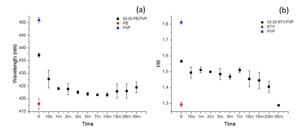
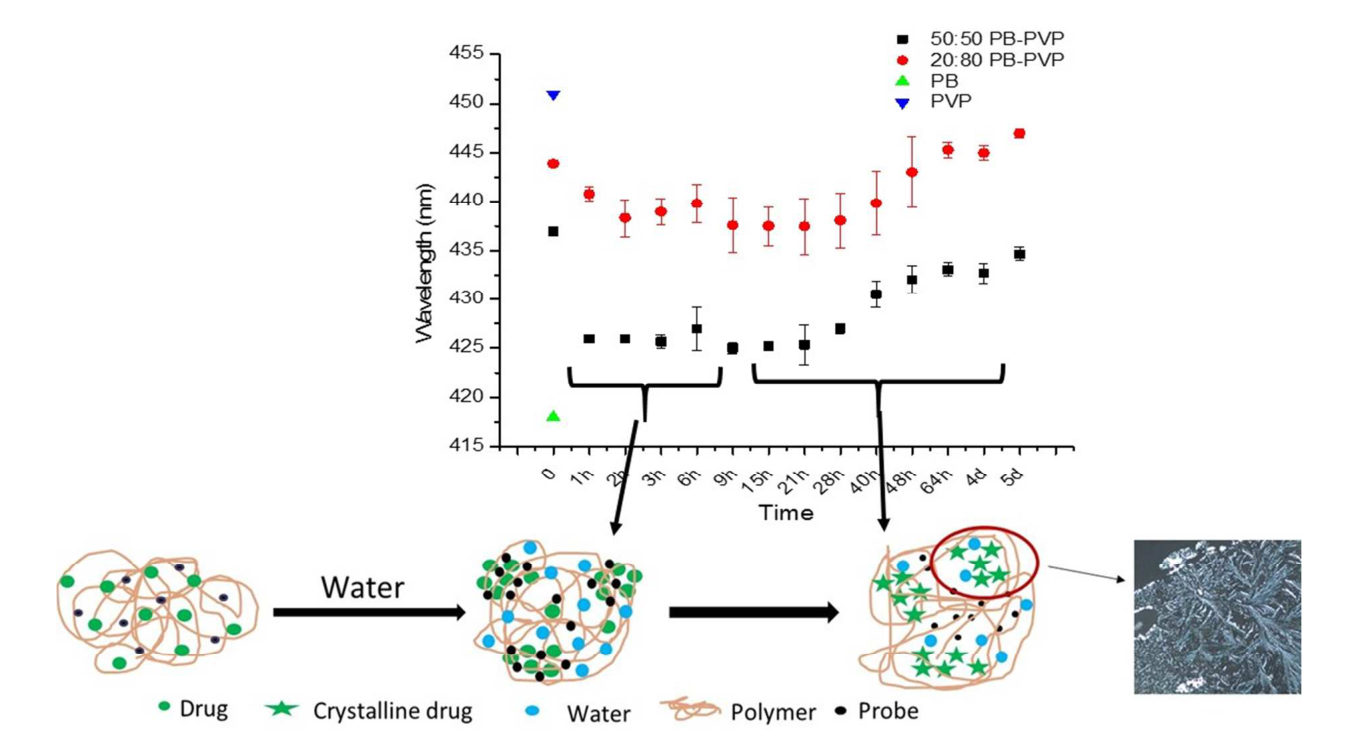
Fluorescent probes shift their emission when they are in less polar surroundings. In films and dispersions, and in supersaturated solutions, the probe signal moved toward the value it gives in the pure drug once a drug-rich region formed. For example, the ratio of the pyrene probe, which falls as the surroundings become less polar, went from 1.60 to 1.30 in ritonavir–PVP films at 97% RH purohit2015b R6, R15; purohit2017a R7; raina2015a R1, R6.
moderateModerate: the direction is the same in both states and was checked by other methods in the films, but the location of the probe was not measured purohit2015b K2, K3, X8; raina2015a K1, X17.
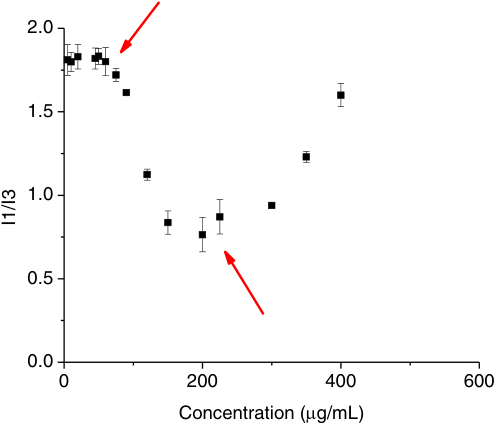
Nifedipine with HPMC reached about 0.76, past the benzene value; buffer and benzene are the solvent references of the same paper. raina2015a R1, R6, R9, R16
The powders read like the pure drug, not somewhere between drug and polymer. purohit2017a R7
For your drug
- Record the probe in films of the pure drug and the pure polymer prepared the way you prepare the dispersion; a miscible dispersion should lie between them and a separated one near the drug value.
- In solution, record the probe in a solvent series on your own instrument, read a result beyond the benzene value as "non-aqueous environment" and not as a polarity, and confirm the phase state by one probe-free method.
purohit2017a R24, R7; raina2015a R1, X6
How it works and key numbers
- A probe emits at a shorter wavelength (or, for pyrene, a lower ratio) in a less polar surrounding purohit2015b C4, C5, R4; raina2015a C25, C26.
- A lipophilic (poorly water-soluble) probe moves from the polar phase into a drug-rich region as soon as one forms purohit2015b C4, C5; raina2015a C25, C26.
- In the films the region is made when sorbed water replaces drug–polymer contacts in PVP, and pyrene "will preferentially partition" into it "given sufficient mobility" purohit2015b C24, C27; purohit2017a C7.
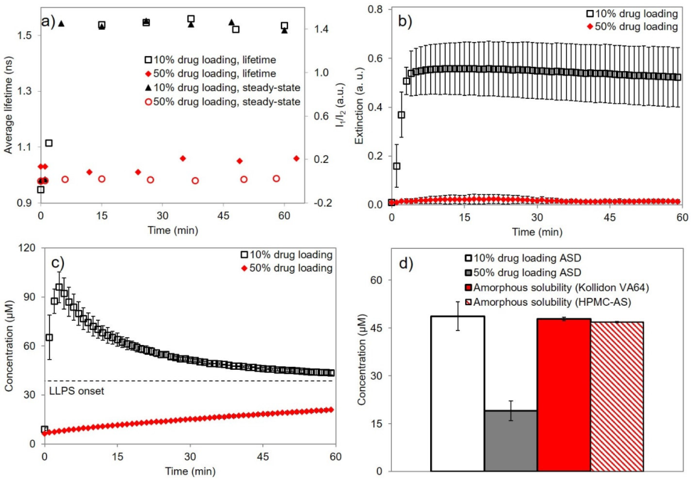
- In solution the region forms above the amorphous solubility (the drug concentration above which a drug-rich liquid phase forms), and more drug adds more phase without raising the free concentration, so more probe moves in raina2015a C26, C36; tres2017 C34.
- The location of the probe and its partition coefficient were not measured in either state purohit2015b C3; raina2015a X17. (synthesis: so a shift shows that a non-aqueous, drug-like region exists and, inside one drug–probe pair, at most a rank.)
Evidence: data, conditions and papers
Finding F1. The same lipophilic probes move toward the pure-drug value when a drug-rich region forms, in amorphous films and dispersions as in supersaturated solutions, but the value is a rank within one drug, probe and medium and not a polarity
In short. In spin-coated films of amorphous probucol and procaine pyrene read 1.2 against 1.4 and prodan 418 against 448 nm, and prodan across ten polymers ran from 460.0 nm in PAA to 427.0 nm in PVAc in the order of their solubility parameters purohit2015b R1, R3 (Solid-state methods F16). In separated dispersions the pyrene ratio sat at the value of the drug: ritonavir–PVP at 97% RH fell from 1.60 to 1.30, and itraconazole–HPMC powders read 1.38–1.39, equal to itraconazole alone (1.39) and not between itraconazole and HPMC (1.54) purohit2015b R6; purohit2017a R7 (Solid-state methods F17, Solid-state methods F19). In supersaturated solutions the same direction holds from buffer to benzene (pyrene 1.6 to 1.07), and in a felodipine or nifedipine drug-rich phase three probes went past their benzene values raina2015a R1, R6, R9, R16 (Solution methods F4). (synthesis: a scale built in one state cannot be carried to the other.)
Type. observation in two states, films and dispersions of 4 drugs and 10 polymers, and solutions of 10 drugs Strength. the direction is the same in both states and was checked against probe-free methods in the films (IR, DSC, imaging, XRD) and against extinction and a thermodynamic estimate in solution, but probe partitioning was inferred and not measured in either purohit2015b K2, K3, X8 (Solid-state methods F17); raina2015a K1, X17 (Solution methods F5).
How it was shown
Purohit and Taylor spin coated amorphous probucol and procaine with probes, ten polymers with prodan, and drug–polymer films with 0.1% probe stored at 97% RH purohit2015b P2, P4, P1, P5. Raina and co-workers diluted six probes in water, buffer and organic solvents, then read each within 1 min after stepwise drug additions raina2015a P8, P9.
What the data show
The pyrene ratio of ritonavir–PVP 50:50 at 97% RH fell from 1.60 to 1.30, whereas ritonavir–HPMCAS went from 1.42 to 1.47 purohit2015b R6, R15. Itraconazole–HPMC powders read 1.38–1.39, equal to the drug alone and not between drug and HPMC (1.54) purohit2017a R7. In solution, three probes in a felodipine or nifedipine drug-rich phase went past their benzene values raina2015a R1, R6, R9, R16.
| system | method and conditions | result | ids |
|---|---|---|---|
| amorphous probucol / procaine films | four probes in spin-coated films | pyrene I1/I3 1.2 / 1.4; prodan 418 / 448 nm | purohit2015b R1 |
| ten polymer films | prodan emission maximum | 460.0 nm (PAA) to 427.0 nm (PVAc), in the order of the solubility parameter | purohit2015b R3 |
| ritonavir–PVP 50:50, 97% RH | pyrene I/III on the film | 1.60 to 1.30, the value of amorphous ritonavir | purohit2015b R6, R15 |
| itraconazole–HPMC 40:60, three drying routes | pyrene I1/I3 | 1.38 / 1.39 / 1.39 against itraconazole 1.39 and HPMC 1.54 | purohit2017a R7 |
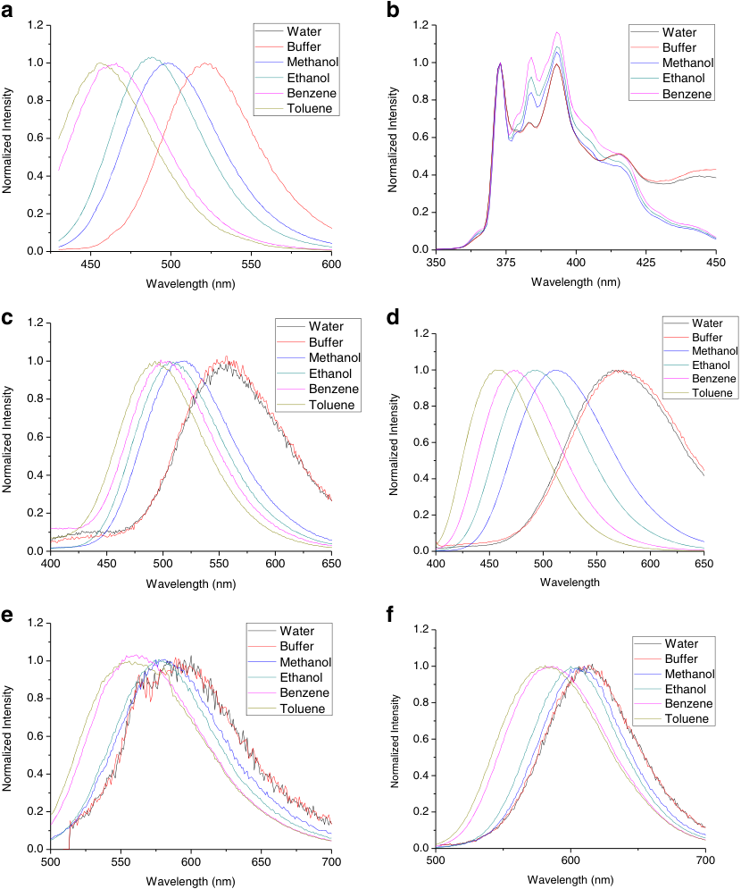
| six probes, buffer to benzene | emission maximum in solvents | pyrene 1.6 / 1.2 / 1.07 (buffer / methanol / benzene); PRODAN 520 / 498 / 463 nm | raina2015a R1 |
| felodipine, 37 °C; nifedipine with HPMC | DASPEI, 4-Di-2-ASP and pyrene, stepwise additions | pyrene about 1.8 to 0.76 (benzene 1.07) | raina2015a R6, R9, R16, R1 |
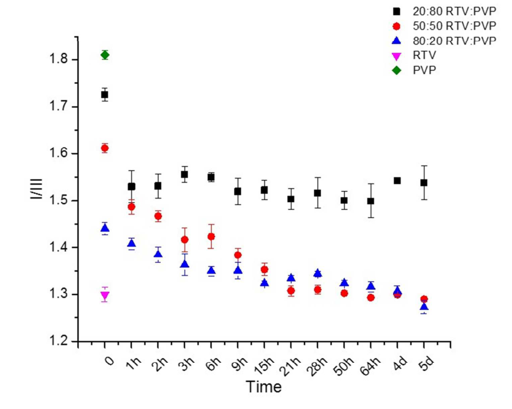
A lower pyrene ratio means less polar surroundings, and the 50:50 and 80:20 curves approach the value of pure ritonavir purohit2015b R4, R6.
Why
Both groups of authors read a shorter wavelength or a lower ratio as a lipophilic probe moving into a drug-rich, less polar region purohit2015b C4, C5; raina2015a C25, C26. Neither the probe's location nor a partition coefficient was measured purohit2015b C3; raina2015a X17. (synthesis: a probe shift is a presence test for a non-aqueous, drug-like region and, inside one drug–probe pair, at most a rank purohit2015b R3; raina2015a R6, R9, R16.)
Limits and transfer
The same HPMC 606 gave 442.5 nm in one paper and about 451 nm in another, so each drug–probe pair needs its own references purohit2015b R3; purohit2017a R13, X9. Mugheirbi and co-workers found itraconazole–HPMC films spun at 7% RH homogeneous by TEM mugheirbi2017 R10, C1. Record the probe in pure-drug and pure-polymer films prepared as the dispersion is, and confirm the phase state by one probe-free method.
How it was shown
Purohit and Taylor spin coated amorphous probucol and procaine with a trace of probe, ten polymers with prodan, and drug–polymer films with 0.1% probe (prodan for probucol–PVP, pyrene for the ritonavir films), and stored the dispersion films at 97% RH purohit2015b P2, P4, P1, P5 (Solid-state methods F16, Solid-state methods F17). Purohit and co-workers dried one 40:60 itraconazole–HPMC solution by four routes and read pyrene on films and powders purohit2017a P1, P2, P3, P4, P5 (Solid-state methods F19). In solution, Raina and co-workers diluted each of six probes to 1 µg/mL in water, buffer and organic solvents to record the emission maximum, then read each probe within 1 min after stepwise additions of drug from methanol raina2015a P8, P9 (Solution methods F4). Tres and co-workers used PRODAN at 0.9 µM to read lifetimes tres2017 M4, P5 (Solution methods F4).
What the data show
In films probucol was the less polar host with all four probes, and the prodan maxima of ten polymers fell in exactly the order of their solubility parameters purohit2015b R1, R3 (Solid-state methods F16). At 97% RH the pyrene ratio of ritonavir–PVP fell from 1.60 to 1.30 at 50:50 and from 1.44 to 1.29 at 80:20, but only from 1.72 to 1.48 at 20:80, whereas ritonavir–HPMCAS went from 1.42 to 1.47 purohit2015b R6, R15 (Solid-state methods F17). The itraconazole powders of three drying routes all read like the pure drug purohit2017a R7 (Solid-state methods F19). In solution the emission moved from buffer to benzene by 16 nm (DASPEI) to 103 nm (dapoxyl) raina2015a R1 (Solution methods F4), and the probe onsets for five of six dihydropyridines lay within a factor of 1.0–1.7 of the extinction onset and of the estimated amorphous solubility raina2015a R11, X9 (Solution methods F5).
| chapter finding | system | method and conditions | result | ids |
|---|---|---|---|---|
| Solid-state methods F16 | amorphous probucol / procaine films | four probes in spin-coated films | pyrene I1/I3 1.2 / 1.4; prodan 418 / 448 nm; 4-Di-2-ASP 548 / 570 nm; dansyl glycine 473 / 501 nm | purohit2015b R1 |
| Solid-state methods F16 | ten polymer films | prodan emission maximum | 460.0 nm (PAA) to 427.0 nm (PVAc), in the order of the solubility parameter | purohit2015b R3 |
| Solid-state methods F17 | ritonavir–PVP 50:50, 97% RH | pyrene I/III on the film | 1.60 to 1.30, the value of amorphous ritonavir | purohit2015b R6, R15 |
| Solid-state methods F17 | ritonavir–HPMCAS 50:50, 97% RH | pyrene I/III; IR | 1.42 to 1.47; IR spectrum unchanged | purohit2015b R13, R15, R18 |
| Solid-state methods F19 | itraconazole–HPMC 40:60, three drying routes | pyrene I1/I3 | 1.38 / 1.39 / 1.39 against itraconazole 1.39 and HPMC 1.54 | purohit2017a R7 |
| Solution methods F4 | six probes, buffer to benzene | emission maximum in solvents | pyrene 1.6 / 1.2 / 1.07 (buffer / methanol / benzene); PRODAN 520 / 498 / 463 nm | raina2015a R1 |
| Solution methods F4 | felodipine, 37 °C; nifedipine with HPMC, 37 and 25 °C | DASPEI, 4-Di-2-ASP and pyrene, stepwise additions | DASPEI about 578 to 558 nm (benzene 564); 4-Di-2-ASP about 610.5 to 579 nm (585); pyrene about 1.8 to 0.76 (1.07) | raina2015a R6, R9, R16, R1 |
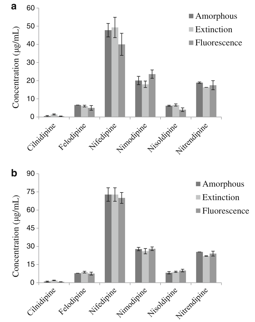
| Solution methods F5 | six dihydropyridines, 25 and 37 °C | DASPEI emission, extinction at 450 nm, thermodynamic estimate | highest over lowest of the three onsets 1.0–1.7 for five drugs, about 3 for cilnidipine | raina2015a R11, X9 |
Figure
Why
The authors of both groups of papers give the same mechanism as their interpretation: an environment-sensitive probe emits at shorter wavelength in a less polar surrounding, and a lipophilic probe moves from the polar phase into a drug-rich region purohit2015b C4, C5 (Solid-state methods F16); raina2015a C25, C26 (Solution methods F4). In the films the drug-rich region is made when sorbed water replaces drug–polymer contacts in PVP (a "classic case of solvent-induced immiscibility"), and pyrene "will preferentially partition" into it "given sufficient mobility" purohit2015b C24, C27; purohit2017a C7 (Solid-state methods F17, Solid-state methods F19). In solution the region forms above the amorphous solubility, and further drug adds phase without raising the free concentration, so more probe moves in raina2015a C26, C36; tres2017 C34 (Solution methods F5). Neither the location of the probe nor a partition coefficient was measured in either state purohit2015b C3 (Solid-state methods F16); raina2015a X17 (Solution methods F5). (synthesis: the same unmeasured step stands under both read-outs, so a probe shift is a presence test for a non-aqueous, drug-like region and, inside one drug–probe pair, at most a rank. The film values lie between those of pure-component films, while a solution drug-rich phase reads beyond the benzene value purohit2015b R3 (Solid-state methods F16); raina2015a R6, R9, R16 (Solution methods F4).)
Conditions and limits
In the films the extent of separation compares probe shifts only within a drug–probe pair, because prodan was used for probucol and pyrene for ritonavir, and only one HPMCAS dispersion was run at 97% RH purohit2015b P5, X7, X12 (Solid-state methods F17). The same probe gave 442.5 nm in one paper and about 451 nm in another for nominally the same HPMC 606, so each drug–probe pair needs its own references purohit2015b R3; purohit2017a R13, X9 (Solid-state methods F16). Water alone moves the probe the other way in single-component films, for example prodan in PVP from about 450.5 to 455.3 nm purohit2015b R9 (Solid-state methods F17). In solution the pyrene baseline differs between experiments (about 1.8 in the nifedipine series, 1.6 in the solvent table), so ratios are compared only within one experiment raina2015a X6, R1, R16 (Solution methods F4), and the drug's own fluorescence can mask a probe signal: with felodipine four of six probes gave an onset of 5.00–6.00 µg/mL, while the emission of dansyl glycine and dapoxyl was masked by the felodipine band raina2015a R12, R13 (Solution methods F6).
Counterexamples and conflicts
The itraconazole result is contested: Mugheirbi and co-workers found itraconazole–HPMC films spun at 7% RH homogeneous by TEM and films at 20–40% RH phase separated, so a pure-drug probe value shows that a drug-rich region exists in that sample, not that the pair is inherently immiscible mugheirbi2017 R10, C1 (Solid-state methods F19). The authors of the solution paper rank the styryl dyes as most polarity-sensitive, but these two dyes show the smallest solvent shifts in their own table raina2015a C29, R1, X4 (Solution methods F4). None found that contradicts the direction of the shift in either state.
Transfer check
(1) For a dispersion, record the probe in films of the pure drug and the pure polymer prepared as the dispersion is prepared; a miscible dispersion should lie between them and a separated one near the drug value. (2) For a solution, record the probe in a solvent series on your own instrument and take the aqueous value from a probe-only solution measured the same day; read a result beyond the benzene value as "non-aqueous environment" and not as a polarity. (3) Include pure-component films as the control for water alone in humidity tests. (4) Check that the drug does not emit in the probe window. (5) Confirm the phase state by one probe-free method before quoting it.
History
2014: pyrene I1/I3 had been used for danazol solutions jackson2014 R6, R8 (Solution methods F4). 2015: Purohit and Taylor calibrated prodan and pyrene in solid films and then applied them to dispersions, and Raina and co-workers published the solution-state calibration of six probes in the same year raina2015a C8 (Solid-state methods F16). 2017: Tres and co-workers extended PRODAN from spectra to lifetime tres2017 C33, C37 (Solution methods F4), and Purohit and co-workers used the pyrene ratio of drug and polymer references as the test for a miscible dispersion and carried it to powders purohit2017a R24, R7 (Solid-state methods F16, Solid-state methods F19). 2018: Tres and co-workers kept PRODAN for ASD dissolution tres2018 M5 (Solution methods F4).